Vol. 44, No. 2, 2006 pp. 111-116
Stromal myofibroblasts in breast cancer: relations
between their occurrence, tumor grade and expression of some tumour markers
Paweł Surowiak
1,2,3, Sławomir Suchocki
4, Balazs Györffy
2,5, Tserenchunt Gansukh
2,6, Andrzej Wojnar
3, Adam Maciejczyk
3, Marek Pudełko
3and Maciej Zabel
1,71
Department of Histology and Embryology, University School of Medicine, Wrocław, Poland;
2
Institute of Pathology, Charité Campus Mitte, Berlin, Germany;
3
Lower Silesian Centre of Oncology, Wrocław, Poland;
4
Department of Obsterics and Gynaecology, Voivodship Hospital, Wałbrzych, Poland;
5
Joint Laboratory of the Hungarian Academy of Sciences and the Semmelweis University, 2nd Department of Internal Medicine, Semmelweis University, Budapest, Hungary;
6
Health Sciences University of Mongolia, Ulaanbaatar, Mongolia;
7
Department of Histology and Embryology, University School of Medicine, Poznan, Poland
Abstract: It is suggested that tumour stromal myofibroblasts exert an unfavourable effect on the biology of breast cancer. We
are aware of only a single study which examined relationships between manifestation of myofibroblasts in the stroma of breast cancer and clinicopathological data of the patients. The present study was aimed at estimation of the effect exerted by myofibroblasts present in the tumour stroma on principal pathological parameters and on expression of Ki67 , P53 and HER-2 proteins in the group of the most frequent breast cancers, the ductal cancers. In paraffin sections of 60 ductal breast cancers (20 cases in G1, 20 in G2 and 20 in G3), immunohistochemical reactions were performed to detect expression of smooth muscle actin (SMA) in order to visualize myofibroblasts, Ki67, P53 and HER-2. The studies demonstrated that the most numerous myofibroblasts were present in G3 cases and they were the least frequent in G1 cases (P=0.02). Positive correlations were observed between the presence of myofibroblasts in tumour stroma and expression of Ki67 and HER-2 in breast cancer cells in the entire group (P <0.001 and P=0.001, respectively), in G2 cases (P=0.003 and P=0.03) and in G3 cases (P=0.01 and
P=0.03). Considering that the higher grade, Ki67 and HER-2 are thought to represent unfavourable prognostic factors, the elevatedcontent of myofibroblasts in tumour stroma is probably typical for cases with worse prognosis. (www.cm-uj.krakow.pl/FHC)
Key words: Myofibroblasts - Breast cancer - Grade - Ki67 - P53 - HER-2Introduction
Breast cancer is the most common malignant tumour of females in the western world, being responsible for about 32% of the estimated new female cancer cases.
The incidence of breast cancer remains high, and the clinical courses are highly variable. For years, a growing incidence of the disease has been documented and the risk of dying of breast cancer continues to increase in consecutive cohorts of generations [5]. Therefore, in numerous centres intense efforts develop to detect new
prognostic factors which would permit to intensify the therapy in high risk cases. Moreover, such prognostic factors might provide an interesting topic for studies on new therapeutic approaches. Conventional prognostic and predictive markers for breast cancer are stage of the advancement (nodal status, tumour size and presence of the metastases), tumour grade and tumour size [4]. Ex- pression of receptors for female sex hormones [10] and HER-2 is also of importance [4]. In addition, expression of the proliferation-associated antigen, Ki67 and of the suppressor protein, P53 are frequently estimated in order to establish the dynamics of tumour growth [12]. Signific- ance of several other proteins and of their expression in breast cancer cells continues to stimulate studies world- wide. Until now, however, only expression of receptors for
Correspondence: P. Surowiak, Dept. Histology and Embryology, University School of Medicine, Chałubinskiego 6a,
50-356 Wrocław, Poland ; e-mail: pawel.surowiak@interia.pl
female sex hormones and of HER-2 have found appli- cation in a routine diagnosis of breast cancer [4].
In recent years, numerous publications have de- scribed effects of tumour-associated tissues on the dynamics of neoplastic disease. Invasion of tumour cells is slowed down by myoepithelial cells and basal membranes [9]. The immune system has been found to protect the body from neoplastic cells. On the other hand, tumour cells produce, i.a. metalloproteinases and growth factors which may facilitate invasion of tumour cells. Tumour-associated vessels provide path- way for spread of tumour cells [6]. The interactions between tumour cells and stromal myofibroblasts re- main, however, poorly recognized. Myofibroblasts pro- duce several factors which may stimulate proliferation of cancer cells and facilitate their infiltration. They have been found to secrete, i.a., insulin-like growth factor-2 (IGF-2) and hepatocyte growth factor (HGF) [2]. Recog- nition of relationships between myofibroblasts and breast cancer cells may facilitate definition of new prognostic factors and provide targets for novel therapeutic ap- proaches.
The present study was aimed at examination of rela- tionships between the presence of myofibroblasts in tumour stroma and principal pathological parameters as well as expression of Ki67, P53 and HER-2 proteins in a group of most frequent breast cancers, the ductal cancers.
Materials and methods
Patients. Immunocytochemical analysis was performed retrospec- tively on tissue samples collected for routine diagnostic purposes.
Based on histology (invasive ductal breast cancer) and grade (equal groups for each grade), 60 patients with primary invasive breast cancer diagnosed in the years 1999 to 2000 in the Lower Silesian Centre of Oncology (Wroclaw, Poland) were included in the study.
The mean age of the patients was 55.62 years ± 9.86 SD (age range:
43 to 72 years). Every grade (G1, G2 and G3) was represented by 20 cases. In 34 patients stage 2a, in 22 patients stage 2b and in 4 patients stage 3 of the tumour was diagnosed. Fragments sampled from the studied tumours were fixed in 10% buffered formalin and embedded in paraffin. In every case, hematoxylin and eosin stained sections were examined by two pathologists. The stage of tumours was assessed according to TNM classification system [8]. Tumour grade was estimated according to Bloom-Richardson, according to the modification of Elston and Ellis [3] (Table 1).
Immunohistochemistry. Formalin-fixed, paraffin-embedded sec- tions (4 µm) were mounted on Superfrost slides (Menzel Gläser, Germany), dewaxed with xylene, and gradually rehydrated. Activity of endogenous peroxidase was blocked by 30 min incubation in 1%
H2O2. The studied sections were boiled 15 minutes in Target Retrie- val Solution (DakoCytomation, Poland) , in a microwave oven at 250 W. Then, immunohistochemical reactions were performed using monoclonal antibodies detecting smooth muscle actin (SMA, clone 1A4, DakoCytomation, Poland) diluted 1:200, Ki67 (clone MIB-1, DakoCytomation, Poland) diluted 1:100, P53 (clone DO-7, Dako- Cytomation, Poland) diluted 1:100 and polyclonal anti-HER-2 anti- bodies (DakoCytomation, Poland) diluted 1:350. The antibodies were diluted in the Antibody Diluent, Background Reducing (Dako-
Cytomation, Denmark). The sections were incubated with antibodies for 1 h at room temperature. Subsequent incubations included bioti- nylated secondary antibodies (15 min, room temperature) and strep- tavidin-biotinylated peroxidase complex (15 min, room temperature) (LSAB+, HRP, DakoCytomation, Denmark). NovaRed (Vector La- boratories, UK) was used as chromogen (10 min, at room tempera- ture). All sections were counterstained with Meyer’s hematoxylin.
In every case, control reactions were included, in which specific antibody was substituted by the Primary Mouse Negative Control (DakoCytomation, Denmark).
Scoring of immunostaining results. Myofibroblasts visualized by SMA expression were assessed semiquantitatively with the use of a simplified scale based on the percentage of myofibroblasts in the population of tumour stromal cells (0 - no myofibroblasts, 1 - <10%
of myofibroblasts in tumour stroma, 2 - 10-30% of myofibroblasts in tumour stroma, 3 - >30% of myofibroblasts in tumour stroma).
Expression of P53 and Ki67 was quantitated by calculating the percentage values of immunopositive cells. For evaluation of HER-2 reactivity, the DakoCytomation scoring system was used (0 = nega- tive; + = partial membranous; ++ = complete membranous, weak;
+++ = complete membranous, strong). The intensity of immunohis- tochemical reactions was assessed independently by two patholog- ists; in doubtful cases a re-evaluation was performed using a double-headed microscope. In each case we analyzed 6 microscopic fields at × 200 magnification.
Statistical analysis. Statistical analysis of the obtained results was conducted using Statistica 98 PL software (Statsoft, Poland). At the first stage of the calculations, the presence of myofibroblasts was compared in individual groups (G1, G2, G3) using Kruskal-Wallis rank ANOVA test. Subsequently, relationships were examined (em- ploying the same test) between the presence of myofibroblasts and expression of the studied variables in all groups together (G1 to G3) and in each group (G1, G2 and G3).
Results
Immunostaining
Detection of SMA yielded reaction of a cytoplasmic localization and of variable intensity in individual cases.
SMA was present in myofibroblasts (Fig. 1), myoepithe-
Table 1. Characteristics of patients and tumours
Characteristics No. (%)
Patients 60 (100)
Age (mean 55.62 years ± 9.86 SD)
≤50 5 (8.3)
50-60 32 (53.3)
>60 23 (38.3)
Tumour grade
1 20 (33.3)
2 20 (33.3)
3 20 (33.3)
Tumour stage
2a 34 (56.7)
2b 22 (36.7)
3 4 (6.6)
Histology Invasive ductal
breast cancer 60 (100)
lial cells and in muscles of blood vessels. According to the applied scale, the mean manifestation of myofibro- blasts amounted to 2.01±0.69.
Application of antibodies against P53 or Ki67 yielded in tumour cells colour reaction of nuclear local- ization and of a variable intensity in individual cases.
The mean percentage of P53-positive and Ki67-positive breast cancer cells was 40.71±26.69 and 28.56±17.7, respectively.
In the case of HER-2, membrane reaction was ob- tained of various intensity in individual cases, localized in the tumour cells. The mean reaction intensity in DakoCytomation 0-3 score was 1.84±0.74.
Relationships between the presence
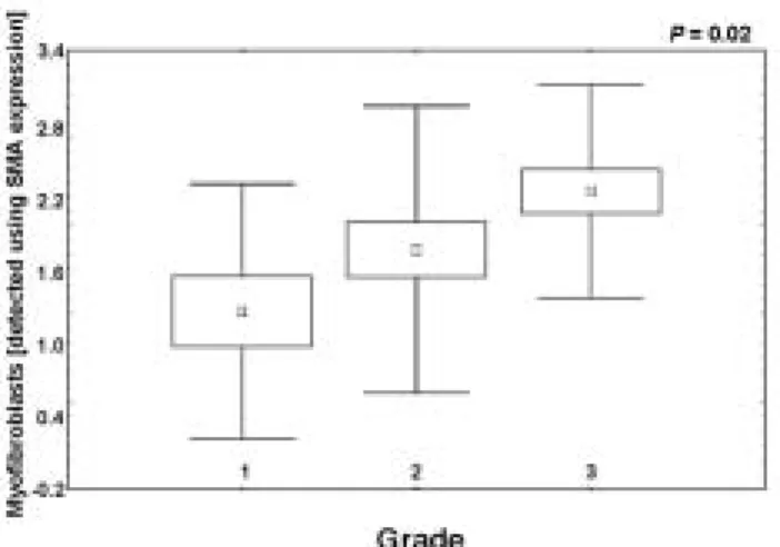
of myofibroblasts and histopathological parameters The analysis performed using the Kruskal-Wallis rank ANOVA test demonstrated no relationships between the presence of myofibroblasts in tumour stroma and stage or age of the studied patients (P=0.67 and P=0.54, respectively). Nevertheless, a significant positive corre- lation was disclosed between the presence of myofibro- blasts in tumour stroma and grade of the studied tumors (P=0.02, Fig. 2). Cases of higher grade manifested augmented content of myofibroblasts in tumour stroma.
Relationships between the presence of myofibroblasts and expression of P53, Ki67 and HER-2
Using also the Kruskal-Wallis rank ANOVA test, rela- tionships were examined between the presence of myofibroblasts in the tumours and expression of P53, Ki67 and HER-2 in individual grades (G1, G2 and G3) or in the entire group (G1-3). The test demonstrated no significant relationships in the G1 group (P>0.05, Table 2).
In none of the examined groups could a relationship be disclosed between the presence of myofibroblasts in the
Fig. 1. Immunohistochemical localization of smooth muscle actin in breast cancer tumors. A. Reaction in individual myofibroblasts (<10%, score: 1) in tumour stroma (gray arrow) and in blood vessel (black arrow). × 200. B. Reaction in numerous myofibroblasts (>30%, score: 3) in tumour stroma (gray arrow). × 200. C. Reaction in numerous myofibroblasts (>30%, score: 3) in tumour stroma (gray arrow). × 400.
Fig. 2. The presence of myofibroblasts in tumour stroma related to tumour grade (Kruskal-Wallis rank ANOVA test). Myofibroblasts are the most numerous in stroma of G3 tumours.
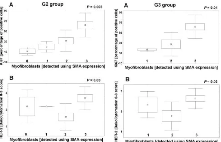
tumour stroma and expression of P53 in cancer cells (P>0.05, Table 2). However, in groups G2 (Fig. 3), G3 (Fig. 4) and in the entire studied material (G1-3, Fig. 5) a significant positive correlation was noted between the presence of myofibroblasts in tumour stroma and ex- pressions of Ki67 and HER-2 in cancer cells (P<0.05, Table 2). In cases with the highest content of myofibro- blasts in tumour stroma, the most pronounced express- ion of Ki67 and HER-2 was also observed in cancer cells.
Discussion
The present study demonstrates the significance of myofibroblast presence in tumour stroma, as detected by smooth muscle actin (SMA) expression [1]. We have documented that myofibroblasts are present in tumour stroma in a proportion of breast cancers. This result has confirmed results of other authors. Yazhou et al. [11]
investigated the relation between the presence of myofi- broblasts in tumour stroma and clinico-pathological variables in the breast cancer patients. In the present study we have shown that cases of higher grade demon- strate higher content of myofibroblasts in tumour stro-
ma. Similarly to Yazhou et al. [11], we have noted no relationship between manifestation of myofibroblasts and age of the studied patients. Also, in line with the data of the cited authors, the content of myofibroblasts in tumour stroma has not been related to stage of the studied tumours.
We have also examined potential relationships be- tween manifestation of myofibroblasts in tumour stroma and expression of Ki67 , P53 and HER-2 in breast cancer cells. Until now, only the relationship between myofi- broblast content and expression of Ki67 in breast cancer cells has been investigated [11] and no significant rela- tionship has been detected. In the present study, how- ever, cases with higher content of myofibroblasts in tumour stroma have been found to contain higher pro- portions of cancer cells with Ki67 expression. Since myofibroblasts, both in tumours and in healing pro- cesses, produce numerous growth factors [2], we sug- gest that myofibroblast-released growth factors might stimulate proliferation of breast cancer cells.
In this study, no relationship has been documented between the presence of myofibroblasts in tumour stro- ma and expression of P53 in breast cancer cells. How- ever, cases with higher content of myofibroblasts have
Fig. 4. In G3group, cases with the highest content of myofibroblasts exhibit: A. Higher content of cancer cells with Ki67 expression; B.
Higher expression of HER-2 in cancer cells (Kruskal-Wallis rank ANOVA test).
Fig. 3. In G2 group, cases with the highest content of myofibroblasts exhibit: A. Higher content of cancer cells with Ki67 expression; B.
Higher expression of HER-2 w in cancer cells (Kruskal-Wallis rank ANOVA test).
been found to exhibit more intense expression of HER-2 in breast cancer cells. Myofibroblasts may secrete in- sulin-like growth factor-2 (IGF-2) and hepatocyte growth factor (HGF) [7]. They represent ligands for receptors of the Epidermal Growth Factor Receptor group, which includes, i.a., HER-2. The ligands, most probably in a paracrine way, stimulate expression of HER-2 in breast cancer cells. Since HER-2 represents one of the best recognised negative prognostic factors [4] we suggest that the presence of myofibroblasts in tumour stroma may be linked to unfavourable prognosis in breast cancer patients.
In summary, the present investigations have demon- strated that breast cancer cases with higher content of myofibroblasts in tumour stroma are characterised by higher grade of the tumour, more intense proliferation of neoplastic cells and more intense expression of HER- 2 in cancer cells. To our knowledge, this paper describes for the first time a positive correlation between the presence of myofibroblasts in tumour stroma and intens- ity of Ki67 and HER-2 expression. Taking into account that higher grade, Ki67 and HER-2 are regarded to represent negative prognostic indicators, the intensified content of myofibroblasts in tumour stroma may be
typical for cases with a worse prognosis. The signific- ance of myofibroblast presence in the stroma of breast cancers should be investigated in detail, in order to detect new prognostic indicators and novel targets for thera- peutic approaches.
References
[ 1] Chaponnier C, Gabbiani G (2004) Pathological situations char- acterized by altered actin isoform expression. J Pathol 204:
386-395
[ 2] De Wever O, Mareei M (2003) Role of tissue stroma in cancer cell invasion. J Pathol 200: 429-447
[ 3] Elston CW, Ellis IO (1991) Pathological prognostic factors in breast cancer. The value of histological grade in breast cancer:
experience from a large study with long-term follow-up. Histo- pathology 5: 403-410
[ 4] Fitzgibbons PL, Page DL, Weaver D, Thor AD, Allred DC, Clark GM, Ruby SG, O‘Malley F, Simpson JF, Connoly JL, Hayes DF, Edge SB, Lichter A, Schnitt SJ (2000) Prognostic factors in breast cancer. College of American Pathologists Con- sensus Statement 1999. Arch Pathol Lab Med 124: 966-978 [ 5] Jemal A, Thomas A, Murray T, Thun M (2002) Cancer statistics.
CA Cancer J Clin 52: 23-47
[ 6] Mori I, Yang Q, Kakudo K (2002) Predictive and prognostic markers for invasive breast cancer. Pathol Inst 52: 186-194 [ 7] Offersen BV, Nielsen BS, Hoyer-Hansen G, Rank F, Hamilton-
Dutoit S, Overgaard J, Andreasen PA (2003) The myofibroblast is the predominant plasminogen activator inhibitor-1-express- ing cell type in human breast carcinomas. Am J Pathol 163:
1887-1899
[ 8] Sobin LH, Wittekind C (2002) TNM classification of malignant tumours. 5th edition. Wiley-Liss INC., New York
[ 9] Surowiak P, Dziegiel P, Matkowski R, Kornafel J, Wojnar A, Zabel M (2002) Immunocytochemical evaluation of metallo- thionein (MT) expression in myoepithelial cells of ductal mam- mary carcinoma and its relation to survival time: analysis of Table 2. Correlations between the presence of myofibroblasts in tumour stroma (SMA) and expression intensity of the studied anti- gens in breast cancer cells in the entire group (G1-3) and in the G1, G2 and G3 groups (Kruskal-Wallis rank ANOVA test).
Studied pair
of variables Grade P value
SMA and P53
G1-3 0.5399
G1 0.2625
G2 0.2521
G3 0.3957
SMA and Ki67
G1-3 <0.001
G1 0.1111
G2 0.0031
G3 0.0114
SMA and HER-2
G1-3 0.0011
G1 0.2083
G2 0.0299
G3 0.0349
Fig. 5. In the entire material (G1-3), cases with higher content of myofibroblasts exhibit: A. Higher proportion of cancer cells with expression of Ki67; B. Higher expression of HER-2 in cancer cells (Kruskal-Wallis rank ANOVA test).
7-year course of the disease. Folia Histochem Cytobiol 40:
199-200
[10] Surowiak P, Dziegiel P, Zabel M, Matkowski R, Kornafel J (2001) Prognostic value of immunocytochemical estimation of estrogen receptor (ER) and of pS2 estrogen-dependent protein in cells of mammary ductal carcinoma. Analysis of five-year course of the disease. Folia Histochem Cytobiol 39: 143-144 [11] Yazhou C, Wenlv S, Weidong Z, Licun W (2004) Clinicopa-
thological significance of stromal myofibroblasts in invasive ductal carcinoma of the breast. Tumor Biol 25: 290-296
[12] Zasławski R, Surowiak P, Paluchowski P, Dziegiel P, Ma- ciejczyk A, Pudełko M, Wojnar A, Zabel M (2005) Differences in oestrogen and progesterone receptors, HER-2, p53 express- ion and proliferation in ductal breast cancers in relation to histopathological grade. Folia Morphol 64: 9-15
Received: October 14, 2005 Accepted after revision: December 14, 2005