hjic.mk.uni-pannon.hu DOI: 10.33927/hjic-2019-22
ASSESSMENT OF THE ECOTOXICITY OF NANOPLASTICS
NÓRAKOVÁTS*1, BETTINAECK-VARANKA1, ZSÓFIABÉKÉSSY1, DORINADIÓSI1, KATALIN
HUBAI1,ANDJÁNOSKORPONAI2
1Institute of Environmental Sciences, University of Pannonia, 8200 Veszprém, Egyetem u. 10, HUNGARY
2Department of Environmental Science, Sapientia Hungarian University of Transylvania, 400193 Cluj-Napoca, Calea Turzii 4, ROMANIA
The presence of micro- and nanoplastics in aquatic environments (including freshwater and marine ecosystems as well as their sediments) is becoming an increasingly serious problem worldwide. A wide range of studies have addressed the ecological effects these particles pose on biota. The main exposure pathway are food chains, e.g. under laboratory conditions these particles accumulate in the brain tissues of fish that feed on zooplankton causing brain damage. These studies, however, report mainly on the physical effects. In order to establish actual ecotoxicological effects, nanoplastics (50 nm in diameter) were assessed using theVibrio fischeribioluminescence inhibition bioassay (VFBIA). Our results showed that even environmentally relevant concentrations might trigger ecotoxicological effects. This study can be con- sidered to be a first screening, however, results indicate the need for more complex testing on a battery of aquatic test organisms.
Keywords: nanoplastic; ecotoxicity;Vibrio fischeri; kinetic assay
1. Introduction
Aquatic environments contaminated by plastic litter are an emerging problem. Remote, pristine mountainous ar- eas are even contaminated by atmospheric microplastic deposition [1]. Polymer particles < 5 mm in diameter are defined as microplastics (MP) and may be derived directly from the use of industrial pellets or indirectly from the degradation and fragmentation of plastic parti- cles [2]. Polystyrene was proven to degrade into micro- and nanoplastics under laboratory conditions [3]. High levels of contamination have been reported in both ma- rine and freshwater habitats [4,5]. Micro- and nanoplas- tics (NP) can float freely in bodies of water or be de- posited as sediments.
The highest risk associated with these particles is their ingestion, which occurs at different levels in the aquatic food chain. Jabeenet al.[6], for example, listed approximately 150 different fish species where ingestion and accumulation have been reported. Particles can also progress upwards in the trophic levels of the food chain, i.e. fish can be exposed to the ingestion of zooplankton which is not able to discriminate between different food sources and consumes micro- and nanoplastics [7]. An experimental study showed that in fish exposed to NPs via the food chain, these particles caused brain damage and behavioural disorders as a result of accumulation in
*Correspondence:kovats@almos.uni-pannon.hu
brain tissues [8]. Biomagnification may also affect food safety and human health, though certain knowledge gaps exist in this field [9].
Ingestion may actually lead to starvation and even- tually the impairment of their physical condition. Un- der laboratory conditions, Daphnia magna exposed to polystyrene nanoparticles (PS-NP) exhibited reduced body size and severe alterations in terms of reproduc- tion [10].D. magnais a widely studied species due to its key role in the aquatic food chain. It was shown to ingest nano- and microplastics (20nm to70µm in diam- eter) from water [11]. In a laboratory study by Mattsson et al.[8], particles52nm in diameter elucidated the most severe effects. Cuiet al.[12] exposedD. galeatato PS- NPs (5mg/l,52nm in diameter) and detected a significant mortality rate after2days of exposure until the end of the study which lasted5days. Although a standard ecotoxi- cological test was conducted in this case, the mechanisms of mortality are still unclear: physical contact might have led to a reduction in the survival rate.
In general, most ecotoxicological studies have used relatively high concentrations. Manfraet al.[13] investi- gated the impact of green fluorescently labeled carboxy- lated polystyrene nanoparticles of40nm in diameter with various surface charges on the marine rotiferBrachionus plicatilis. It was found that anionic PS-NPs did not elu- cidate mortality within the range of concentrations tested (0−50µg/ml), while cationic PS-NPs caused mortality at
concentrations≥2.5µg/ml. Changes in oxidative stress enzymes were detected within the concentration range of 10−20µg/ml in different organisms, e.g. the rotiferBra- chionus koreanus and the marine copepodParacyclop- ina nana[14]. The same concentration,10 µg/ml, was reported to cause40% growth inhibition in the green mi- croalgaDunaliella tertiolecta[15].
In order to distinguish real (eco)toxicological effects from physical damage, a test based on the biolumines- cence inhibition of the marine bacteriumVibrio fischeri was selected. The species has been reclassified as Ali- ivibrio fischeri[16], however, as most standards and even recent papers from the literature still use the nameV. fis- cheri, it will be used hereinafter.
Bioluminescence is regulated by the enzyme system NAD(P)H:FMN oxidoreductase-luciferase. In toxic en- vironments, enzyme inhibition is reflected by a rapid de- crease in the luminous emittance of the bacterium. The reduction in light intensity is easy to measure as it is pro- portional to the strength of the toxicant, therefore, pro- vides a quantifiable endpoint. This test has been used in various environmental matrices [17–19].
Lappalainenet al.[20,21] developed a special version of the test which was later standardised (ISO 21338:2010:
Water quality - Kinetic determination of the inhibitory ef- fects of sediment, other solids and coloured samples on the light emission ofVibrio fischeri(kinetic luminescent bacteria test)) in which bacteria are kept in suspension in direct contact with potentially toxic solid particles. Lumi- nescence readings were taken when the test commenced and the light intensity continuously monitored over the first 30secs after the sample had been mixed with the bacteria. The light output pattern, therefore, might al- ready provide some indication of the expected toxicity of the sample [22]. The light intensity was measured once more after the pre-set exposure time (5,15or 30 mins as per standard). Toxicity values are normally expressed as EC50 and EC20, i.e. concentrations causing lumines- cence inhibitions of50 and20% in this assay, respec- tively.
2. Materials and Methods
In our experiments, the Ascent luminometer (Flash sys- tem, marketed by Aboatox, Finland) was used. A suspen- sion of the test bacteria (NRRL B-11177) was prepared in accordance with manufacturer instructions (Hach Lange GmbH).
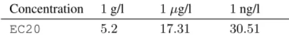
Polystyrene particles with a nominal diameter of50 nm were used as a sample (supplier Thermo Fisher Sci- entific). As no comparative data were available on the potentially toxic concentration, a range-finding concen- tration series was set [23]. Three initial sample concen- trations were selected (1 g/l,1 µg/l and 1 ng/l), which were further diluted, the number of dilutions was11(the number of concentrations the96-multiwell plate permits) and the dilution ratio1 : 2.
Table 1:The measuredEC20 values of the polystyrene nanoparticles.
Concentration 1g/l 1µg/l 1ng/l
EC20 5.2 17.31 30.51
TheVibrio fischeristrain NRRL B-11177 was recon- stituted by adding the contents of one vial of+4◦C 1243- 551 Reagent Diluent. The reconstituted reagent was equi- librated at+4◦C for30min. Then the reagent was sta- bilised at+15◦C for30mins before being pipetted into the wells.
Luminescence readings were taken when the test commenced,Time0, and after the pre-set exposure time of 30 mins, Time30. The luminescence inhibition of each sample was calculated as follows:
CF = IC30/IC0
INH \% = 100 $-$ 100 x (IT30 / CF x IT0)
where
CF = correction factor
IC30 = luminescence intensity of the control sample after the contact time (30 mins) in the RLU
IC0 = initial luminescence intensity of the control sample in the RLU
IT30 = luminescence intensity of the test sample after the contact time (30 mins) in the RLU
IT0 = initial luminescence intensity of the test sample in the RLU
EC20values were calculated using the Ascent software, also developed by Aboatox Oy.
3. Results and Discussion
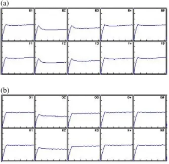
Table 1shows the ecotoxicity expressed inEC20, i.e. the calculated concentration of the sample that caused20% bioluminescence inhibition.Fig. 1illustrates the biolumi- nescence inhibition during the first30secs for the sam- ples of1g/l and1ng/l in concentration.
EC20 (or in some cases, EC10) are considered thresholds for the estimation of the lowest observed effec- tive concentration [24], i.e. the sample is normally con- sidered (eco)toxic if the elucidated effect exceeds20%.
These results show that the V. fischeri bioassay de- tected a measurable degree of toxicity even at a concen- tration of1 ng/l. Boothet al. [25] used the non-kinetic version of this bioassay (Microtox®), however, in their study, the calculated toxic concentration exceeded the range of concentrations studied (0.001−1000mg/l). The same negative effect was reported by Casadoet al.[26].
The higher degree of (detectable) toxicity in our study might be explained by the differences in the test system used. While Microtox® is a non-kinetic test, the Flash system (Ascent luminometer) was especially developed to test the toxicity of different suspensions or samples containing solid particles. The Ascent luminometer uses a 96-multiwell microplate. A specific feature of it is that
(a)
(b)
Figure 1:Kinetic diagram of the1g/l (a) and1ng/l (b) samples. The light output is recorded over the first30sec- onds. After the peak, toxicity causes a rapid reduction in the light output, on the other hand, it remains constant dur- ing the control. The two columns show the two replicates.
E1-F1/G1-H1 (left): control. E2-F2/G2-H2 (right): sam- ple, maximum concentration.
during luminescence readings, the microplate is continu- ously shaken by the instrument, resulting in the resuspen- sion of particles.
According to our results, environmentally relevant concentrations might already pose ecotoxic effects. Ac- tual environmental concentrations are relatively difficult to compare and assess, mostly due to difficulties in sam- pling and the lack of standardized sampling methodolo- gies [27,28]. Indicative data are available: e.g. microplas- tic concentrations of0.4−34ng/l in bodies of freshwa- ter in the USA [29] or0.51mg/l in marine environments [10].
However, in real-world environments, even higher levels of toxicity can be expected as particles might absorb organic pollutants from the surrounding water [30], including highly toxic pesticides or polychlorinated biphenyls (PCBs) [31]. Though their bioavailability is still questionable [32], Batelet al.[33] conducted a lab- oratory study on microplastics and one polycyclic aro- matic hydrocarbon (PAH), benzo[a]pyrene (BaP). It was demonstrated that BaP adsorbed on microplastics and was transferred via an artificial food chain. These par- ticles might also possess inherent toxicity due to the use of additives during manufacturing processes [34].
4. Conclusions
It is a well-known paradigm in ecotoxicology that the sensitivity of different test organisms to a particular chemical varies, therefore, theV. fischeritest can be re- garded as a first screening. The bioluminescence inhibi-
tion assay is an acute test that uses a maximum expo- sure of only30minutes. Naturally, chronic effects can- not be extrapolated from these results. However, the fact that the tested nanoplastics have already elucidated eco- toxicological effects in environmentally relevant concen- trations emphasises the need for more complex ecotoxi- cological testing involving a properly selected battery of test organisms. In addition to widely used aquatic test or- ganisms such as the aforementionedDaphnia magna, an ideal candidate could be theCaenorhabditis eleganstest.
It is a standardised bioassay using a sediment-dwelling, widely distributed nematode. However, in order to distin- guish physical damage from toxic effects, the measure- ment of changes in oxidative stress enzymes can be use- ful no matter which test organism is applied.
Acknowledgement
This work was funded by the BIONANO_GINOP-2.3.2- 15-2016-00017 project.
REFERENCES
[1] Allen, S.; Allen, D.; Phoenix, V.R.; Le Roux, G.;
Jiménez, P.D.; Simonneau, A.; Binet, S.; Galop, D.:
Atmospheric transport and deposition of microplas- tics in a remote mountain catchment,Nature Geo- science2019,12(5), 339–344DOI: 10.1038/s41561-019- 0335-5
[2] Mintenig, S.M.; Dris, R.; Imhof, H.; Sanchez, W.;
Gasperi, J.; Galgani, F.; Tassin, B.; Laforsch, C.:
Beyond the ocean: Contamination of freshwater ecosystems with (micro-)plastic particles, Environ.
Chem.2015,12(5) 539–550DOI: 10.1071/en14172
[3] Lambert, S.; Wagner, M.: Characterisation of nanoplastics during the degradation of polystyrene, Chemosphere 2016, 145 265–268
DOI: 10.1016/j.chemosphere.2015.11.078
[4] Cole, M.; Lindeque, P.; Halsband, C.; Galloway, T.S.: Microplastics as contaminants in the marine environment: a review, Mar. Pollut. Bull.2011,62 2588–2597DOI: 10.1016/j.marpolbul.2011.09.025
[5] Dris, R.; Gasperi, J.; Rocher, V.; Saad, M.; Renault, N.; Tassin, B.: Microplastic contamination in an ur- ban area: a case study in Greater Paris, Environ.
Chem.201512(5), 592–599DOI: 10.1071/en14167
[6] Jabeen, K.; Su, L.; Li, J.; Yang, D.; Tong, C.; Mu, J.;
Shi, H.: Microplastics and mesoplastics in fish from coastal and fresh waters of China, Environ. Pollut.
2017,221, 141–149DOI: 10.1016/j.envpol.2016.11.055
[7] Chae, Y.; An, Y.-J.: Effects of micro- and nanoplas- tics on aquatic ecosystems: Current research trends and perspectives, Mar. Pollut. Bull. 2017, 124(2), 624–632DOI: 10.1016/j.marpolbul.2017.01.070
[8] Mattsson, K.; Johnson, E.V.; Malmendal, A.; Linse, S.; Hansson, L.-A.; Cedervall, T.: Brain damage and behavioural disorders in fish induced by plastic nanoparticles delivered through the food chain,Sci.
Rep.2017,7(1), 11452DOI: 10.1038/s41598-017-10813-0
[9] Barboza, L.G.A.; Vethaak, A.D.; Lavorante, B.R.B.O.; Lundebye, A.-K.; Guilhermino, L.:
Marine microplastic debris: An emerging is- sue for food security, food safety and human health,Mar. Pollut. Bull.2018,133, 336–348DOI:
10.1016/j.marpolbul.2018.05.047
[10] Besseling, E.; Wang, B.; Lürling, M.; Koel- mans, A.A.: Nanoplastic Affects Growth of S.
obliquus and Reproduction of D. magna, Envi- ron. Sci. Technol. 2014,48(20), 12336-12343DOI:
10.1021/es503001d
[11] Rosenkranz, P.; Chaudhry, Q.; Stone, V; Fernandes, T.F.: A comparison of nanoparticle and fine particle uptake byDaphnia magna,Environ. Toxicol. Chem.
2009,28(10), 2142–2149DOI: 10.1897/08-559.1
[12] Cui, R.; Kim, S.W.; An, Y.-J.: Polystyrene nanoplastics inhibit reproduction and induce abnor- mal embryonic development in the freshwater crus- taceanDaphnia galeata,Sci. Rep.2017,7(1), 12095
DOI: 10.1038/s41598-017-12299-2
[13] Manfra, L.; Rotini, A.; Bergami, E.; Grassi, G.;
Faleri, C.; Corsi, I.: Comparative ecotoxicity of polystyrene nanoparticles in natural seawater and reconstituted seawater using the rotiferBrachionus plicatilis,Ecotoxicol. Environ. Saf.2017,145, 557–
563DOI: 10.1016/j.ecoenv.2017.07.068
[14] Proki´c, M.D.; Radovanovi´c, T.B.; Gavri´c, J.P.; Fag- gio, C.: Ecotoxicological effects of microplastics:
Examination of biomarkers, current state and future perspectives,Trends. Analyt. Chem.2019,111, 37–
46DOI: 10.1016/j.trac.2018.12.001
[15] Gambardella, C.; Morgana, S.; Bramini, M.; Ro- tini, A.; Manfra, L.; Migliore, L.; Piazza, V.; Gar- aventa, F.; Faimali, M.: Ecotoxicological effects of polystyrene microbeads in a battery of ma- rine organisms belonging to different trophic lev- els, Mar. Environ. Res. 2018, 141, 313–321 DOI:
10.1016/j.marenvres.2018.09.023
[16] Urbanczyk, H.; Ast, J.; Higgins, M.J.; Carson, J.;
Dunlap, P.V.: Reclassification of Vibrio fischeri, Vibrio logei,Vibrio salmonicida andVibrio woda- nis as Aliivibrio fischeri gen. nov., comb. nov., Aliivibrio logeicomb. nov.,Aliivibrio salmonicida comb. nov. andAliivibrio wodaniscomb. nov.,Int.
J. Syst. Evol. Microbiol.2007,57(12), 2823–2829
DOI: 10.1099/ijs.0.65081-0
[17] Girotti, S.; Ferri, E.N.; Fumo, M.G.; Maiolini, E.:
Monitoring of environmental pollutants by biolumi- nescent bacteria,Anal. Chim. Acta2008,608(1), 2–
29DOI: 10.1016/j.aca.2007.12.008
[18] Ma, X.Y.; Wang, X.C.; Ngo, H.H.; Guo, W.; Wu, M.N.; Wang, N.: Bioassay based luminescent bac- teria: Interferences, improvements, and applica- tions,Sci. Total Environ. 2014,468–469, 1–11DOI:
10.1016/j.scitotenv.2013.08.028
[19] Abbas, M.; Adil, M.; Ehtisham-ul-Haque, S.; Mu- nir, B.; Yameen, B.; Ghaffar, A. et al.: Vibrio fis- cheri bioluminescence inhibition assay for ecotoxi-
city assessment: A review,Sci. Total Environ.2018, 626, 1295–1309DOI: 10.1016/j.scitotenv.2018.01.066
[20] Lappalainen, J.; Juvonen, R.; Vaajasaari, K.; Karp, M.: A new flash method for measuring the toxicity of solid and colored samples,Chemosphere1999, 38(5), 1069–1083DOI: 10.1016/s0045-6535(98)00352-x
[21] Lappalainen, J.; Juvonen, R.; Nurmi, J.; Karp, M.:
Automated color correction method forVibrio fis- cheritoxicity test. Comparison of standard and ki- netic assays,Chemosphere2001,45(4–5), 635–641
DOI: 10.1016/s0045-6535(00)00579-8
[22] Mortimer, M.; Kasemets, K.; Heinlaan, M.; Kurvet, I.; Kahru, A.: High throughput kinetic biolumines- cence inhibition assay for study of toxic effects of nanoparticles,Toxicol. in Vitro2008,22(5), 1412–
1417DOI: 10.1016/j.tiv.2008.02.011
[23] USEPA (2000) Method Guidance and Recommen- dations for Whole Effluent Toxicity (WET) Testing (40 CFR Part 136). EPA 821-B-00-004. U.S. Envi- ronmental Protection Agency, Office of Water [24] Ventura, S.P.M.; Silva, F.A.; Gonçalves, A.M.M.;
Pereira, J.P.; Gonçalves, J.P.; Coutinho, J.A.P.: Eco- toxicity analysis of cholinium-based ionic liquids to Vibrio fischerimarine bacteria,Ecotoxicol. Environ.
Saf.2014,102, 48–54DOI: 10.1016/j.ecoenv.2014.01.003
[25] Booth, A.M.; Hansen, B.H.; Frenzel, M.;
Johnsen, H.; Altin, D.: Uptake and Toxicity of Methylmethacrylate-Based Nanoplastic Particles in Aquatic Organisms,Environ. Toxicol. Chem.2016, 35(7), 1641–1649DOI: 10.1002/etc.3076
[26] Casado, M.P.; Macken, A.; Byrne, H.J.: Ecotox- icological assessment of silica and polystyrene nanoparticles assessed by a multitrophic test battery, Environ. Int. 2013, 51, 97–105 DOI:
10.1016/j.envint.2012.11.001
[27] Rocha-Santos, T.; Duarte, A.C.: A critical overview of the analytical approaches to the occurrence, the fate and the behavior of microplastics in the en- vironment,Trends Analyt. Chem.2015,65, 47–53
DOI: 10.1016/j.trac.2014.10.011
[28] Twiss, M.R.: Standardized methods are required to assess and manage microplastic contamination of the Great Lakes system,J. Great Lakes Res. 2016, 42(5), 921–925DOI: 10.1016/j.jglr.2016.07.032
[29] Eriksen, M.; Mason, S.; Wilson, S.; Box, C.;
Zellers, A.; Edwards, W.et al.: Microplastic pollu- tion in the surface waters of the Laurentian Great Lakes, Mar. Pollut. Bull. 2013, 77(1–2), 177–182
DOI: 10.1016/j.marpolbul.2013.10.007
[30] Mato, Y.; Isobe, T.; Takada, H.; Kanehiro, H.;
Ohtake, C.; Kaminuma, T.: Plastic resin pellets as a transport medium for toxic chemicals in the marine environment, Environ. Sci. Technol. 2001, 35(2), 318–324DOI: 10.1021/es0010498
[31] Cedervall, T.; Hansson, L.A.; Mattsson, K.: Nano- plastics in the aquatic environment, Environ. Sci.
Process. Impacts 2015, 17(10), 1712–1721 DOI:
10.1039/c5em00227c
[32] Beckingham, B.; Ghosh, U.: Differential bioavail- ability of polychlorinated biphenyls associated with environmental particles: Microplastic in compari- son to wood, coal and biochar,Environ. Poll.2017, 220, 150–158DOI: 10.1016/j.envpol.2016.09.033
[33] Batel, A.; Linti, F.; Scherer, M.; Erdinger, L.;
Braunbeck, T.: The transfer of benzo[a]pyrene from microplastics toArtemianauplii and further to ze-
brafish via a trophic food web experiment–CYP1A induction and visual tracking of persistent organic pollutants, Environ. Toxicol. Chem. 2016, 35(7), 1656–1666DOI: 10.1002/etc.3361
[34] Avio, C.G.; Gorbi, S.; Regoli, F.: Plastics and mi- croplastics in the oceans: From emerging pollutants to emerged threat,Mar. Environ. Res.2017,128, 2–
11DOI: 10.1016/j.marenvres.2016.05.012