N A R C O T I C S A N D A L C O H O L J. H. QUASTEL
Research Institute, Montreal General Hospital, Montreal, Canada The physiological and psychological consequences of administration of narcotics, including alcohol, to the intact animal resemble in many respects those associated with the changes of cerebral metabolism during anoxia. I would like to recall a few observations bearing on the results of anoxic conditions. McFarland (1952) has pointed out that one of the most sensitive and direct ways of demonstrating that psychological phenomena are directly dependent on the underlying physiological processes is to deprive the organism of oxygen. His experiments on dark adaptation have revealed that one's ability to see dim objects against a completely dark background is markedly impaired by oxygen lack. Under conditions equivalent to an altitude of only 5,000 ft the change in visual sensitivity is rather significant. "In so far as visual changes may reflect other alterations in the central nervous system, any significant degree of anoxia might be expected to impair maximum performance: . . . The changes in behavior occurring in normal subjects under conditions of anoxia can be attributed to a diminished tension of oxygen being available to the nervous system, the changes being of cellular origin, not circulatory. The final result is an impair
ment of sensory and mental function and integration, the cortical cells apparently suffering more than other parts of the central nervous sys
tem." (McFarland, 1952). There are many references to be found in the studies of anoxia to the similarities of behavior of a person suffering from oxygen want to one under the influence of alcohol. Under both conditions a person becomes irrational and uninhibited and loses capac
ity for self-criticism, memory, and motor control. In fact Peters and Van Slyke (1931) hazarded the opinion that in alcoholism "the tissue cells are poisoned in such a manner that they cannot use the oxygen properly."
The oxygen consumption of brain tissue is extremely high. Thanks to the work of Kety (1950, 1948) and his colleagues we know that this oxygen consumption may amount to almost 25% of the total oxygen taken up by the body under basal metabolic conditions. Per gram of tissue
209
210 J . Η. QUASTEL
this consumption is about fifty times that of peripheral nerve. Grey mat
ter has four times the oxygen consumption, per gram of tissue, of white matter. If this oxygen consumption takes place only in the neurones, these cells must be among the most active in the body so far as oxygen consumption is concerned. It is reasonable to conclude that any impair
ment of this utilization of oxygen by the nerve cell, and thereby its high metabolic activity, will result in an impairment of its functional activity.
The considerable metabolic activity of the brain is also shown by the fact that in the waking state the temperature of the brain is 0.5° C higher than that of the arterial blood. The metabolic activity varies with the extent of functional activity of the brain; brain temperature rises sharply when an animal is excited and falls when the animal rests; the tempera
ture rise is apparently due to an increase in the metabolic activity of the cells of brain cortex (Richter, 1952).
Equally important with oxygen, so far as the activity of the nerve cell is concerned, is the presence of glucose which is the major substance burned by the brain. Many other substances are, of course, consumed by the brain, including L-glutamic acid, amines, and even fatty acids at a low rate. But glucose and its breakdown products, such as pyruvic acid, represent the major fuel of the nervous system. Diminution of the availability of glucose to the brain results in effects resembling those produced by oxygen deprivation. The decrease in cerebral oxygen con
sumption associated with diabetic coma amounts, according to Kety (1952), to about 40% reduction from the normal value. This reduction of oxygen consumption by the brain is not due to a decrease in cerebral blood flow. The patients suffer from an intrinsic defect in the oxygen uptake by the brain cells. Kety points out that the brain must consume its own carbohydrate store to account for the amount of oxygen still being taken up in diabetic coma.
These facts are intended to convey what must be apparent to most of us, the conclusion that oxygen availability and carbohydrate meta
bolism in the brain represent factors of the greatest importance in our understanding of the mechanism of action of neurotropic drugs.
A word or two may be said at this juncture about the effects of exposure to high tensions of oxygen. It is now well known that exposure of this description may result in severe mental disturbances. In studies that I made a few years ago (Mann and Quastel, 1946), just before the war, on the effects of high oxygen tensions on metabolism in the central nervous system it became clear that the presence of 100% oxygen has a toxic effect on brain metabolism, a reduction of the rate of oxygen
consumption by the isolated brain tissue taking place. Results of investi
gations of this phenomenon indicated that one enzyme was particularly affected by the presence of a high oxygen tension, viz. pyruvic oxidase, an enzyme that holds a pivotal position in cell metabolism. This enzyme is a thiol enzyme, the thiol groups being sensitive to the presence of a high tension of oxygen. This work was confirmed and expanded by Dickens (1946). Thus it would appear that the physiological and psychological effects of high oxygen tension may well be due to a poison
ing of the nerve cell, whereby its metabolism is depressed in a manner similar to that occurring in conditions of oxygen want or of glucose deprivation, or, as will be shown, in the presence of narcotics or ethanol.
The narcotics may be divided roughly into two groups of substances.
The first group includes barbiturates and such substances as chloral, chloretone (chlorobutanol), ether, and ethanol. The second group includes the quaternary nitrogen bases such as morphine and heroin.
I propose to deal with the first group and to acquaint you with new results on their effects on brain respiration in vitro.
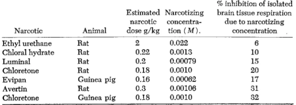
The barbiturates and chloretone group is characterized by the fact that with the small concentrations of narcotics that produce narcosis in the animal, inhibitions of oxidations brought about the isolated brain tissue take place (Table 1). Drugs such as luminal (phenylethylbarbit- urate), evipan, and chloretone, at concentrations that may bring about deep narcosis, also bring about specific inhibitions of the oxidations by brain tissue of substances important in carbohydrate metabolism, namely, glucose, pyruvic acid, and lactic acid. For example, evipan at a concentration of 6 X 10_ 4M inhibits respiration of guinea pig brain cortex slices in a glucose phosphate medium by over 10%. Its
TABLE 1. Narcotizing Concentration and Effects on the Respiration of Brain Cortex Slices in a Glucose Medium (Jowett, 1 9 3 8 )
% inhibition of isolated Estimated Narcotizing brain tissue respiration
narcotic concentra due to narcotizing Narcotic Animal dose g/kg tion ( M ) . concentration
Ethyl urethane Rat 2 0 . 0 2 2 6
Chloral hydrate Rat 0 . 2 2 0 . 0 0 1 3 1 0
Luminal Rat 0 . 2 0 . 0 0 0 7 9 1 5
Chloretone Rat 0 . 1 8 0 . 0 0 1 0 2 0
Evipan Guinea pig 0 . 1 6 0 . 0 0 0 6 2 1 7
Avertin Rat 0 . 3 0 . 0 0 1 0 6 3 1
Chloretone Guinea pig 0 . 1 8 0 . 0 0 1 0 3 2
212 J . Η. QUASTEL
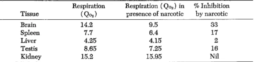
TABLE 2. Effect of 0.033% Evipan * on Respiration of Guinea Pig Tissues in Presence of Glucose (Jowett and Quastel, 1937)
Respiration Respiration (Qo2) in % Inhibition Tissue (Qo2) presence of narcotic by narcotic
Brain 14.2 9.5 33
Spleen 7.7 6.4 17
Liver 4.25 4.15 2
Testis 8.65 7.25 16
Kidney 15.2 15.95 Nil
* Sodium N-methyl cyclo hexenylmethylbarbiturate.
effect on brain exceeds that on other organs (Table 2 ) . The inhibi
tion rises rapidly with the concentration of narcotic, the curve being sigmoid in character. The inhibition with most narcotics is practically completely reversble, indicating that, at the concentrations used, the narcotics do not damage the cells. The oxidation, however, of certain substances such as sodium succinate or p-phenylenediamine, which are oxidized freely by the brain, is not affected by the narcotics. Neither are the oxidations of L-glutamate and α-glycerophosphate by brain af
fected by narcotics to the same extent as those of glucose and pyruvate.
These results indicate that narcotics do not interfere with the access of oxygen to the nerve cell but that they interfere specifically at low con
centrations with the oxidative breakdown of glucose or of pyruvic acid.
These facts acquire a special significance when considered in relation to the dominant position held by carbohydrate breakdown in the meta
bolism of brain. They have led to the view that narcosis may result from a suppression of certain respiratory events at the nervous centers where the narcotics are absorbed.
It is important to emphasize that the narcotics at the low concentra
tions that bring about their pharmacological activities do not affect enzymic processes in any general sense but that their biochemical effects are restricted to certain metabolic events. At high concentrations, nar
cotics exert nonspecific, over-all inhibitions on very many enzyme sys
tems, but with these we are not at present concerned.
Recent evidence obtained by Dr. Johnson and myself (1953) makes it clear that the narcotics (barbiturates and chloretone) at the low con
centrations that produce narcosis inhibit, in isolated nerve tissue, biolog
ical acetylations including the formation in brain of acetylcholine (Table 3 ) . We have been able to demonstrate that the narcotics only interfere with the process of acetylation so long as this is dependent upon oxidative reactions (Tables 4, 5 ) . The inhibitory effect of the
TABLE 3. Effects of Narcotics on Respiration and Acetylation of Choline by Rat Brain Mince at 37° C
Per cent Per cent
Pyruvate Oxygen inhibition of Choline inhibition of concen ATP uptake respiration acetylated acetylation
tration Narcotic cone. μΐ by narcotic μ%/g tissue by narcotic
0 — — 420 - 10 —
0.01 Μ — — 866 - 17 —
0.01 Μ Narconumal — 498 43 11 35
0.01 Μ - 0.002 Μ 745 - 20 —
0.01 Μ Narconumal 0.002 Μ 539 28 18 10
0.03 Μ — — 928 - 12 —
0.03 Μ Chloretone — 170 81 5 59
0.03 Μ - 0.002 Μ 1028 - 18 —
0.03 Μ Chloretone 0.002 Μ 307 70 10 44
Narconumal concentration, 0.004 Μ; Chloretone concentration, 0.005 Μ. Time, 2 hr. Gas phase, air.
TABLE 4. Effects of Chloretone (0.004 M) on Sulfanilamide Acetylation by Tissue Extracts (Rat Brain an d Pigeon Liver)
Aerobic conditions: 200 /ig sulfanilamide initially present
Sulfanilamide % inhibition Oxygen uptake acetylated of acetylation
Sodium pyruvate (0.02 Μ) 326 146 —
« + Chloretone 95 22 85
Sodium fumarate (0.02 M) 536 101 —
tc " + Chloretone 103 18 82
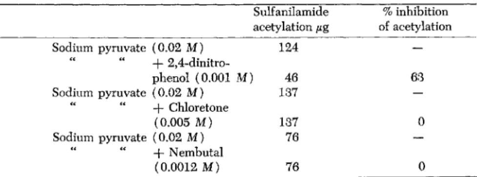
TABLE 5. Effects of 2,4-Dinitrophenol and Narcotics on Sulfanilamide Acetylation by Pigeon Liver Extracts
Anaerobic conditions: 200 μ% sulfanilamide initially present Sulfanilamide % inhibition acetylation μ% of acetylation
Sodium pyruvate (0.02 M) 124 —
" " -f 2,4-dinitro-
phenol (0.001 M) 46 63
Sodium pyruvate (0.02 M) 137 —
-f Chloretone
(0.005 M) 137 0
Sodium pyruvate (0.02 M) 76 —
+ Nembutal
(0.0012 M) 76 0
narcotic can be neutralized in vitro by the addition to the system of adenosinetriphosphate (ATP). The narcotic does not interfere with the
214 J. Μ. QÜASTEL
acetylation process per se nor with the utilization of ATP. It interferes seriously, however, with the oxidative synthesis of ATP, and, therefore, with those many reactions, of which acetylation is simply one, which are dependent upon the presence of ATP. These effects of narcotics occur at low concentrations where very small inhibitory effects on oxygen uptake may be observed. Eiler and McEwen (1949) have already shown that pentobarbital inhibits the generation of high energy phos
phate (e.g. ATP) to the extent that it interferes with oxygen utilization.
Brody and Bain (1951) have, indeed, postulated that uncoupling of phosphorylation from oxidation may be connected with the narcotic action of barbiturates. Dr. Johnson and I (Johnson and Quastel, 1953) have shown, however, that the behavior of narcotics in vitro differs from that of a typical uncoupling agent such as 2,4-dinitrophenol, and the mechanisms of action of the two types of drugs are dissimilar. One notable example of difference is that, though 2,4-dinitrophenol prevents phosphorylation occurring as a result of anaerobic pyruvic acid dismuta- tion, narcotics such as barbiturates have no such effect. Yet both sub
stances are equally effective in inhibiting phosphorylations due to oxida
tion by the cell of pyruvate or glucose. The evidence would indicate that the main effects of narcotics such as the barbiturates or chloretone at the nerve cell is to impede oxidative processes (especially that of pyruvate) leading to ATP synthesis. This conclusion, however, is con
tested by those (e.g. Richter, 1952) who hold that the narcotics affect primarily, in an unknown manner, synaptic transmissions, with a conse
quent drop in cerebral activity, and produce secondarily falls in oxygen consumption. This view is largely based on the observation that the low concentrations of narcotic that are pharmacologically active may exercise only small (or no) inhibition of the oxygen uptake of brain cortex in vitro. For example, the results of the experiments of Larrabee and his colleagues (1950), and of Bronk and Brink (1951) and Brink (1951) show that the small oxygen uptake shown by isolated nerve is not suppressed when transmission of an impulse is blocked by the presence of a narcotic such as chloretone. If the nerve is stimulated, however, there is an increased oxygen consumption and in the presence of a narcotic this oxygen consumption is greatly depressed. Thus Bronk and Brink (1951) and Brink (1951) have shown that the rate of oxygen uptake by resting frog nerve is reduced 15% by 2 mM chloretone, but that the increment in the rate of oxygen uptake by nerves carrying impulses at the rate of 50 impulses per second is decreased 50% by the same quantity of narcotic.
Why, however, is there no drop in oxygen consumption in the rest
ing nerve in the presence of concentrations of the narcotic that serve to block transmission of impulses? The answer, as I see it, is that the total oxygen consumption of the nerve cell, like that of any cell, is due to the operation of many oxidative processes, each of which contributes its moiety to the whole. But out of these many processes, only a few are specifically affected by the narcotic at the low concentrations that are pharmacologically active. In the resting nerve cell these may con
tribute but little to the total oxygen uptake. When stimulation of the nerve cell takes place, however, the activities of these processes become greatly increased, and, in consequence, the inhibitory effect of the nar
cotic becomes more clearly seen. Larrabee (1952), who, on the basis of his early results, concluded that low concentrations of narcotics do not affect nerve metabolism, has himself shown recently that even with the resting nerve cell the presence of a narcotic, at a concentration which impedes transmission but which seems not to affect respiration, increases the rate of glucose breakdown. This would be expected if the narcotic acts as inhibitor of an oxidative process in the nerve cell, for it is well known that such inhibitors bring about a disturbance of the Pasteur effect, i.e., the equilibrium between oxidative events and glucose breakdown, the latter increasing in rapidity when the former are sup
pressed. Such changes in the Pasteur effect may take place with little or no noticeable drop in total oxygen consumption.
I would like to mention, now, recent results obtained by Dr. Ghosh and myself that emphasize the important effects of narcotics and ethanol on the respiration and metabolism of brain.
Following the demonstration of Bronk and Brink and of Larrabee of the high sensitivity of stimulated nerve respiration to the action of narcotics, Mcllwain (1953) has shown that narcotics such as the bar
biturates and chloral inhibit the respiration of electrically stimulated slices of cerebral cortex at concentrations that have but little perceptible effect on the respiration of unstimulated tissue. He has also shown that stimulation of the respiration of cerebral cortex by potassium ions and by 2,4-dinitrophenol renders the respiration more sensitive to the action of barbiturates and chloral.
Dr. Ghosh and I (1954) have found that when brain cortex respiration is stimulated by the presence of high concentrations of potassium ions
(0.1 Μ KCl), the presence of Luminal brings about an enhanced inhibi
tion of the rate of respiration (Table 6 ) . The percentage inhibition by 0.0017 Μ luminal is increased over 90% under these circumstances. A
216 J. Η. QUASTEL
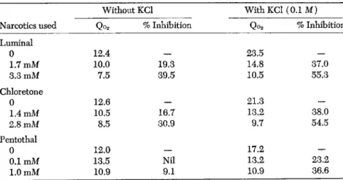
TABLE 6. Effects of Potassium Chloride on the Sensitivity of Rat Brain Cortex Respiration to Luminal and Chloretone
Without KCl With KCl (0.1 Μ)
Narcotics used Qo2 % Inhibition Qo2 % Inhibition Luminal
0 12.4 — 23.5 —
1.7 mM 10.0 19.3 14.8 37.0
3.3 mM 7.5 39.5 10.5 55.3
Chloretone
0 12.6 — 21.3 —
1.4 mM 10.5 16.7 13.2 38.0
2.8 mM 8.5 30.9 9.7 54.5
Pentothal
0 12.0 — 17.2 —
0.1 mM 13.5 Nil 13.2 23.2
1.0 mM 10.9 9.1 10.9 36.6
Vessel contents: slices of rat brain cortex (approx. 90 mg fresh tissue) in 3 ml Ringer- phosphate-glucose (0.1 M) medium, containing additions shown in table. The respiratory activities cited are those obtained 30 minutes after addition of the narcotic.
similar phenomenon is observed when chloretone is used as the narcotic.
These results confirm those of Mcllwain and make it clear that brain cortex respiration is made much more sensitive to the inhibitive action of narcotics by the presence of a stimulant such as 0.1 Μ KCl.
Another conclusion, however, not yet commented upon, emerges from our results. It would appear that the apparent increased sensitivity of brain respiration to narcotics is due largely to the fact that the narcotic inhibits, or eliminates, the stimulating effect of the added potas
sium ions (Table 7 ) . If the effect of added potassium ions is simply to
TABLE 7. Effects of Luminal on Sensitivity of Rat Brain Cortex Respiration to Potassium Chloride
Expt.
Additions
1 2 3
Without added KCl
1 2 3
With added KC10.1M
Average % increase of Qo2 due to KCl
Glucose 14.5 12.6 11.5 24.5 20.0 21.0 70
Glucose -f-
Luminal 3.3 mM 9.5 7.4 6.9 10.5 8.5 7.8 12
render the brain more sensitive to the action of the narcotic, it would be expected that respiratory rates lower than those found in the absence pf added potassium would be found. We have not yet found this to be
Qo2 Percent No Chloretone In presence of diminution Substrates present 3.3 mM Chloretone in Qo2
Glucose
Without KCl 12.5 8.3 35
With KCl 20.5 9.2 56
Fructose
Without KCl 12.6 8.5 35
With KCl 20.0 9.5 57
Sodium pyruvate
Without KCl 13.2 10.0 24
With KCl 18.8 11.2 42
Sodium lactate
Without KCl 11.8 9.5 22
With KCl 16.8 10.2 40
Sodium L-glutamate
Without KCl 7.5 5.0 30 30
With KCl 7.0 5.0 28
Sodium succinate
Without KCl 10.8 10.5 3
With KCl 9.0 8.5 5
the case. All the results indicate that the inhibitive action of the nar
cotics, at the low concentrations that are pharmacologically active, is restricted to that aspect of brain cortex respiration that is stimulated by potassium ions. Another way of expressing the same result is to state that the stimulating action of potassium ions only operates on that aspect of brain respiration that is inhibited by narcotics at low concentrations.
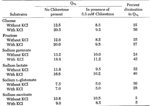
The stimulating effect of the presence of 0.1 Μ KCl on brain cortex respiration is seen not only with glucose, as combustible substrate, but also with fructose, pyruvate, and lactate. With all these substances the increased effect of the narcotic in inhibiting respiration is observable.
Moreover, in presence of the narcotic but little stimulatory action of added potassium on brain respiration is observable.
When sodium L-glutamate or sodium succinate is present as com
bustible substrate, there is no stimulating effect due to added potassium ions and no increased sensitivity to the presence of a narcotic (Table 8 ) . A possible explanation of these results is that the presence of high concentrations of potassium ions catalyzes the processes of formation of
TABLE 8. The Effect of Chloretone on the KCl-Stimulated Respiration of Rat Brain Cortex Slices in Presence of Different Substrates (0.01 Μ)
218 J . Η. QUASTEL
energy rich phosphates, such as ATP (Boyer et al., 1943; Stanbury and Mudge, 1953), which in turn enhance the rates of oxidation of glucose or pyruvate (Banga et al., 1939; Ochoa, 1941; Mcllwain, 1952; Wenner et al., 1953). Granting this assumption and the correctness of the con
clusion (Johnson and Quastel, 1953) that narcotics at low concentrations block certain oxidative events leading to synthesis in the brain of ATP, and pyruvate oxidation (Michaelis and Quastel, 1941; Greig, 1946;
Persky et al., 1950), it is apparent that, in the presence of narcotics, the stimulating effect of potassium ions will be reduced or eliminated.
It follows, too, if these views are correct, that a portion of the respiration of brain cortex in vitro is neither narcotic-sensitive (for low concentra
tions of narcotics) nor able to respond to the presence of high concen
trations of potassium ions.
In the light of these results, it would appear that narcotics exercise relatively large inhibitory effects on the total respiration of the nerve cell only when this is in a stimulated phase. Clearly such stimulation is always operating in the conscious animal. With the resting nerve cell, carrying no impulses, the respiration is partly due to processes that are neither narcotic-sensitive nor capable of stimulation of potassium ions.
It is for this reason so little effect on the respiration of the nerve in vitro is seen by a narcotic at pharmacologically active concentrations. It is for further research to discover the exact nature of the chemical processes that make up the narcotic-insensitive and potassium-unresponsive frac
tion of nerve respiration.
It is already known that the potassium stimulated respiration of brain cortex is highly malonate-sensitive (Kimura and Niwa, 1953), a fact that is consistent with the conclusion that the stimulated phase involves the oxidation of carbohydrate (or pyruvate) through the operation of the citric acid cycle.
In view of the decided inhibition exerted by narcotics on potassium stimulated brain cortex in vitro, our general conclusion that narcotics, at pharmacologically active concentrations, act on the nerve cell by the suppression of oxidative events, particularly those involved in glucose or pyruvate oxidation, is upheld. There is clearly no reason to suppose that such an oxidative suppression is secondary to some other unknown phenomenon.
As an example of a barbiturate that has been claimed (Bain and Brody, 1954) to exert its effects by uncoupling of phosphorylation and not by oxidative inhibition, pentothal (thiopental sodium) may be cited.
Yet this narcotic, which has so little effect at low concentrations on the
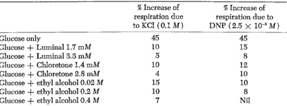
TABLE 9. The Effects of Potassium Ions and of 2,4-Dinitrophenol on Respiration of Rat Brain Cortex in Presence of Narcotics
% Increase of % Increase of respiration due respiration due to to KCl (0.1 Μ) DNP (2.5 X 10"5M)
Glucose only 45 45
Glucose + Luminal 1.7 mM 10 15
Glucose -f- Luminal 3.3 mM 5 8
Glucose + Chloretone 1.4 mM 10 12
Glucose + Chloretone 2.8 mM 4 10
Glucose + ethyl alcohol 0.02 Μ 15 10
Glucose + ethyl alcohol 0.2 Μ 10 8
Glucose + ethyl alcohol 0.4 Μ 7 Nil
The values given in this table refer to respiratory activities (Qo2) calculated during the 30-60 minute interval of the experiment.
respiration of brain cortex in vitro and may even show a small stimula
tion, is a potent inhibitor, at the same concentrations, when the brain cortex is stimulated by potassium ions. Conceivably the stimulation shown by pentothal of brain cortex respiration in absence of 0.1M KCl is due to oxidation of, or a catalysis by, the thiol group of this narcotic
(Table 6 ) .
Another narcotic showing a phenomenon similar to that obtaining with pentothal is ethyl alcohol. It is known that brain tissue is capable of oxidizing ethanol when this is present in small concentrations, and possibly this fact has made it difficult to study narcotic effects of ethanol in vitro. Depression of brain metabolism in vivo by ethanol has been often observed (Himwich, 1951).
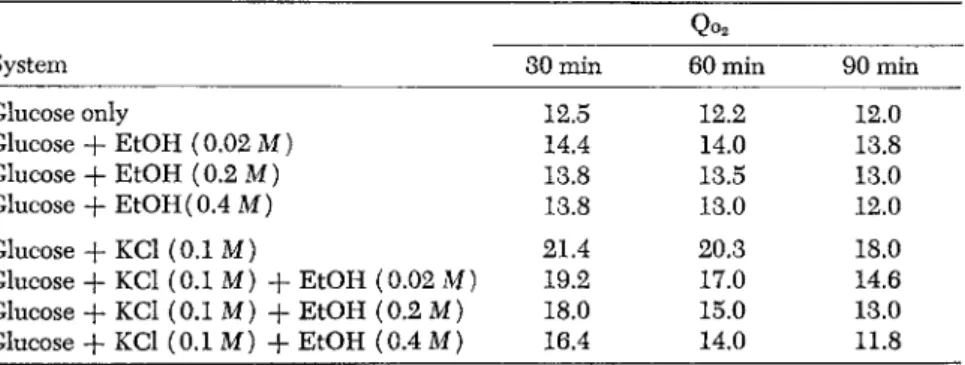
Our results show that the addition of ethanol at small concentrations to slices of rat brain in cortex respiring in a glucose phosphate medium brings about an increase in the rate of respiration, the increase being diminished with increase of ethanol concentration.
Ethanol, however, even at the lowest concentration tested (0.02M = 92 mg per 100 ml.) exercises a definite inhibition of rat brain cortex respiration in presence of 0.1 Μ KCl. The presence of ethanol brings about inhibitions of the stimulated respiration of rat brain cortex that increase with increase of ethanol concentration. The same phenomenon holds with 2,4-dinitrophenol-stimulated respiration of rat brain cortex (Tables 9, 10, 11). These observations indicate that ethanol behaves in a manner similar to luminal or pentothal or chloretone on the stim
ulated respiration of rat brain.
220 J. Η. QUASTEL
TABLE 10. The Effects of Ethyl Alcohol on the Normal and KCl-Stimulated Respiration of Rat Brain Cortex Slices
Qo2
System 30 min 60 min 90 min
Glucose only 12.5 12.2 12.0
Glucose + EtOH (0.02 Μ) 14.4 14.0 13.8
Glucose + EtOH (0.2 Μ) 13.8 13.5 13.0
Glucose + EtOH (0.4 Μ) 13.8 13.0 12.0
Glucose + KCl (0.1 Μ) 21.4 20.3 18.0
Glucose + KCl (0.1 Μ) + EtOH (0.02 Μ) 19.2 17.0 14.6 Glucose + KCl (0.1 Μ) + EtOH (0.2 Μ) 18.0 15.0 13.0 Glucose + KCl (0.1 Μ) + EtOH (0.4 Μ) 16.4 14.0 11.8
TABLE 11. The Effects of Ethyl Alcohol on the Normal and 2,4-Dinitrophenol- Stimulated Respiration of Rat Brain Cortex Slices
Average Qo2 20 min 40 min 60 min
Glucose (0.01 M) only 12.6 12.0 12.0
Glucose (0.01 M) + EtOH (0.02 Μ) 15.0 14.8 14.0
Glucose (0.01 M) + EtOH (0.2 Μ) 15.0 14.5 13.8
Glucose (0.01 Μ) + EtOH (0.4 Μ) 14.7 14.0 12.0
Glucose (0.01 Μ) + DNP (2.5 χ 10~5 M) 24.0 22.8 20.8 Glucose (0.01 M) + DNP (2.5 χ 10~5 M) + E t O H (0.02 M) ) 22.0 17.0 15.0 Glucose (0.01 M) + DNP (2.5 X 10~5 Μ) + EtOH (0.2 Μ) 20.0 16.0 13.2 Glucose (0.01 M) + DNP (2.5 χ ΙΟ"5 M) + E t O H (0.4 Μ) 17.4 13.8 10.0
It is likely, though not yet proved, that the increase of respiration of unstimulated rat brain found on the addition of small concentrations of ethanol is due to oxidation of the ethanol itself and that such increase is quite obscured by the stimulation of oxidation that occurs in a glucose medium in presence of 0.1 Μ potassium chloride or 2,4-dinitrophenol.
These results make it apparent that, in the intact animal, narcotics such as luminal and thiopental, or chloretone or ethanol exert inhibi
tory effects on the stimulated respiratory events of the nerve centers, where they are absorbed, suppressing pyruvate oxidation and the oxida
tive synthesis of adenosinetriphosphate.
The consequence of this paralysis of the nerve centers in question is a suppression of the metabolic activity of those portions of the nervous system that are controlled by the inhibited nerve centers. Such a sup
pression of metabolic activity of a considerable part of the nervous sys
tem may manifest itself by gross biochemical changes, not necessarily
identical with those that take place at the specific nerve centers affected by the narcotic. Thus the increased formation of acetylcholine, phos- phocreatine, or ATP that may be found (Richter, 1952) in the brains of sleeping or narcotized animals is, on this view, a reflection of the gen
erally depressed metabolic activity of the brain consequent upon the suppression of the functional activites of the regulating nerve centers.
To summarize briefly, the biochemical effect in vitro of administra
tion of narcotics such as the barbiturates or chloretone or ethanol is to suppress at specific nerve centers, at pharmacologically active concen
trations, oxidative events linked with the oxidation of carbohydrate or pyruvate, and the oxidative synthesis of ATP. This suppression is most marked, or only manifest, with stimulated brain cortex, in which phase the respiratory events in question become dominant and contribute most to the metabolic activity of the brain. In the conscious animal, in which the brain is stimulated, the oxidative activities of specific nerve centers are suppressed, their functional activities are diminished, and the meta
bolism of the nervous system controlled by these centers becomes adjusted to a new equilibrium.
References
Banga, I., Ochoa, S., and Peters, R. A. (1939). Biochem. J. 33, 1980.
Boyer, P. D., Lardy, Η. Α., and Phillips, P. H. (1943). /. Biol. Chem. 149, 529.
Brink, F. (1951). Trans. 2nd. Josiah Macy, Jr., Conf. on Nerve Impulse, New York, p. 124.
Brody, Τ. M., and Bain, J. A. .(1951). Proc. Soc. Exptl Biol. Med. 77, 50.
Brody, Τ. M., and Bain, J. A. (1954). J. Pharmacol. Exptl. Therap. 110, 148.
Bronk, D. W., and Brink, F . (1951). Federation Proc. 10, 19.
Dickens, F . (1946). Biochem. J. 40, 145.
Edwards, C , and Larrabee, M. G. (1953). Federation Proc. 12, 37.
Eiler, J . J., and McEwen, W. K. (1949). Arch. Biochem. 20, 163.
Ghosh, J. J., and Quastel, J. H. (1954). Nature 174, 28.
Greig, Μ. E . (1946). J. Pharmacol. Exptl Therap. 87, 185.
Himwich, Η. E., "Brain Metabolism and Cerebral Disorders." Williams and Wilkins, 1951.
Johnson, W. J., and Quastel, J. H. (1953). Nature 171, 602.
Johnson, W. J., and Quastel, J. H. (1953). /. Biol. Chem. 205, 163.
Jowett, M. (1938). /. Physiol. 92, 322.
Jowett, M., and Quastel, J. H. (1937). Biochem. J. 31, 565.
Kimura, Y. and Niwa, T. (1953). Nature 171, 881.
Kety, S. S., "Biology of Mental Health and Disease," p. 28. P. B. Hoeber, 1952.
Kety, S. S. (1950). Am. J. Med. 8, 205.
Kety, S. S., and Schmidt, C. F . (1948). J. Clin. Invest. 27, 476.
222 J. Η. QUASTEL
Larrabee, Μ. G , in "Biology of Mental Health and Disease," p. 384. P. H. Hoeber, 1952.
Larrabee, M. G., Ramos, J . G., and Bülbring, E . (1950). Federation Proc. 9, 75.
McFarland, R. Α., in "Biology of Mental Health and Disease," p. 334. P. B. Hoeber, 1952.
Mcllwain, H. (1952). Biochemical Society Symposia No. 8, Metabolism and Func
tion in Nervous Tissue, p. 27.
Mcllwain, H. (1953). Biochem. J. 53, 403.
Mann, P. J. G, and Quastel, J. H. (1946). Biochem. J. 40, 139.
Michaelis, Μ., and Quastel, J. H. (1941). Biochem. J. 35, 518.
Ochoa, S. (1941). /. Biol. Chem. 138, 751.
Persky, H., Goldstein, M. S., and Levine, R. (1950). /. Pharmacol. Exptl. Therap.
100, 273.
Peters, J. P., and Van Slyke, D. D., "Qualitative Clinical Chemistry," Vol. 1. Wil
liams and Wilkins, 1931.
Richter, D. (1952). Biochemical Society Symposia No. 8, Metabolism and Function in Nervous Tissues, p. 62.
Stanbury, S. W., and Mudge, G. H. (1953). Proc. Soc. Exptl. Biol. Med. 82, 675.
Wenner, C. E., Dunn, D. F., and Weinhouse, S. (1953). /. Biol. Chem. 205, 409.