Injury 52 (2021) S3–S6
ContentslistsavailableatScienceDirect
Injury
journalhomepage:www.elsevier.com/locate/injury
Periosteal and endosteal microcirculatory injury following excessive osteosynthesis ✩
Ferenc Greksa
a, Edina Butt
b, Endre Csonka
b, Péter Jávor
b, Eszter Tuboly
c, László Török
b, Andrea Szabo
c, Endre Varga
b, Petra Hartmann
c,∗aDepartment of Orthopaedics, University of Szeged, 6725 Szeged, Semmelweis u.6, Szeged, Hungary
bDepartment of Traumatology, University of Szeged, 6725 Szeged, Semmelweis u.6, Szeged, Hungary
cInstitute of Surgical Research, University of Szeged, 6724 Szeged, Pulz u. 1, Szeged, Hungary
a rt i c l e i n f o
Article history:
Accepted 16 November 2020
Keywords:
Endosteum Periosteum Osteosynthesis Microcirculation Laser-Doppler
a b s t r a c t
Introduction: Weexaminedtheendostealandperiostealcirculationsinapatientwithfracturenon-union whohadundergoneexcessiveosteosynthesisapplications(twolongplateshadbeenplacedmediallyand laterallyonthelefttibiaextendingfromtheproximal 2/7tothedistal6/7 partsofthebone,whilea tibialcomponentofatotalkneeprosthesiswithalongstemhadbeeninsertedatthesametime).
Methods: Concomitantperfusion changesweredetermined inthe anterolateral and anteromedialpe- riostealsheath of the non-united boneends and intramedullary nearest the osteosynthesis materials duringtheir surgicalremoval onre-operation.The bloodflow inthe periosteumand endosteum was recordedbyalaser-Dopplerflowmetricdeviceusinganovelapproach.Controlmeasurementsweremade atidenticalpointsoftherighttibia.
Results: Considerably lowerbloodflowvaluesweremeasuredalongthetibial periostealregionofthe re-operatedlimbthanonthecontralateralside(theaverageperfusionunit(PU)was76vs.106PU,re- spectively).Perfusionvaluesweremarkedlylowerintheendostealregion(averagevaluesofapprox.30 PU)inthecontroltibiaandwereevenmorediminishedinthere-operatedtibialendosteum(average9 PU).
Conclusions: Ourstudy was conductedtocharacterizethe microcirculatorychangesofalongbonein responseto intramedullaryimplantationand toprovidequantitativedataonthe insufficiencyoflocal perfusioninapatientwithfracturenon-union.Ourresultshighlighttheassociationbetweenlocalper- fusionfailureandtheunfavorableoutcome(i.e.fracturenon-union),confirmingthatthevitalaspectsof themicrocirculationshouldnotbedisregardedwhenaimingformechanicalstability.
Microcirculatorymeasurements constituteanewarea ofimprovement inplanningthe adequatetreat- mentforfracture non-unionswithanunclear aetiology.Furtherrefinement ofthe laser-Dopplertech- niquecouldhavepotentialbenefitsforbonesurgeryandpostoperativetraumacareinthefuture.
© 2020PublishedbyElsevierLtd.
Introduction
Fracture healing is a complex,multifactorial process requiring theanatomicalandfunctionalintegrityofthesurroundingbiolog- icalmembranes[1].Experimentaldatasuggestthatosteosynthesis
✩This paper is part of a supplement supported by the Hungarian Trauma Society.
∗ Corresponding author.
E-mail addresses: dr.greksaferenc@gmail.com (F. Greksa), butt.edina@gmail.com (E. Butt), ecsonka2@med.u-szeged.hu (E. Csonka), javor.peter.janos@med.u- szeged.hu (P. Jávor), eszter.tuboly4@gmail.com (E. Tuboly), torok.laszlo@med.u- szeged.hu (L. Török), szabo.andrea.exp@med.u-szeged.hu (A. Szabo), hartmann.petra@med.u-szeged.hu (P. Hartmann).
techniquesappliedtothe fixationoffractures(plateosteosynthe- sisandintramedullarynailing)caninducecirculatorydisturbances ofthebone;forexample,plateosteosynthesis(e.g.DCplates)im- pairsperiostealcirculation,whileintramedullaryreamingdestroys theendostealcirculation,resultinginnecrosis intheinner 2/3of thecorticalbone[2–6].Althoughtheimportanceoftheintegrityof themicrocirculationhasbeenemphasizedinnumeroustextbooks, noclinicalevidencehasbeenprovidedontheextentofmicrocircu- latoryinjury intheseconditions,mostlyowing tomethodological limitations.
Both experimental and clinical observations suggest that dis- ruptionof the continuityofthe periosteumor endosteumdelays https://doi.org/10.1016/j.injury.2020.11.053
0020-1383/© 2020 Published by Elsevier Ltd.
S4 F. Greksa, E. Butt and E. Csonka et al. / Injury 52 (2021) S3–S6
bone healing, but impairment of only one or the other of these structures does not hinder overall fracture healing [7–11]. The anatomicalconnection betweenthetwosystemsisnot wellchar- acterizedinhumans, butitis extensivein younganimals, where thearterialsupplyandvenousdrainagetraverseboththeendosteal andperiostealsurfaces;therefore,eitherofthetwosystemsissuf- ficienttoprovideadequatebonecirculation[9,11].
The destruction ofthe endosteumwithnailingusing different materials causesdistinct changes inthe periosteal vascular orga- nization [10,12]. Furthermore, the dependenceof the healing ca- pacity of the cortex on the endosteal and periosteal blood sup- ply following osteosynthesis has been demonstrated in different species[11,13].Perfusionchangesoftheperiosteumhavebeenin- vestigatedusingthelaser-Dopplertechniqueinanimals; however, thereis limitedexperiencewith humanmodels [12–15].In addi- tion,nosuch dataare available regardingthe humanendosteum.
Therefore,a clinical study wasconducted to provide evidence of themagnitude oflocal blood perfusion deficitin response to in- tramedullary implants. The microcirculation of the tibial perios- teum andendosteum was examined using a laser-Doppler flow- metricapproach ina patient withnon-union. Twoyearsprior to ouranalysis, the patient had a tibial fracture at the level of the endpointofthestemofacementedtotalkneeendoprosthesis.We wereunabletoachievehealingeitherwithDCplateosteosynthesis orwithsubsequently appliedangular stableplates onthemedial andlateral sidesofthetibia.Consequently,non-union developed.
Weassumed therole ofmicrocirculatory disturbances inthe de- velopment ofnon-union; measurements were thus performedto detect concomitant microcirculatory perfusion deficit nearest the osteosynthesismaterialsatthetimeofimplantremoval.
Methods
Intraoperative microcirculatory measurements were performed withthepermission andsignedconsent ofthepatient. Thestudy wasapprovedbytheEthicalCommitteeoftheUniversityofSzeged Clinical Center and the Regional Ethical Committee on Human MedicalResearch(No.0623.2014).
The following surgical interventions were performed: Two years priortoouranalysis,thepatienthadatibialfractureatthelevelof theendpointofthestemofacementedtotalkneeendoprosthesis.
WewereunabletoachievehealingeitherwithDCplateosteosyn- thesis orwith subsequently applied angular stable plateson the medialandlateralsidesofthetibia.Consequently,pseudarthrosis developed.Atthetimeofre-operation,thepatientwasexperienc- ingpain,andbothplateswerebroken.
As the initial step of the re-operation,the two angular stable plateswere removedtogether withtheir partially broken screws.
Subsequently,thetibialcomponentofthekneeprosthesiswasex- tracted.Aspecialtotalkneeprosthesiswasdesignedandmanufac- turedto manage the non-union. Its tibial componenthad an ex- tended stem with distal holes that bridged the fracture. Locking screwswereplacedinthedistalholessothattheboneendswere heldcloselyopposed.Cementfixationwasnotused.Thepolyethy- lene component (15 mm in size) of the tibial tray was also re- placed. The bone ends were refreshed andthe fracture gap was tightly filled with autologous spongiosa from the left iliac crest toinduce osteogenesis.Microcirculatory measurements were per- formedduringtheremovaloftheoldimplants.
Measurement of the tibial periosteal and endosteal microcircu- latory variables with laser-Doppler flowmetry: The blood flow in theperiosteumandendosteumwasrecordedwithalaser-Doppler flowmetric device (supplied witha 780-nm laser diode; PeriFlux System5000,Perimed,Järfälla,Sweden)withasterilizedfibreop- ticprobe (#416, “dental probe”; fibre separation: 0.25 mm, pen- etrationdepth ~ 1 mm). The tibial periosteum was explored via
Fig. 1. Scheme of the laser-Doppler flowmetry approach to the observation of per- fusion of the periosteum (A) and the endosteum (B). The periosteum ( p ) was ob- served through a small incision made in the skin and the underlying tissues, while the endosteal region ( e ) was reached through a small hole drilled in the anterior cortex, providing access to the endosteal region at the opposite (inner surface of the posterior) medullary wall.
a conventional anterior incision in the re-operated limb and via a small(~ 2cm) skinincision inthecontralateral limb.The flow probe washeld perpendicularly to the surfaceofthe periosteum witha plasticholder, whichreducedthe contactpressureon the observedareaandrestrictedtheangularmovementsoftheprobe.
The endoprosthesis was then removed, and the endosteal mem- brane compartment was approached through the bone cavity. A smallhole wasdrilled in theanterior cortex, providing accessto the endosteum at the opposite (inner surface of the posterior) medullarywall (Fig.1).Thesizeofthedrilledholeallowed aper- fectfitfortheflowprobe.Sinceanendoprosthesiswaspresenton the contralateral (non-operated) side, it waspossible to measure theendostealcirculationdistallytothestemoftheendoprosthesis, somewhatbelowthelevelofthemeasurementonthere-operated side;the difference was~ 1cm.Giventhe examination depthof the laser-Doppler device andthe thickness of the periosteal and endosteal membranes, it wasmostly the periosteum and endos- teumthatcouldbeexaminedwithaportionoftheunderlyingcor- tex.
Characteristic flow curvessynchronized with the heart circles were reproducibly detected in the
τ
= 0.2s mode, showing that pressure artefacts were avoided. After the requiredsignal quality hadbeen reached, recordings were made in30s periods andre- peated three times. Tissue perfusion was expressed arbitrarily in perfusionunits(PU);beforethemeasurements,theprobewascal- ibrated withthespecialmotility standardsuppliedby themanu- facturer.Data were collectedandstoredon a computer andsub- sequentlyanalyzedwiththecomputer softwaresuppliedwiththe device.Throughoutthe observationperiod,the roomtemperature (20±2°C) and core temperature of the patient were maintained;thestablemacrohaemodynamicparameterswererecordedcontin- uously.
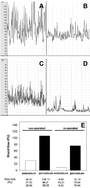
Since thisstudyrelates toonly one patient andthe measure- ments wereconductedrepeatedly atbasicallythesamelocations, nostatisticalcomparisonswereperformed.Rawdata(expressedin arbitraryunits)arepresentedinFig.2Eto illustratepotentialdif- ferences.
Results
Considerablylowerperiostealbloodflowvaluesweremeasured inthere-operatedtibialperiosteum(Fig.2B)thaninthecontralat- eral,non-operatedlimb(Fig.2A)(averagelevelsof76and106PU, respectively) (Fig. 2E). A great deal of lower perfusion was ob- served in the proximity of the endosteum; however, an average endostealperfusionof30PUwasfound inthenon-operatedtibia (Fig. 2C). Aneven lower perfusion levelwasmeasured in there-
F. Greksa, E. Butt and E. Csonka et al. / Injury 52 (2021) S3–S6 S5
Fig. 2. Original laser-Doppler flowmetric recordings of the periosteal (A and B) and endosteal regions (C and D) in the non-operated (A and C) and operated (B and D) tibia. Data related to the perfusion of the above compartments are presented in part (E). The mean values of three measurements constitute the presented data.
operatedtibialendostealregion(average9PU;Fig.2D)eveninthe presenceofcharacteristicallygoodsignalquality(Fig.2E).
Radiographstakensixweeksaftertheoperationrevealedcallus formationinthedorsalpartofthefractureandgoodalignmentof thefractureends(Fig.3).Thepatientnowwalkswiththeaidofa knee-ankle-toeorthosiswithoutloadingtheosteosynthesis.
Discussion
Our patient underwent a seriesof internal fixationsthat pro- videdadequatestability.Thediamondmodelofbonefractureheal- ing describes the interactions between the extrinsic mechanical
Fig. 3. Anteroposterior and lateral views of the left tibia prior to the re-operation (A) and 6 weeks after surgery (B). In addition to the proper alignment of the bone ends, callus formation was detected in the dorsal part of the fracture.
environment andendogenous factors; however, the microcircula- tionofthebiologicalmembranesofthebonearealsoessentialand sometimesoverlooked,asdemonstratedinourstudy[16,17].
From a physiological/biological aspect, the endosteal and pe- riostealbloodflowwasdiminishedinthefracturedlimb(ascom- paredtothecontralaterallimb),and,asaresult,non-uniondevel- oped.Inlongbones,endochondralbonerepairistheprimaryform ofbone healingwhenmicro-motionsexist betweenthefractured parts of bones covered with the periosteum [14,6]. This process is based on the ability of periosteum-derived cells to proliferate andto formperiostealcallusintheinitial phaseoffracture heal- ing[15,18].Inlinewiththis,inanovine“periostealsleeve” model, theperiosteumalonewasshowntobecapableofregeneratingthe longbone defect whenthe endosteumwascompletelydestroyed [16,19]. Probably because of the decisive role of the periosteum, theimportance oftheendosteuminbonehealingisgenerallyun- derestimated[15,18]. Similarlyto the periosteum,the endosteum covering all interior surfacesof bones(including all of theinner surfacesofthecortex andthespongiosa)alsoprovidesarichvas- cular environment and harbors osteoprogenitor cells for fracture healing,butcellularmechanismsdiffergreatly[18,20,21].Selective disruptionoftheendosteumoccursinclinical practicemostlyvia thereamingofthemedullarycavity.As aresult, avascularityand eventhepartialnecrosisofthecortexcanbedetected,suggesting thatasignificantproportionofthecorticalbloodflowisprovided bytheintramedullaryvessels.
Our patient underwent a sequence of distinct osteosynthesis techniquesthatprovidedexcellentmechanicalstabilityintheshort term. However, the integrity of the periosteum and endosteum were affected simultaneously, resulting in inhibition of adequate fracturehealingandformationofnon-union.
Doppler flowmetry is a suitable method for the analysis of microvascular blood flow, which could be utilized in bone surgeryandpostoperative traumacare.However, furtherresearch isneededtorefineitspracticalityandinvasiveness.
Theethicalandtechnicallimitationsofperformingahighnum- ber of periosteal and endosteal circulatory measurements under clinicalcircumstancesconstituteourgreatestlimitation.
Therefore,furtherclinicalstudiesarewarrantedtoassesslong- term circulatory derangements after osteosynthesis using nonin- vasive techniques. There are promising results with a combined laser-Dopplerandspectrophotometrysystem(Oxygen-to-see)that allowsmicrocirculatorymeasurementsbothintraoperativelyandat thebedside [22]. Therefore,it could be used ina comprehensive clinicalstudytoevaluatethemicrocirculatoryconsequencesofdif- ferentosteosynthesistechniques.
S6 F. Greksa, E. Butt and E. Csonka et al. / Injury 52 (2021) S3–S6 Conclusions
Thepresentstudyprovidesthefirstevidence-baseddemonstra- tion of the consequences of iatrogenic cessation of the interior andexterior bloodsupplyofalongbone.Aconcurrentdeteriora- tionofbothendostealandperiostealcomponentsofthemicrocir- culationmayexplain theunfavourable clinicaloutcome. Although theremaybe other reasonsforthenon-union ofa fracture,such asmechanicalorendogenousfactors[23–26] – seethe“diamond concept” noted above – the laser-Doppler data from the current studysuggest that the cessation of the periosteal andendosteal microcirculationwasthemaincauseoffracturenon-unioninthis particularpatient.
Appropriate preoperative planningand preservation ofthe lo- calmicrocirculationmaybothcontributetothefinalfavorableout- comeafterre-operation.Therefore,microcirculatorymeasurements constituteanewareaofimprovementinplanningadequatetreat- mentforfracturenon-unionswithanunclearaetiology.Furtherre- finementofthelaser-Dopplertechniquecouldhavepotentialben- efitsforbonesurgeryandpostoperativetraumacareinthefuture.
DeclarationofCompetingInterest
Noneoftheauthorshasanyconflictofinterestsrelatedtothis manuscript.
Acknowledgment
Supported by grants fromthe NationalResearchDevelopment andInnovation OfficeNKFI K120232,FIKP programTUDFO/47138- 1/2019-ITMandEFOP-3.6.2-16-2017-00006.
Supplementarymaterials
Supplementary material associated with this article can be found,intheonlineversion,atdoi:10.1016/j.injury.2020.11.053. References
[1] Macnab I , Dehoas WG . The role of periosteal blood supply in the healing of fractures of the tibia. Clin Orthop 1974;105:27–33 .
[2] Barron SE , Robb RA , Taylor WF , et al. The effect of fixation with intramedullary rods and plates on fracture-site blood flow and bone remodeling in dogs. J Bone Joint Surg Am 1977;59:376–85 .
[3] Kowalski MJ , Schemitsch EH , Kregor PJ , et al. Effect of periosteal stripping on cortical bone perfusion: a laser Doppler study in sheep. Calcif Tissue Int 1996;59:24–6 .
[4] Pazzaglia UE . Periosteal and endosteal reaction to reaming and nailing: the possible role of revascularization on the endosteal anchorage of cementless stems. Biomaterials 1996;17:1009–14 .
[5] Mueller CA , Schlegel V , Hoegel F , et al. Cortical perfusion and local fat oc- clusion after intramedullary nailing of the ovine tibia–comparison of different surgical procedures. Injury 2009;40:760–6 .
[6] Koo H , Hupel T , Zdero R , et al. The effect of muscle contusion on cortical bone and muscle perfusion following reamed, intramedullary nailing: a novel canine tibia fracture model. J Orthop Surg Res 2010;5:89 .
[7] Kregor PJ , Senft D , Parvin D , et al. Cortical bone perfusion in plated fractured sheep tibiae. J Orthop Res 1995;13:715–24 .
[8] Seibold R , Schlegel U , Kessler SB , et al. Healing of spiral fractures in the sheep tibia comparing different methods-osteosynthesis with internal fixation, inter- locking nailing and dynamic compression plate. Unfallchirurg 1995;98:620–6 . [9] Gustilo RB , Merkow RL , Templeman D . The management of open fractures. J
Bone Joint Surg Am 1990;72:299–304 .
[10] Esterhai JL Jr , Gelb I . Adult septic arthritis. Orthop Clin North Am 1991;22:503–14 .
[11] Whiteside LA , Ogata K , Lesker P , et al. The acute effects of periosteal stripping and medullary reaming on regional bone blood flow. Clin Orthop Relat Res 1978;131:266–72 .
[12] Greksa F , Tóth K , Boros M , et al. Periosteal microvascular reorganization after tibial reaming and intramedullary nailing in rats. J Orthop Sci 2012;17:477–83 . [13] Lippuner K , Vogel R , Tepic S , et al. Effect of animal species and age on plate-induced vascular damage in cortical bone. Arch Orthop Trauma Surg 1992;111:78–84 .
[14] Kowalski MJ , Schemitsch EH , Kregor PJ , et al. Effect of periosteal stripping on cortical bone perfusion: a laser Doppler study in sheep. Calcif Tissue Int 1996;59:24–6 .
[15] Schemitsch EH , Kowalski MJ , Swiontkowski MF , et al. Comparison of the effect of reamed and unreamed locked intramedullary nailing on blood flow in the callus and strength of union following fracture of the sheep tibia. J Orthop Res 1995;13:382–9 .
[16] Giannoudis PV , Einhorn TA , Marsh D . Fracture healing: the diamond concept.
Injury 2007;4:S3–6 .
[17] Douras P , Tosounidis T , Giannoudis PV . Application of the ’diamond concept’
with fast bone marrow aspirate concentration for the treatment of medial malleolus non-union. Injury 2018;12:2326–30 .
[18] Shapiro F . Bone development and its relation to fracture repair. The role of mesenchymal osteoblasts and surface osteoblasts. Eur Cell Mater 2008;15:53–76 .
[19] Knothe Tate ML , Ritzman TF , Schneider E , et al. Testing of a new one-stage bone-transport surgical procedure exploiting the periosteum for the repair of long-bone defects. J Bone Joint Surg Am 2007;89:307–16 .
[20] Kojimoto H , Yasui N , Goto T , et al. Bone lengthening in rabbits by callus distraction. The role of periosteum and endosteum. J Bone Joint Surg Br 1988;70:543–9 .
[21] Colnot C , Zhang X , Knothe Tate ML . Current insights on the regenerative po- tential of the periosteum: molecular, cellular, and endogenous engineering ap- proaches. J Orthop Res 2012;30:1869–78 .
[22] Kalbas Y , Qiao Z , Horst K , Teuben M , Tolba RH , Hildebrand F , Pape H-C , Pfeifer R , Research Group T . Early local microcirculation is improved after in- tramedullary nailing in comparison to external fixation in a porcine model with a femur fracture. Eur J Trauma Emerg Surg 2018;5:689–96 .
[23] Simpson A . Tsang STJ. Non-union after plate fixation. Injury 2018;49(Suppl 1):S78–82 Jun .
[24] Morwood MP , Gebhart SS , Zamith N , Mir HR . Outcomes of fixation for periprosthetic tibia fractures around and below total knee arthroplasty. Injury 2019;4:978–82 .
[25] Haubruck P , Heller R , Apitz P , Kammerer A , Alamouti A , Daniel V , Schmid- maier G , Moghaddam A . Evaluation of matrix metalloproteases as early biomarkers for bone regeneration during the applied Masquelet therapy for non-unions. Injury 2018;10:1732–8 .
[26] Yılmaz O , Özmeriç A , Alemdaro ˘glu KB , Celepli P , Hücümeno ˘glu S , ¸S ahin Ö.
Effects of concentrated growth factors (CGF) on the quality of the induced membrane in Masquelet’s technique–an experimental study in rabbits. Injury 2018;8:1497–503 .