R E S E A R C H A R T I C L E
Theileria highjacks JNK2 into a complex with the macroschizont GPI (GlycosylPhosphatidylInositol) ‐ anchored surface protein p104
Perle Latré De Laté
1,2*
|Malak Haidar
1,2,3*
|Hifzur Ansari
3 |Shahin Tajeri
1,2 |Eszter Szarka
4 |Anita Alexa
4 |Kerry Woods
5 |Attila Reményi
4 |Arnab Pain
3,6 |Gordon Langsley
1,21Laboratoire de Biologie Cellulaire Comparative des Apicomplexes, Faculté de Médecine, Université Paris Descartes, Sorbonne Paris Cité, Paris 75014, France
2Inserm U1016, CNRS UMR8104, Cochin Institute, Paris, France
3Pathogen Genomics Laboratory, Biological and Environmental Sciences and Engineering (BESE) Division, King Abdullah University of Science and Technology (KAUST), Thuwal, Jeddah 23955‐6900, Kingdom of Saudi Arabia
4Institute of Enzymology, Research Centre for Natural Sciences, Hungarian Academy of Sciences, Budapest, Hungary
5Vetsuisse Faculty, University of Bern, Bern, Switzerland
6Global Station for Zoonosis Control, Global Institution for Collaborative Research and Education (GI‐CoRE), Hokkaido University, Sapporo, Japan
Correspondence
Gordon Langsley, Laboratoire de Biologie Cellulaire Comparative des Apicomplexes, Faculté de Médecine, Université Paris Descartes, Sorbonne Paris Cité, Paris 75014, France.
Email: gordon.langsley@inserm.fr Funding information
Agence Nationale de la Recherche, Grant/
Award Number: ANR‐11‐LABX‐0024; King Abdullah University of Science and Technol- ogy, Grant/Award Numbers: BAS/1/1020‐01‐ 01, URF/1/2610‐01‐01; Swiss National Sci- ence Foundation, Grant/Award Number:
PZ00P3_154689; Hungarian NKFIH, Grant/
Award Number: NN114309; CNRS; INSERM;
Labex ParaFrap, Grant/Award Number: ANR‐ 11‐LABX‐0024
Abstract
Constitutive c
‐Jun N
‐terminal kinase (JNK) activity characterizes bovine T and B cells infected with
Theileria parva, and B cells and macrophages infected withTheileria annulata. Here, we show thatT. annulatainfection of macrophages manipulates JNK activation by recruiting JNK2 and not JNK1 to the parasite surface, whereas JNK1 is found predominantly in the host cell nucleus. At the parasite's surface, JNK2 forms a complex with p104, a GPI
‐(GlycosylPhosphatidylInositol)
‐anchor
T. annulataplasma membrane protein. Sequestration of JNK2 depended on Protein Kinase
‐A (PKA)
‐mediated phosphorylation of a JNK
‐binding motif common to
T. parvaand a cell pen- etrating peptide harbouring the conserved p104 JNK
‐binding motif competitively ablated binding, whereupon liberated JNK2 became ubiquitinated and degraded.
Cytosolic sequestration of JNK2 suppressed small mitochondrial ARF
‐mediated autophagy, whereas it sustained nuclear JNK1 levels, c
‐Jun phosphorylation, and matrigel traversal. Therefore,
T. annulatasequestration of JNK2 contributes to both survival and dissemination of
Theileria‐transformed macrophages.
K E Y W O R D S
Dissemination, JNK2, penetrating peptide, PKA,Theileria
1
|I N T R O D U C T I O N
In mammals c‐Jun N‐terminal kinase (JNK) is encoded by three genes, mapk8, mapk9, and mapk10; mapk8 and mapk9 code,
respectively, for the ubiquitously expressed JNK1 and JNK2 pro- teins andmapk 10codes for JNK3, whose expression is restricted to cardiac, nervous, and testicular tissues (Davis, 2000). JNKs are activated by environmental stress including extracellular insults such as radiation, redox, osmotic and temperature shocks, and intracellu- lar stress such as misfolded proteins (Davis, 2000). Biological
*Co‐first authors with equal contribution.
DOI: 10.1111/cmi.12973
Cellular Microbiology. 2019;21:e12973.
https://doi.org/10.1111/cmi.12973
© 2018 John Wiley & Sons Ltd
wileyonlinelibrary.com/journal/cmi 1 of 12
responses transduced through JNK‐dependent pathways encompass proliferation, migration, survival, differentiation, and inflammation (Davis, 2000), and some of the JNK substrates participating in these processes have been identified (Bogoyevitch & Kobe, 2006).
Changes in gene expression resulting from JNK activation may be accounted for by the phosphorylation of several transcription fac- tors and the ensuing alteration in their transcriptional activity (Bogoyevitch & Kobe, 2006). A well‐characterized substrate of JNK is c‐Jun, a component of the AP‐1 transcription factor that is essen- tial for proliferation and cell survival. JNK can affect c‐Jun both pos- itively and negatively: N‐terminal phosphorylated c‐Jun displays increased trans‐activating activity (Hibi, Lin, Smeal, Minden, & Karin, 1993), whereas in neurons, the E3 ubiquitin ligase SCFFbw7specifi- cally targets phosphorylated c‐Jun for proteasome degradation, thereby controlling the JNK/c‐Jun apoptotic pathway (Nateri, Riera‐Sans, Da Costa, & Behrens, 2004). Additionally, in T lympho- cytes, c‐Jun turnover is regulated by the E3 ubiquitin ligase Itch, whose activity increases upon JNK‐dependent phosphorylation (Gao et al., 2004). JNK can promote cell motility via alteration of focal adhesion dynamics following JNK‐mediated phosphorylation of the focal adhesion adaptor paxillin (Huang, Rajfur, Borchers, Schaller, & Jacobson, 2003; Ueno et al., 2015; Wang et al., 2013).
Importantly, loss ofjnk2in mouse embryonic fibroblasts increases cell proliferation, whereas a loss ofjnk1leads to decreased prolifera- tion, and these contrasting effects are attributed to differential regulation of the mitogenic transcription factor c‐Jun. JNK1 increases c‐Jun stability via phosphorylation of serine 73, whereas JNK2 decreases c‐Jun stability by promoting its ubiquitination (Bode &
Dong, 2007; Fuchs, Dolan, Davis, & Ronai, 1996). JNK2 also promotes ubiquitination‐dependent proteasomal degradation of small mitochon- drial ARF (smARF), as in jnk2−/−mouse embryonic fibroblasts levels of smARF rise to induce autophagy (Budina‐Kolomets, Hontz, Pimkina,
& Murphy, 2013; Zhang et al., 2015a). SmARF is a short isoform of the tumour suppressor p19ARF, and interestingly, suppression of smARF did not require the kinase activity of JNK2 to be consistent with JNK2 acting as a scaffold protein (Zhang et al., 2015b). The above examples highlight the disparate functions of JNK isoforms and underscore the necessity to study them individually and together to properly grasp the cellular impact of JNK activation.
Parasites of the genus Theileria are intracellular protozoans belonging to the Apicomplexa phylum.Theileria annulataandTheileria parva are two particularly pathogenic species that cause bovine lymphoproliferative diseases, respectively named tropical theileriosis and the East Coast Fever. The target host cells ofT. parvaare T and B lymphocytes, whereas monocytes/macrophages and B cells are preferentially infected byT. annulata. East Coast Fever and tropical theileriosis display similarities to human lymphomas and myeloid leukemias, respectively. A vaccine exists to tropical theileriosis based onT. annulata‐infected macrophages attenuated in their capacity to disseminate (Darghouth, 2008), which is generated by multiple passages of infected monocytes/macrophages, which become attenu- ated after having lost their hyper‐disseminating virulence trait (Baylis, Megson, & Hall, 1995).Theileria‐infected leukocytes behave as trans- formed cell lines, and as they no longer require exogenous growth or survival factors, they can form colonies in soft agar and give rise to
disseminated tumours in immuno‐compromised mice (Fell, Preston, &
Ansell, 1990; Lizundia et al., 2006). Known as the only eukaryote pathogen to transform a eukaryote host cell,Theileriaachieves this by manipulating host cell signalling pathways, reviewed in B. Shiels et al.
(2006). Several different signalling pathways have been implicated, including TGF‐β(Chaussepied et al., 2010; Haidar et al., 2015; Haidar, Echebli, Ding, Kamau, & Langsley, 2015) and JNK kinase leading to con- stitutive phosphorylation of c‐Jun and activation of AP‐1 (Adamson, Logan, Kinnaird, Langsley, & Hall, 2000; Baylis et al., 1995; Chaussepied et al., 1998; Lizundia et al., 2007).
Remarkably,Theileria‐induced transformation is strictly dependent on the presence of live parasites, as the transformed host cell pheno- type is fully reversible upon drug‐induced parasite death; drug‐treated transformed leukocytes return to a quiescent nonactivated state and eventually die (Dobbelaere & Heussler, 1999). Theileria‐dependent JNK1 activity is required for survival ofT. parvatransformed B lym- phocytes, as demonstrated by overexpression of a dominant negative kinase‐dead mutant of JNK1 and/or via the use of pan‐JNK inhibitor (pan‐JNKi; Lizundia et al., 2005, 2006). Whereas the parasite‐derived molecular mechanism(s) underlying JNK activation is unknown, JNK1‐mediated survival ofTheileria‐transformed leukocytes has been attributed to AP‐1‐driven expression of the anti‐apoptotic genes Mcl‐1andc‐IAP(Lizundia et al., 2006), and uncontrolled proliferation is linked to AP‐1‐driven expression of transferrin receptor and cyclin D1 (Lizundia et al., 2007).
One of the characteristics ofTheileria‐transformed leukocytes is that they display heightened oxidative stress due in part to uncon- trolled proliferation‐related production of H2O2 (Metheni et al., 2014; Metheni, Lombes, Bouillaud, Batteux, & Langsley, 2015). This raises the possibility that exposure to H2O2contributes to induction of JNK activity, as JNK activation occurs in response to stress. Taken together, JNK activation seems a key event inTheileria‐induced leuko- cyte transformation, and the aim of this study was to examine whether Theileria infection affects differentially JNK1 versus JNK2 and whether the two isoforms play similar or different roles in parasite‐induced leukocyte transformation.
2
|R E S U L T S
2.1
|Cytosolic localisation of JNK2 versus nuclear localisation of JNK1 in Theileria‐ infected macrophages
To understand how the two major JNK isoforms participate in Theileria‐induced leukocyte transformation, we ascertained the subcellular distribution of JNK1 versus JNK2 in Theileria‐infected macrophages. JNK1 partitions into the cytosolic and nuclear fractions, and expression levels are parasite dependent, decreasing upon Bw720c‐induced parasite death, and this is particularly obvious for nuclear JNK1 (Figure 1a). In contrast to JNK1, JNK2 partitions principally in the cytosolic fraction and again levels decrease upon Bw720c. Immunofluorescence analysis revealed JNK2 in the cytosol decorating the intracellular macroschizont (Figure 1b), which is highlighted with a monoclonal antibody (1C12) toT. annulatap104 (Woods et al., 2013).
2.1.1
|T. annulata p104 is a putative macroschizont surface JNK ‐ binding protein
As JNK2 appears associated with the macroschizont, we searched for a parasite surface protein predicted to have a JNK‐binding motif (Bogoyevitch & Kobe, 2006). In silico analyses were performed on three different species ofTheileria(T. annulata, T. parva, andTheileria orientalis). A D‐Finder scan of the whole predicted proteomes of three Theileria species in search of D‐motifs (Zeke et al., 2015) led to identification of 26, 24, and 25 proteins, respectively (Supporting Information S1). Next, we asked which of these 75 putativeTheileria JNK‐binding proteins also had a predicted signal peptide, and this criterion identified only one protein inT. annulata(TA08425), two pro- teins inT. parva(TP04_0437 orthologue of TA08425, and a hypothet- ical protein TP02_0553), and no protein inT. orientalis. TA08425 codes for a GPI‐anchoredT. annulatamacroschizont surface protein called
p104 (Woods et al., 2013), which has three putative decapeptide D‐motifs (KNESMLRLDL, KSPKRPESLD, and KRSKSFDDLT) located between amino acids 331–341, 606–616, and 804–814, respectively.
However, only the decapeptide motif between amino acids 804 and 814 is conserved in theT. parvap104 orthologue (TP04_0437). The amino acid sequence in this region of p104 is not conserved in the nontransformingT. orientalis(TOT_040000478).
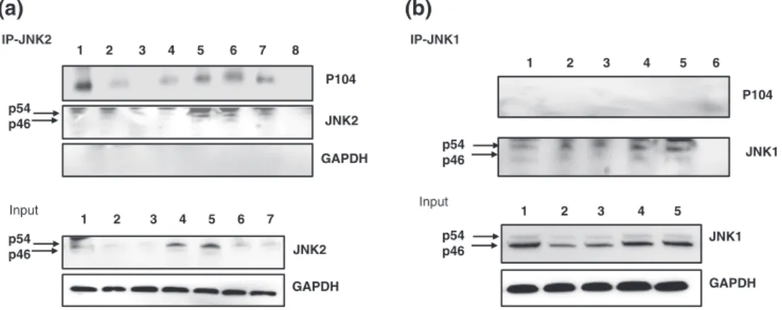
We examined, therefore, whether the conserved D‐motif (KRSKSFDDLT) mediated JNK2 binding to the T. annulata macroschizont surface protein p104. First, a pan‐JNK antibody precipitated endogenous p104 from extracts ofT. annulata‐infected TBL3 B cells but not from uninfected BL3 B cells (Figure 2a). As expected (see Figure 1), p104 was preferentially found in pan‐JNK pre- cipitates (Figure 2b, left) and specifically in JNK2 precipitates (Figure 2b, right, and Figure 3b). Altogether, these results suggest that JNK2 is associated with p104 at the surface of theTheileriamacroschizont.
N=30
Vc V Merge Dapi p104 JNK2
(a) (b)
- + - +
Cytosol Nucleus Bw720c
JNK1
PARP JNK2
pan JNK
p46
p54 p46
p54 p46
FIGURE 1 JNK2 is predominantly in the infected macrophage cytosol associated with the parasite. (a) Localisation of JNK1 and JNK2 in Theileria‐infected macrophages treated or not with parasiticide drug BW720c. Western blot analysis of nuclear and cytosolic extracts probed with specific JNK1, JNK2, and pan‐JNK antibodies. PARP antibodies were used to control for the specificity of the nuclear extracts. (b, top panel) Immunofluorescence images (V) showing association of JNK2 with the parasite decorated with a parasite monoclonal antibody (1C12) to P104. (b, bottom panel) No immunofluorescence was observed (Vc) when just secondary antibodies were used as a negative control. JNK2/P104 colocalization was analysed by the Manders method of pixel intensity correlation measurements using ImageJ/Fiji‐Coloc2 plugin, and an average for 30 independent cells is given. DNA was stained with DAPI (blue)
FIGURE 2 p104 interacts with JNK2 inTheileria‐infected leukocytes. (a) Immunoprecipitation with a pan‐anti‐JNK (JNK‐IP) antibody using whole cell lysates derived from infected (TBL3) and noninfected B cells (BL3), with the precipitate probed with the anti‐p104 monoclonal antibody 1C12.
Inputshows JNK and P104 protein levels in BL3 and TBL3 cells revealed by respective antibodies. 1, BL3; 2, TBL3 1μM; 3, IgG; 4, V; and 5, input form V. (b, right) Immunoprecipitation with pan‐JNK, specific anti‐JNK2, and irrelevant IgG control antibodies with the precipitate from infected macrophages (V) probed with 1C12.Inputshows the levels of the p46 JNK1 and p54 JNK2 isoforms revealed with the pan‐JNK antibody. An anti‐ actin antibody was used as a loading control
2.1.2
|Phosphorylation of the JNK ‐ binding motif increases the affinity of p104 for JNK2
To understand the consequences of JNK2 association with p104, we specifically ablated their interaction. Located on the macroschizont surface GPI‐(Glycosylphosphatidylinositol)‐anchored, p104 has been described as being phosphorylated in vivo at several sites (Wiens et al., 2014). We noticed that two phosphorylated residues occurred in the conserved JNK‐binding motif, specifically phospho‐S806 and phospho‐S808 (TA08425 numbering). Consequently, penetrating pep- tides harbouring the conserved p104 decapeptide JNK‐binding motif and a mutant peptide, where S806 and S808 had been replaced by ala- nine together with an irrelevant peptide, were synthesized (Table 1).
All peptides were FITC conjugated, and penetration into T. annulata‐infected macrophages was confirmed by immunofluores- cence (Figure S1). The peptide (P) corresponding to the wild‐type JNK‐binding motif was able to ablate in a dose‐dependent manner JNK2 association with p104 (Figure 3). Importantly, the mutant (S > A) peptide (mP) did not abrogate JNK2 binding to p104 (Figure 3a, Tracks 6 and 7), consistent with phosphorylation of S806 and/or S808 promoting p104 binding to JNK2. Following peptide‐ mediated abrogation of the JNK2/p104 complex, the level of p54 JNK2 decreased due to ubiquitination of JNK2, but no drop in JNK2
levels was observed following proteasome blockade by MG132 (Figure 3a, Tracks 4 and 5, and Figure 4a). Although peptide‐induced complex disruption reduced JNK2 levels, no effect was observed on cytosolic JNK1 (Figure 4b), but loss of cytosolic JNK2 led to an increase in the amount of nuclear JNK1 (Figure 4b).
2.1.3
|Protein Kinase ‐ A (PKA), but not c ‐ Jun N ‐ terminal Kinase (JNK), contributes to p104 association with JNK2
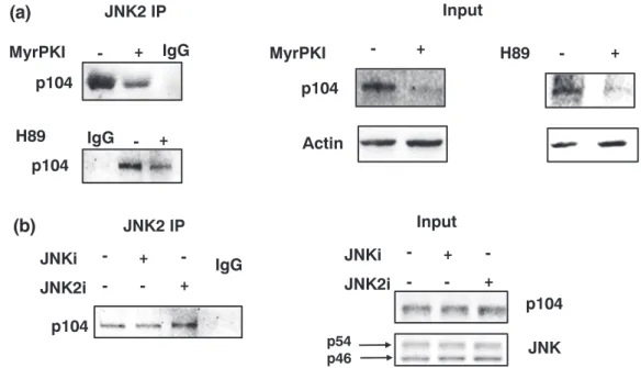
Both S806 and S808 in p104 occur in a context (KRS*KS*FD) consis- tent with them being potentially phosphorylated by PKA (Wiens et al., 2014). Consequently,T. annulata‐infected macrophages were treated for 2 hr with myristoylated PKI (MyrPKI) or the ATP analogue H89, and both treatments dampened the association of p104 with JNK2 (Figure 5a). By contrast, treatment with a pan‐JNKi or a JNK2‐specific inhibitor (JNK2i) did not alter the ability of p104 to bind JNK2 (Figure 5b). We conclude that cAMP‐dependent PKA likely phosphor- ylates S806 and/or S808, and their phosphorylation favours binding of p104 to bind JNK2. The kinase activity of JNK2 does not appear nec- essary for complex formation, leaving open that JNK2 could have a scaffold function (see below).
IP-JNK1
Input
JNK1
GAPDH p54
p46
P104
p54 JNK1 p46 P104
JNK2 GAPDH
JNK2 GAPDH IP-JNK2
Input
1 2 3 4 5 6
1 2 3 4 5 6 7 8
p54 p46
p54 p46
(a) (b)
1 2 3 4 5 6 7 1 2 3 4 5
FIGURE 3 Abrogation of JNK2/p104 interaction leads to proteasome‐mediated JNK2 degradation. Immunoprecipitation analyses with specific anti‐ JNK2 and anti‐JNK1 antibodies using whole cell lysates derived fromTheileria annulata‐infected macrophages treated or not with 1 or 5μM of the penetrating JNK‐binding motif peptide and treated or not with MG132 (P), mutant (S > A) peptide (mP), or irrelevant peptide. (a) JNK2‐IP shows western blot of the JNK2 precipitate probed with the anti‐p104, anti‐JNK2, and anti‐GAPDH antibodies. Bottom panel shows western blot analysis of immunoprecipitations probed with anti‐JNK2 and anti‐GAPDH antibodies. 1, V; 2, V + P 1μM; 3, V + P 5μM; 4, V + P 1μM + MG132; 5, V + P 5μM + MG132; 6, V + mP 1μM; 7, V + mP 5μM; 8, IgG. (b) JNK1‐IP: Immunoprecipitation analyses with anti‐JNK1 using whole cell lysates derived fromT. annulata‐infected macrophages treated or not with 1 or 5μM of P or mP peptides. JNK1 protein expression was decreased following the treatment with JNK‐binding motif competitive peptide, whereas no effect was observed with the mP peptide. Bottom panel shows western blot analysis of immunoprecipitations probed with anti‐JNK1 and anti‐GAPDH antibodies. 1, V; 2, V + P 1μM; 3, V + P 5μM; 4, V + mP 1μM; 5, V + mP 5μM; 6, IgG
TABLE 1 Synthesized peptides harbouring JNK‐binding motif
Peptides Sequences Phospho‐epitope
Predicted kinases phosphorylation sites P (peptide with conserved
JNK‐binding motif)
HVKKKKIKREIKITGKIVKLKRSKSFDDLTTK‐(FITC) S806, S808 PKA
mP (mutant peptide) HVKKKKIKREIKITGKIVKLKRAKAFDDLTTK‐(FITC) Mutations of S806, S808 in alanine
PKA
IrrP (irrelevant peptide) HVKKKKIKREIKIAAGRYGRELRRMADEFHV‐K (FITC)
Note. The different FITC‐labelled penetrating peptides synthesized. (P) is the conserved wild‐type amino acid sequence with S806 and S808 shown underlined. The mutant peptide (mP) has S806 and S808 changed to A806 and A808 (underlined). An irrelevant peptide (IrrP) used as a negative control to compete for JNK‐binding to p104. The JNK‐binding motif is in bold, and the sequence of the fused penetrating peptide is indicated at the N‐terminus.
2.2
|Abrogation of JNK2/p104 association reduces matrigel traversal of Theileria‐ transformed
macrophages
Matrigel traversal is used as a measure of dissemination potential and virulent (V) T. annulata‐transformed macrophages traverse matrigel better than attenuated (A) macrophages. Matrigel transversal is signifi- cantly decreased when T. annulata‐transformed macrophages are treated with the wild‐type JNK‐binding motif peptide (P), whereas the (S > A) mutant peptide mP and the irrelevant peptide (irrP) had no signif- icant effect (Figure 6a). It is well established that Theileria‐infected macrophages are characterized by AP‐1‐driven transcription ofmmp9 and increased matrix metallopeptidase 9 (MMP9) activity promotes matrigel traversal and dissemination (Adamson et al., 2000; Cock‐Rada et al., 2012; Echebli et al., 2014). JNK2 association with p104 was therefore ablated, and loss of MMP9 activity was revealed by gelatin gel assay (Figure 6b, left). Peptide‐induced disruption of JNK2/p104 binding slightly but significantly inhibited mmp9 transcription as estimated by qRT‐PCR (Figure 6b, right). Peptide‐induced complex dis- sociation also specifically reduced nuclear c‐Jun phosphorylation (Figure 6c) consistent with the drop in nuclear JNK1 levels (Figure 4b).
2.3
|Abrogation of the JNK2/p104 association leads to up ‐ regulation of ARF levels
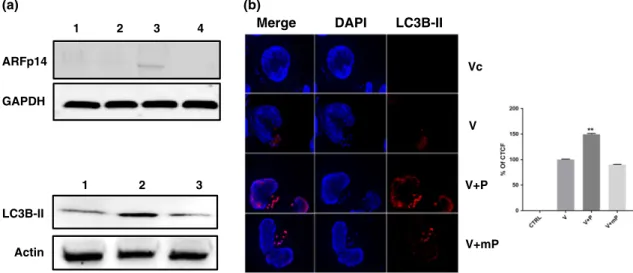
Because a nonkinase scaffold protein function for JNK2 has been described to regulate smARF levels and induce autophagy (Zhang et al., 2015a), we monitored ARF levels following disruption of the p104/JNK2 complex and subsequent JNK2 degradation (Figure 7). Loss of JNK2 provoked by peptide treatment resulted in up‐regulation of p14ARF. The rise in ARF levels was accompanied with an increase in amount of processed autophagosome membrane protein LC3B‐II and appearance of LC3B‐II‐positive foci (Figure 7a, bottom panel, and b).
3
|D I S C U S S I O N
Constitutively active JNK1 has a nuclear localisation that is dependent on liveT. annulatamacroschizonts, because nuclear JNK1 levels were ablated upon drug (Bw720c)‐induced parasite death. By contrast, JNK2 is mainly in the infected macrophage cytosol associated with the macroschizont with only a minor fraction of JNK2 being nuclear, and cytosolic JNK2 levels also depend on the live parasite being
JNK2
Actin p54
Input
P MG132
+ +
- + -
- -
+
JNK2 JNK1
Actin H3 Histone
JNK2 JNK1 Cytosolic extract Nuclear extract
p54 p46 p54 p46 IP-JNK2
P MG132
- +
+ + -
- +
- 250
130 95
55 Kd
(a)
(b)
FIGURE 4 Association with p104 protects JNK2 from ubiquitination and proteosomal degradation. (a) Immunoprecipitation analyses with anti‐ JNK2 antibodies using whole cell lysates derived from virulentTheileria annulata‐infected macrophages treated or not with 5μM of JNK‐binding motif peptide P and treated or not with MG132. Western blot of the JNK2 precipitate probed with an anti‐ubiquitin and JNK2 antibodies. (b) Western blot analysis of nuclear and cytosolic extracts probed with specific anti‐JNK1 and anti‐JNK2 antibodies using whole cell lysates derived fromT. annulata‐infected macrophages treated or not with 5μM of P and treated or not with MG132 and virulent treated with irrelevant peptide.
Actin and H3 histone antibodies were used as loading control. Input (a), JNK2 levels in the extracts were estimated compared with actin levels used as loading control. (b) JNK1 and JNK2 levels in nuclear extracts were compared with histone H3 levels
present (Figure 1). Simply by their different subcellular localisations, one can surmise that JNK1 and JNK2 likely play different roles in Theileria‐induced transformation of host leukocytes.
The close association between JNK2 and the parasite revealed by immunofluorescence led us to search for a parasite‐encoded JNK‐binding protein located on the macroschizont surface. Interrogat- ing the predicted Theileria proteomes with a consensus D‐motif revealed the presence of three potential JNK‐binding motifs in the GPI‐anchored macroschizont T. annulata protein known as p104 (Woods et al., 2013). We decided to characterize the putative JNK‐binding site located between amino acids 804 and 814, as this is conserved in theT. parvap104 orthologue (TP04_0437) and absent in the nontransforming T. orientalis (TOT_040000478) protein.
Although not characterized here, it remains possible that one or both of the other two T. annulata p104‐specific D‐motifs might also contribute to species‐specific JNK2 binding by p104. In vitro recombi- nantT. annulatap104 clearly binds to recombinant JNK2 (Figure S2A), making it possible to study the contribution of the two other D‐motifs to species‐specific JNK2 binding. We note that a secretedT. annulata protein called Ta9 is predicted to contain a D‐motif and can activate AP‐1‐driven transcription in HEK293T cells, but whether Ta9 does this inT. annulata‐infected macrophages by binding to JNK was not demonstrated (Unlu et al., 2018).
The conserved JNK‐binding motif in p104 is distal to the previ- ously described EB1‐binding motif (SKIP) that is located between amino acids 566 and 569 (Woods et al., 2013). It is highly unlikely therefore that the JNK‐binding motif penetrating peptide competes for EB1‐binding. Furthermore, EB1 colocalizes with p104 on the macroschizont surface in a cell cycle‐dependent manner, being more pronounced during cell division (Woods et al., 2013). This contrasts
with JNK2‐binding to p104 that does not require division of infected macrophages. Although p104 acts as an EB1‐binding protein, attempts to interfere with EB1 binding to the macroschizont surface failed (Woods et al., 2013). One explanation could be that JNK2 binding to p104 creates a platform favourable to EB1 association with the macroschizont during host cell mitosis. As we have shown that PKA can phosphorylate in vitro p104 (Figure S2B), this strongly argues that in vivo PKA‐mediated phosphorylation of p104 (Wiens et al., 2014) promotes association with JNK2, and indeed, PKA inhibition reduced the amount of p104 detected in JNK2 immunoprecipitates (Figure 5). This implies that the complex at the surface of the macroschizont contains not only p104 and JNK2 but also PKA. By contrast, Cdk1‐mediated phosphorylation of p104 seems to play a role in EB1 binding during mitosis, although the role of other kinases in regulating the interaction between p104 and EB1 has not been ruled out (Woods et al., 2013).
Importantly, both S806 and S808 have previously described as being phosphorylated in vivo (Wiens et al., 2014), so the strategy we adopted to elucidate the role of the JNK2/p104 complex was to use a penetrating peptide harbouring the conserved JNK‐binding motif, as a competitive p104 substrate for PKA. When S806 and S808 are changed to A806 and A808, the mutant penetrating pep- tide is no longer a competitive substrate for PKA and is incapable of disrupting JNK2 binding to p104. It is noteworthy that binding of JNK2 to p104 in vivo was disrupted by inhibition of PKA activity by two independent PKA inhibitors, MyrPKI and H89. Inter- estingly, however, recruitment of JNK2 to p104 was insensitive to inhibition of JNK kinase activity, suggesting JNK2 acts as a scaffold protein on which the complex is assembled at the macroschizont surface.
FIGURE 5 PKA phosphorylation increases association of p104 with JNK2. (a) Immunoprecipitation analyses with an anti‐JNK2 antibody using whole cell lysates derived from virulentTheileria annulata‐infected macrophages treated or not with the PKA specific inhibitor myristoylated PKI (MyrPKI) and H89. Left panel shows western blot of the JNK2 precipitate from nontreated, or MyrPKI/H89 treated (+) cells probed with the anti‐p104 1C12 monoclonal antibody. Middle and right panels show the input levels of p104 and actin revealed by their respective antibodies. (b) Left panel: Western blot of JNK2 precipitates using extracts of cells treated with a pan‐JNK inhibitor (pan‐JNKi), or a JNK2‐specific inhibitor (JNK2i) probed with the anti‐p104 1C12 monoclonal antibody. Right panel shows the input levels of p104 and the two JNK isoforms revealed by their respective antibodies
We focused on JNK2 to gain insights as to what might be the physiological advantage toTheileria‐induced leukocyte transformation in retaining JNK2 in the infected host cell cytosol sequestered at the macroschizont surface. The ensembles of our results pinpoint at least three advantageous consequences of p104‐mediated JNK2 sequestra- tion: (a) While associated with the macroschizont surface, JNK2 is protected from ubiquitination and proteasomal degradation, and sustained JNK2 levels suppress smARF‐mediated autophagy (Budina‐Kolomets et al., 2013; Zhang et al., 2015b). As such, JNK2 sequestration contributes toTheileria‐infected leukocyte survival; and indeed, 24 hr following peptide‐induced JNK2 degradation, infected macrophages become annexin V positive (Figure S3). (b) As nuclear JNK2 decreases c‐Jun stability by promoting its ubiquitination (Bode
& Dong, 2007; Fuchs et al., 1996), cytosolic retention of JNK2 could contribute to sustained nuclear c‐Jun levels perhaps by preventing DET1‐mediated ubiquitination (Marsolier et al., 2013). Sustaining c‐Jun levels also occurs via secretion of a parasite prolyl isomerase called TaPIN1 that provokes degradation of the host ubiquitin ligase FBW7 (Marsolier et al., 2015). (c) Now, we show that peptide‐induced
complex dissociation led to JNK2 ubiquitination and degradation and a loss of nuclear c‐Jun fluorescence (Figure 6c). Moreover, upon loss of cytosolic JNK2, the nuclear levels of JNK1 also decrease, suggesting an alternative reason for loss of c‐Jun phosphorylation (Figure 4b).
Dampening of nuclear JNK1 levels also likely explains the drop in mmp9transcription and reduced matrigel traversal (Figure 6). Targeting JNK to c‐Jun signalling is clearly a key part of theT. annulata‐induced transformation process that involves both manipulating JNK (this study) and c‐Jun levels (Marsolier et al., 2013, 2015).
The macroschizont surface ofT. annulata‐infected macrophages is known to recruit another host cell tumour suppressor p53, preventing its translocation to the nucleus, inhibiting p53‐mediated apoptosis, and thus contributing to host cell survival (Haller et al., 2010). More- over, ARF participates in the regulation of p53 interaction with MDM2 (Maggi et al., 2014; Trino et al., 2016; Vivo et al., 2015). It is interesting to note that antisense knockdown of jnk2 has been described to dampen phosphorylation of p53 (Buschmann et al., 2001), making it possible that p53 is a substrate of JNK2 at the macroschizont surface inTheileria‐infected leukocytes. It remains to FIGURE 6 Peptide‐provoked disruption of the JNK2/p104 complex diminishes matrigel traversal ofT. annulata‐transformed macrophages. (a) Upper panel: Matrigel traversal of virulent (V) compared with attenuated (a) macrophages and virulent macrophages treated with the JNK‐ motif penetrating peptide, the mutant (S > A) peptide (mP), or control irrelevant peptides (irrP). Peptide (P)‐provoked disruption of the JNK2/p104 complex reduced matrigel traversal of virulent macrophages (V + P) to below attenuated levels, whereas treatment of virulent macrophages with the mutant (S > A) peptide (V + mP) or control peptide (irrP) had no effect. (b, left panel) Zymogram showing MMP9 activity in the supernatants of virulent (V) compared with attenuated (a) macrophages and virulent macrophages treated with the competitive JNK2‐binding peptide (P). (Right panel) Relative expression ofmmp9in virulent and attenuatedTheileria‐infected treated or not with the competitive JNK‐binding peptide (P), or the mutant (S > A) peptide (mP). (c, upper panel) Nuclear c‐Jun phosphorylation displayed by virulent macrophages treated or not with the competitive JNK‐binding peptide (P), the mutant (S > A) peptide (mP), or control peptides (irrP). Scale bar is equivalent to 10μm. (Bottom panel) Percentage of corrected total cell fluorescence due to phospho‐Ser73‐c‐Jun staining based on 30 independent cell images. All experiments were done independently (n= 3). The error bars show SEM values from three biological replicates, *pvalue < 0.05, ***pvalue < 0.001
be seen whether macroschizont recruitment of the IKK signalosome (Heussler et al., 2002) also involves binding to JNK2/p104, or because the number of parasite‐associated IKK signalosomes fluctuates in the course of the host cell cycle, binding occurs indirectly perhaps via EB1, or other cytoskeleton‐associated proteins.
Infection by another apicomplexan parasite Toxoplasma gondii leads to constitutive activation of a MAP/SAP kinase called p38 (Braun et al., 2013). This contrasts withTheileria, wherein different types of leukocytes transformed either byT. annulataorT. parvainfec- tion lead to constitutive activation of JNK rather than p38 (Botteron &
Dobbelaere, 1998; Chaussepied et al., 1998; Galley et al., 1997). Just why JNK2, and not JNK1, binds to p104 in vivo is not clear, because the JNK‐binding motifs identified in p104 do not, in principle, discrim- inate between JNK isoforms. One possibility is that in vivo PKA‐mediated phosphorylation of the conserved D‐motif renders it more specific for JNK2 over JNK1. TheT. gondiip38‐binding protein harbours two MAP‐kinase binding motifs, called KIM1 and KIM2 for kinase interaction motifs (also known as D‐motifs), which occur in a disordered C‐terminal repetitive region of GR24 (Pellegrini et al., 2017). Although the MAP‐kinase binding sites in p104 and GRA24 fit the same loose D‐motif consensus, their amino acid sequences are different. The two D‐motifs present in GRA24 combine to provoke high‐affinity binding of p38, whereas we posit that PKA‐mediated phosphorylation of S806 and S808 in p104 promotes binding to JNK2. It is remarkable that these two pathogenic Apicomplexa both manipulate host cell MAP/SAP kinase signalling but do so in different ways. Secreted GR24 goes into the host cell nucleus and binds and activates p38, whereas GPI‐anchored p104 expressed on the macroschizont surface binds JNK2, preventing it from translocating to the nucleus, whereas activated JNK1 goes to the nucleus and
phosphorylates c‐Jun to drivemmp9transcription. Clearly, the need for a better understanding of both kinase and nonkinase, scaffold‐like functions of JNK2 bound to p104 will animate future studies aimed at dissectingTheileria‐induced leukocyte transformation.
In conclusion,Theileriaparasites infect and transform their host bovine leukocytes into tumour‐like cells that disseminate throughout infected animals, causing a widespread disease called tropical theileriosis. Virulence has been ascribed to the parasite's ability to constitutively activate leukocyte JNK leading to permanent induction of MMP9 that promotes transformed macrophage dissemination.
However, in leukocytes, JNK exists as two isoforms, JNK1 and JNK2; and we have shown that inTheileria‐transformed macrophages, they have different subcellular localisations and perform separate functions. Surprisingly, JNK2 associates with the parasite and is not in the nucleus like JNK1. JNK2 is hijacked by the parasite via binding to a macroschizont surface protein called p104, and upon forced complex dissociation, JNK2 gets degraded, and its loss negatively affects infected macrophage survival and ability to disseminate.
4
|E X P E R I M E N T A L P R O C E D U R E S 4.1
|Chemicals and reagents
Pan‐JNKi (JNK II #420128, Calbiochem, La Jolla) was added at 16μM, and JNK2 inhibitor (JNK IX: #420136, Calbiochem, La Jolla) was added at 50 nM. Synthetised penetrating peptides harbouring JNK‐binding domain were produced by GL Biochem Ltd (Shanghai, China) and was added at 1 or 5μM for 2 hr. MG132 (CAS 1211877‐36‐9) was added at 10μM for 2 hr. PKA inhibitor H89 (Sigma‐Aldrich) was added ARFp14
GAPDH
1 2 3
LC3B-II
Actin
(a) (b)
1 2 3 4 Merge DAPI
V+P V Vc
V+mP LC3B-II
FIGURE 7 Loss of JNK2 provokes appearance of smARF and induction of autophagy. (a) Loss of JNK2 provoked by treating virulent macrophages (Lane 1) with 1μM (Lane 2) and 5μM (Lane 3) of the penetrating JNK‐binding motif peptide causes a dose‐dependent increase in the amount of p14 ARF. No effect was observed with 5μM of mutant peptide (Lane 4). (Bottom) Virulent macrophages (Lane 1) were treated or not with 5μM of penetrating JNK‐binding motif peptide (Lane 2) and cell extracts probed with the specific LC3B‐II antibody; 5μM peptide treatment results in augmented amount of processed LC3B‐II. No effect was observed with 5μM of mP (Lane 3). (b) Immunofluorescence images obtained with anti‐LC3B‐II antibody using virulent (V) macrophages treated or not with JNK‐binding motif peptide (P), or mutant peptide (mP).
Only in peptide treated (V + P) macrophages is an augmentation in LC3B‐II and clustering of LC3B‐II‐positive structures evident. No fluorescence was observed with Alexa‐labelled secondary antibody (Vc). Scale bar is equivalent to 10μm. (Bottom) Percentage of corrected total cell fluorescence due to LC3B‐II staining based on 25 independent cell images. All experiments were done independently (n= 3). The error bars show SEM values from three biological replicates. **pvalue < 0.01
at 10μM for 2 hr, and MyrPKI inhibitor (Sigma‐Aldrich) was added at 50μM for 2 hr.
4.2
|T. annulata‐ infected macrophage culture
T. annulata‐infected monocytes/macrophages used in this study are the Ode virulent corresponding to passage 53 (Singh et al., 2001). All cells were incubated at 37°C with 5% CO2in Roswell Park Memorial Institute medium supplemented with 10% fetal bovine serum, 2 mM L‐glutamine, 100 U penicillin, 0.1 mg/ml streptomycin, and 4‐(2‐hydroxyethyl)‐1‐piperazineethanesulfonic acid (HEPES).
4.3
|Analyses of JNK ‐ binding sites in Theileria proteomes
We obtained the protein sequences of T. annulata, T. parva, and T. orientalisfrom PiroplasmDB. We used“D‐Finder”programme, with default settings (Whisenant et al., 2010), to scan the complete pre- dicted proteomes of the three species in search of D‐sites (docking site for JNK). The search was filtered with cut‐off threshold of 1e−23 as recommended (Whisenant et al., 2010). Signal peptide prediction was done by TOPCONS (Tsirigos, Peters, Shu, Kall, & Elofsson, 2015).
4.4
|Antibodies and western blot analyses
Cells were harvested and lysed by using lysis buffer (20 mM HEPES, 1% Nonidet P‐40, 0.1% SDS, 150 mM NaCl, 2 mM EDTA), phospha- tase inhibitor cocktail tablet (PhosSTOP; Roche), and protease inhibitor cocktail tablet (cOmplete Mini EDTA free; Roche). The protein concentration was determined by the Bradford protein assay.
Cell lysates were subjected to western blot analysis using conven- tional SDS‐PAGE and protein transfer onto nitrocellulose filters (Protran and Whatman). Western blotting was performed as described previously (Haidar, Whitworth, et al., 2015). The membrane was blocked with a solution containing 5% of BSA and Tris‐buffered saline–Tween for 1 hr. The anti‐T. annulata antibodies used and diluted in the blocking solution were the 1C12 monoclonal antibody against p104 (B. R. Shiels, McDougall, Tait, & Brown, 1986) and an antibody to ribonucleotide reductase (Swan et al., 2003). Polyclonal anti‐JNK (sc‐571), polyclonal anti‐JNK2 (sc‐46013), monoclonal anti‐JNK2 (sc‐271133), monoclonal anti‐ubiquitin (sc‐271289), poly- clonal anti‐phospho‐c‐Jun (sc‐7981), and a polyclonal anti‐p14ARF (sc‐8340) were purchased from Santa Cruz Biotechnologies (Santa Cruz, CA, USA), anti‐LC3B‐II (NB600‐1384) from Novus Biologicals, the anti‐PARP antibody (ab194586) from Abcam (Abcam Plc, Cambridge), and anti‐MMP9 (AV33090) from Sigma. After being washed, proteins were visualised with enhanced chemiluminescence western blotting detection reagents (Thermo Scientific) on fusion FX (Vilber Lourmat). The β‐actin level was used as a loading control throughout.
4.5
|Immunofluorescence analysis
Ode macrophages were treated or not with peptide and fixed in buffer containing 4% paraformaldehyde and 3% of sucrose for 15 min.
Permeabilisation was performed using 0.01% Triton in PBS medium for 5 min followed by two washes with 1× PBS. Cells were then blocked with 3% bovine serum albumin for 1 hr, stained with the anti‐LC3B‐II antibody or the anti‐phospho‐c‐Jun for 120 min at room temperature, and washed three times with buffer before incubation with a secondary anti‐rabbit IgG antibody conjugated with, respectively, Alexa 546 or Alexa 647 (Molecular Probes) in darkness for 60 min at room temperature. Cells were stained with 4′,6‐ diamidino‐2‐phenylindole (bisbenzimide H 33258, Sigma) for nucleus labelling. Dako mounting medium was used (Glostrup, Denmark). The immunolabelled cells were examined with a Zeiss observer Z1, camera QICAM.
4.6
|Coimmunoprecipitation
T. annulata‐infected macrophages were harvested and lysed in the lysis buffer containing 20 mM HEPES, 1% Nonidet P‐40, 0.1% SDS, 150 mM NaCl, 2 mM EDTA, phosphatase inhibitor cocktail tablet (PhosSTOP; Roche), and protease inhibitor cocktail tablet (Complete Mini EDTA free; Roche). The protein concentration was determined by the Bradford protein assay. Protein‐G Dynabeads (Invitrogen) were washed twice with 1× PBS solution. After incubation with the antibody of interest for 2.5 hr, 500μg of protein extracts was added overnight. The beads were washed five times with lysis buffer supple- mented with proteases and phosphatase inhibitors and boiled in Laemmli buffer before performing western blotting.
4.7
|Matrigel chambers assay
The invasive capacity ofTheileria‐infected macrophages was assessed in vitro using matrigel migration chambers (Lizundia et al., 2006).
Culture coat 96‐well medium BME cell invasion assay was obtained from Culturex instructions (3482‐096‐K). After 24 hr of incubation at 37°C, each well of the top chamber was washed once in buffer.
The top chamber was placed back on the receiver plate; 100μl of cell dissociation solution/Calcein AM was added to the bottom chamber of each well and incubated at 37°C for 1 hr to fluorescently label cells, and these were dissociated from the membrane before reading at 485‐nm excitation, 520‐nm emission using the same parameters as the standard curve.
4.8
|Zymography (gelatin gel assay)
We used 10% SDS‐PAGE containing 1% copolymerised gelatin to detect secreted gelatinases such as MMP2 and MMP9; 5 × 10⁶cells were washed three times with cold PBS to remove all the serum and were plated in six‐well plates in 5 ml of serum‐free culture medium.
Supernatants from these cultures were collected after 24 hr. Superna- tant samples were mixed with 2× sample buffer containing 0.5 M Tris– HCl pH 6.8, 20% glycerol, 10% SDS, and 0.005% bromophenol blue.
They were left at room temperature for 10 min and then loaded onto the gel. Migration was performed in 1× Tris–glycine SDS running buffer at 125 V. The gels were washed twice for 30 min in renaturing buffer containing 2.5% Triton X‐100, which removed the SDS. To acti- vate the proteases, gels were incubated at 37°C for 18 hr in 30 ml of a solution containing 50 mM Tris–HCl pH 7.6, 5 mM CaCl2, and 0.02%
Triton X‐100. Gels were subsequently stained for 2 hr with a solution containing 0.5% Coomassie Blue R‐250, 40% methanol, 10% acetic acid and de‐stained with 50% methanol, and 10% acetic acid. Areas of digestion appeared as clear bands against a darkly stained back- ground due to the substrate being degraded by the enzyme.
4.9
|RNA extraction, reverse transcription, and qRT ‐ PCR
Total RNA was extracted from cells with the RNeasy® Plus mini kit (QIAGEN) and quantified by the NanoDrop ND1000 Spectrophotom- eter. cDNA was synthesized from 1,000 ng of RNA by using M‐MLV reverse transcriptase enzyme (Promega). The qRT‐PCR reaction mixture included 2.5 μl ABsolute blue qPCR SYBR green (Thermo Scientific), 0.5μl of each forward and reverse primers, 4μl molecular grade water, and 2.5μl of 1:20 diluted cDNA.
Actin left primer: AGAGGCATCCTGACCCTCAA;
Actin right primer: TCTCCATGTCGTCCCAGTTG;
MMP9 left: TGGCACGGAGGTGTGATCTA;
MMP9 right: GACAAGAAGTGGGGCTTCTG.
4.10
|GST pull downs
The C‐terminal disordered region (504‐839) ofT. annulatap104 was subcloned into a modified pET vector enabling expression of GST fusion proteins with a C‐terminal hexa‐histidine tag by PCR. The cDNA of full‐length human JNK2 was subcloned into another modified pET plasmid, allowing the expression of proteins with an N‐terminal hexa‐histidine tag. All protein constructs were expressed inEscherichia coliRosetta (DE) pLysS cells with standard techniques.
Protein expression was induced at 25°C for 3 hr by adding 0.2 mM IPTG, cells were lysed, and the lysate was loaded onto Ni‐NTA resin and eluted by imidazol. GST‐p104 samples were then loaded to glutathione resin and washed with GST wash buffer (20 mM Tris pH 8.0, 150 mM NaCl, 0.05% IGEPAL, 1 mM EDTA, and 5 mM beta‐mercaptoethanol). Ni‐NTA‐eluted JNK2 was further purified by using an ion‐exchange column (resourceQ) and was eluted with a salt gradient (0.1–1 M NaCl). In a typical GST pull‐down experiment, 50μl of glutathione resin loaded with the bait was incubated in 100μM of JNK2 solution in GST wash buffer and washed three times. After addi- tion of SDS loading buffer, the resin was subjected to SDS‐PAGE, and gels were stained by Coomassie Brilliant Blue protein dye, or the gels were subjected to western blots using anti‐His, GE Healthcare (27‐ 4710‐01), or anti‐JNK, Cell Signaling (3708S), antibodies according to the supplier's recommendations. All plasmid DNA sequences were confirmed by sequencing. GST protein with a C‐terminal
hexa‐histidine tag was used as the negative control for the GST pull‐down experiments.
4.11
|In vitro kinase assays
The catalytic domain of PKA with an N‐terminal hexa‐histidine tag was expressed inE. coliusing the pET15b PKA Cat vector (Narayana, Cox, Shaltiel, Taylor, & Xuong, 1997) and purified with Ni‐NTA resin similarly as described above; 0.5μM PKA catalytic subunit was incu- bated with 5 μM GST or GST‐p104 C‐terminal disordered region (504‐839) fusion protein in the presence of radioactively labelled ATP(γ)P32 (~5μCi). Aliquots of the kinase reactions were taken at different time points and run on SDS‐PAGE. Gels were mounted onto filter paper, dried, and subjected to phosphoimaging using a Typhoon Trio+ scanner (GE Healthcare). The kinase buffer contained 20 mM Tris pH 8.0, 100 mM NaCl, 0.05% IGEPAL, 5% glycerol, 2 mM TCEP, 5 mM MgCl2, and 0.25 mM ATP.
4.12
|Flow cytometry
Infected macrophages were treated with 5 μM of nonconstrained CCP; 106Ode cells are prepared in 1 ml of PBS with 10% fetal bovine serum in each test tube. After a centrifugation for 5 min at 200 ×gand 4°C, cells are resuspended in 100μl annexin V binding buffer; 5μl of annexin V and 5μl of 7AAD (7‐aminoactinomycin D) are added to each tube except a single stained control. Ode cells are incubated for 15 min in the dark at room temperature with 400 μl ice‐cold annexin V binding buffer and then analysed on the flow cytometry (Accuri C6‐C flow Plus software).
4.13
|Statistical analysis
Experiments were performed at least three times, and results are presented as mean values ± SEM. p values were determined using Student'sttest. Results were considered significant forp< 0.05.
A C K N O W L E D G E M E N T S
We would like to thank Professor Brian Shiels for the gift of antibodies Theileria p104 (mAb 1C12). This work was supported by the grant Labex ParaFrap (ANR‐11‐LABX‐0024) and core funding from INSERM and the CNRS awarded to G. L. and a CRG4 grant (URF/1/2610‐01‐ 01) from the Office for Sponsored Research (OSR) in King Abdullah University of Science and Technology (KAUST) award to A. P. and G. L. and the faculty baseline fund (BAS/1/1020‐01‐01) awarded to A. P. A. R. acknowledges a Hungarian NKFIH Grant NN114309.
K. W. acknowledges the Swiss National Science Foundation (SNF) Grant PZ00P3_154689. P. L. d. L. and S. T. were recipients of ParaFrap postdoctoral fellowships, and M. H. and H. R. H. were supported by the URF/1/2610‐01‐01 grant from KAUST. There are no competing financial interests in relation to the work described.
O R C I D
Kerry Woods https://orcid.org/0000-0003-0357-2613 Gordon Langsley https://orcid.org/0000-0001-6600-6286
R E F E R E N C E S
Adamson, R., Logan, M., Kinnaird, J., Langsley, G., & Hall, R. (2000). Loss of matrix metalloproteinase 9 activity in Theileria annulata‐attenuated cells is at the transcriptional level and is associated with differentially expressed AP‐1 species. Molecular and Biochemical Parasitology, 106(1), 51–61. https://doi.org/10.1016/S0166‐6851(99)00213‐3 Baylis, H. A., Megson, A., & Hall, R. (1995). Infection withTheileria annulata
induces expression of matrix metalloproteinase 9 and transcription fac- tor AP‐1 in bovine leucocytes.Molecular and Biochemical Parasitology, 69(2), 211–222. https://doi.org/10.1016/0166‐6851(94)00216‐A Bode, A. M., & Dong, Z. (2007). The functional contrariety of JNK.Molecu-
lar Carcinogenesis,46(8), 591–598. https://doi.org/10.1002/mc.20348 Bogoyevitch, M. A., & Kobe, B. (2006). Uses for JNK: The many and varied substrates of the c‐Jun N‐terminal kinases.Microbiology and Molecular Biology Reviews, 70(4), 1061–1095. https://doi.org/10.1128/
MMBR.00025‐06
Botteron, C., & Dobbelaere, D. (1998). AP‐1 and ATF‐2 are constitutively activated via the JNK pathway inTheileria parva‐transformed T‐cells.
Biochemical and Biophysical Research Communications, 246(2), 418–421. https://doi.org/10.1006/bbrc.1998.8635
Braun, L., Brenier‐Pinchart, M. P., Yogavel, M., Curt‐Varesano, A., Curt‐ Bertini, R. L., Hussain, T.,…Hakimi, M. A. (2013). AToxoplasmadense granule protein, GRA24, modulates the early immune response to infection by promoting a direct and sustained host p38 MAPK activa- tion. The Journal of Experimental Medicine, 210(10), 2071–2086.
https://doi.org/10.1084/jem.20130103
Budina‐Kolomets, A., Hontz, R. D., Pimkina, J., & Murphy, M. E. (2013). A conserved domain in exon 2 coding for the human and murine ARF tumor suppressor protein is required for autophagy induction.Autoph- agy,9(10), 1553–1565. https://doi.org/10.4161/auto.25831 Buschmann, T., Potapova, O., Bar‐Shira, A., Ivanov, V. N., Fuchs, S. Y.,
Henderson, S.,…Ronai, Z. (2001). Jun NH2‐terminal kinase phosphory- lation of p53 on Thr‐81 is important for p53 stabilization and transcriptional activities in response to stress.Molecular and Cellular Biology, 21(8), 2743–2754. https://doi.org/10.1128/MCB.21.8.2743‐ 2754.2001
Chaussepied, M., Janski, N., Baumgartner, M., Lizundia, R., Jensen, K., Weir, W.,…Langsley, G. (2010). TGF‐b2 induction regulates invasiveness of Theileria‐transformed leukocytes and disease susceptibility.PLoS Patho- gens,6(11), e1001197. https://doi.org/10.1371/journal.ppat.1001197 Chaussepied, M., Lallemand, D., Moreau, M. F., Adamson, R., Hall, R., &
Langsley, G. (1998). Upregulation of Jun and Fos family members and permanent JNK activity lead to constitutive AP‐1 activation in Theileria‐transformed leukocytes. Molecular and Biochemical Parasitology, 94(2), 215–226. https://doi.org/10.1016/S0166‐6851 (98)00070‐X
Cock‐Rada, A. M., Medjkane, S., Janski, N., Yousfi, N., Perichon, M., Chaussepied, M.,…Weitzman, J. B. (2012). SMYD3 promotes cancer invasion by epigenetic upregulation of the metalloproteinase MMP‐9.
Cancer Research, 72(3), 810–820. https://doi.org/10.1158/0008‐ 5472.CAN‐11‐1052
Darghouth, M. A. (2008). Review on the experience with live attenuated vaccines against tropical theileriosis in Tunisia: Considerations for the present and implications for the future. Vaccine, 26(Suppl 6), G4–G10. https://doi.org/10.1016/j.vaccine.2008.09.065
Davis, R. J. (2000). Signal transduction by the JNK group of MAP kinases.
Cell, 103(2), 239–252. https://doi.org/10.1016/S0092‐8674(00) 00116‐1
Dobbelaere, D., & Heussler, V. (1999). Transformation of leukocytes by Theileria parvaandT. annulata.Annu Rev Microbiol,53, 1–42. https://
doi.org/10.1146/annurev.micro.53.1.1
Echebli, N., Mhadhbi, M., Chaussepied, M., Vayssettes, C., Di Santo, J. P., Darghouth, M. A., & Langsley, G. (2014). Engineering attenuated virulence of aTheileria annulata‐infected macrophage.PLoS Neglected Tropical Diseases, 8(11), e3183. https://doi.org/10.1371/journal.
pntd.0003183
Fell, A. H., Preston, P. M., & Ansell, J. D. (1990). Establishment ofTheileria‐ infected bovine cell lines in scid mice. Parasite Immunology, 12(3), 335–339. https://doi.org/10.1111/j.1365‐3024.1990.tb00959.x Fuchs, S. Y., Dolan, L., Davis, R. J., & Ronai, Z. (1996). Phosphorylation‐
dependent targeting of c‐Jun ubiquitination by Jun N‐kinase.
Oncogene,13(7), 1531–1535.
Galley, Y., Hagens, G., Glaser, I., Davis, W., Eichhorn, M., & Dobbelaere, D.
(1997). Jun NH2‐terminal kinase is constitutively activated in T cells transformed by the intracellular parasiteTheileria parva.Proceedings of the National Academy of Sciences of the United States of America, 94(10), 5119–5124. https://doi.org/10.1073/pnas.94.10.5119 Gao, M., Labuda, T., Xia, Y., Gallagher, E., Fang, D., Liu, Y. C., & Karin, M.
(2004). Jun turnover is controlled through JNK‐dependent phosphory- lation of the E3 ligase Itch.Science,306(5694), 271–275. https://doi.
org/10.1126/science.1099414
Haidar, M., Echebli, N., Ding, Y., Kamau, E., & Langsley, G. (2015).
Transforming growth factor beta2 promotes transcription of COX2 and EP4, leading to a prostaglandin E2‐driven autostimulatory loop that enhances virulence of Theileria annulata‐transformed macrophages.Infection and Immunity,83(5), 1869–1880. https://doi.
org/10.1128/IAI.02975‐14
Haidar, M., Whitworth, J., Noe, G., Liu, W. Q., Vidal, M., & Langsley, G.
(2015). TGF‐beta2 induces Grb2 to recruit PI3‐K to TGF‐RII that activates JNK/AP‐1‐signaling and augments invasiveness ofTheileria‐ transformed macrophages. Scientific Reports, 5, 15688. https://doi.
org/10.1038/srep15688
Haller, D., Mackiewicz, M., Gerber, S., Beyer, D., Kullmann, B., Schneider, I.,
… Seitzer, U. (2010). Cytoplasmic sequestration of p53 promotes survival in leukocytes transformed by Theileria. Oncogene, 29(21), 3079–3086. https://doi.org/10.1038/onc.2010.61
Heussler, V. T., Rottenberg, S., Schwab, R., Kuenzi, P., Fernandez, P. C., McKellar, S.,… Dobbelaere, D. A. (2002). Hijacking of host cell IKK signalosomes by the transforming parasite Theileria. Science, 298(5595), 1033–1036. https://doi.org/10.1126/science.1075462 Hibi, M., Lin, A., Smeal, T., Minden, A., & Karin, M. (1993). Identification of
an oncoprotein‐ and UV‐responsive protein kinase that binds and potentiates the c‐Jun activation domain.Genes & Development,7(11), 2135–2148. https://doi.org/10.1101/gad.7.11.2135
Huang, C., Rajfur, Z., Borchers, C., Schaller, M. D., & Jacobson, K. (2003).
JNK phosphorylates paxillin and regulates cell migration. Nature, 424(6945), 219–223. https://doi.org/10.1038/nature01745 Lizundia, R., Chaussepied, M., Huerre, M., Werling, D., Di Santo, J. P., &
Langsley, G. (2006). c‐Jun NH2‐terminal kinase/c‐Jun signaling promotes survival and metastasis of B lymphocytes transformed by Theileria. Cancer Research, 66(12), 6105–6110. https://doi.org/
10.1158/0008‐5472.CAN‐05‐3861
Lizundia, R., Chaussepied, M., Naissant, B., Masse, G. X., Quevillon, E., Michel, F.,…Langsley, G. (2007). The JNK/AP‐1 pathway upregulates expression of the recycling endosome rab11a gene in B cells transformed by Theileria. Cellular Microbiology, 9(8), 1936–1945.
https://doi.org/10.1111/j.1462‐5822.2007.00925.x
Lizundia, R., Sengmanivong, L., Guergnon, J., Muller, T., Schnelle, T., Langsley, G., & Shorte, S. L. (2005). Use of micro‐rotation imaging to study JNK‐mediated cell survival in Theileria parva‐infected B‐lymphocytes. Parasitology, 130(Pt 6), 629–635. https://doi.org/
10.1017/S0031182004007097
Maggi, L. B. Jr., Winkeler, C. L., Miceli, A. P., Apicelli, A. J., Brady, S. N., Kuchenreuther, M. J., & Weber, J. D. (2014). ARF tumor suppression in the nucleolus. Biochimica et Biophysica Acta, 1842(6), 831–839.
https://doi.org/10.1016/j.bbadis.2014.01.016
Marsolier, J., Perichon, M., DeBarry, J. D., Villoutreix, B. O., Chluba, J., Lopez, T.,…Weitzman, J. B. (2015).Theileriaparasites secrete a prolyl isomerase to maintain host leukocyte transformation. Nature, 520(7547), 378–382. https://doi.org/10.1038/nature14044 Marsolier, J., Pineau, S., Medjkane, S., Perichon, M., Yin, Q., Flemington, E.,
…Weitzman, J. B. (2013). OncomiR addiction is generated by a miR‐
155 feedback loop inTheileria‐transformed leukocytes.PLoS Pathogens, 9(4), e1003222. https://doi.org/10.1371/journal.ppat.1003222 Metheni, M., Echebli, N., Chaussepied, M., Ransy, C., Chereau, C., Jensen, K.,
… Langsley, G. (2014). The level of H(2)O(2) type oxidative stress regulates virulence of Theileria‐transformed leukocytes. Cellular Microbiology,16(2), 269–279. https://doi.org/10.1111/cmi.12218 Metheni, M., Lombes, A., Bouillaud, F., Batteux, F., & Langsley, G. (2015).
HIF‐1alpha induction, proliferation and glycolysis ofTheileria‐infected leukocytes. Cellular Microbiology, 17(4), 467–472. https://doi.org/
10.1111/cmi.12421
Narayana, N., Cox, S., Shaltiel, S., Taylor, S. S., & Xuong, N. (1997). Crystal structure of a polyhistidine‐tagged recombinant catalytic subunit of cAMP‐dependent protein kinase complexed with the peptide inhibitor PKI(5‐24) and adenosine.Biochemistry,36(15), 4438–4448. https://doi.
org/10.1021/bi961947+
Nateri, A. S., Riera‐Sans, L., Da Costa, C., & Behrens, A. (2004). The ubiquitin ligase SCFFbw7 antagonizes apoptotic JNK signaling.
Science, 303(5662), 1374–1378. https://doi.org/10.1126/science.
1092880
Pellegrini, E., Palencia, A., Braun, L., Kapp, U., Bougdour, A., Belrhali, H.,… Hakimi, M. A. (2017). Structural basis for the subversion of MAP kinase signaling by an intrinsically disordered parasite secreted agonist.Struc- ture,25(1), 16–26. https://doi.org/10.1016/j.str.2016.10.011 Shiels, B., Langsley, G., Weir, W., Pain, A., McKellar, S., & Dobbelaere, D.
(2006). Alteration of host cell phenotype byTheileria annulata and Theileria parva: Mining for manipulators in the parasite genomes.
International Journal for Parasitology, 36(1), 9–21. https://doi.org/
10.1016/j.ijpara.2005.09.002
Shiels, B. R., McDougall, C., Tait, A., & Brown, C. G. (1986). Identification of infection‐associated antigens inTheileria annulatatransformed cells.
Parasite Immunology, 8(1), 69–77. https://doi.org/10.1111/j.1365‐ 3024.1986.tb00834.x
Singh, S., Khatri, N., Manuja, A., Sharma, R. D., Malhotra, D. V., & Nichani, A. K. (2001). Impact of field vaccination with a Theileria annulata schizont cell culture vaccine on the epidemiology of tropical theileriosis. Veterinary Parasitology, 101(2), 91–100. https://doi.org/
10.1016/S0304‐4017(01)00502‐7
Swan, D. G., Stadler, L., Okan, E., Hoffs, M., Katzer, F., Kinnaird, J.,…Shiels, B. R. (2003). TashHN, aTheileria annulataencoded protein transported to the host nucleus displays an association with attenuation of parasite differentiation. Cellular Microbiology,5(12), 947–956. https://doi.org/
10.1046/j.1462‐5822.2003.00340.x
Trino, S., De Luca, L., Laurenzana, I., Caivano, A., Del Vecchio, L., Martinelli, G.,
& Musto, P. (2016). P53‐MDM2 pathway: Evidences for a new targeted therapeutic approach in B‐acute lymphoblastic leukemia. Frontiers in Pharmacology,7, 491. https://doi.org/10.3389/fphar.2016.00491 Tsirigos, K. D., Peters, C., Shu, N., Kall, L., & Elofsson, A. (2015). The
TOPCONS web server for consensus prediction of membrane protein topology and signal peptides. Nucleic Acids Research, 43(W1), W401–W407. https://doi.org/10.1093/nar/gkv485
Ueno, H., Tomiyama, A., Yamaguchi, H., Uekita, T., Shirakihara, T., Nakashima, K., … Mori, K. (2015). Augmentation of invadopodia formation in temozolomide‐resistant or adopted glioma is regulated by c‐Jun terminal kinase‐paxillin axis. Biochemical and Biophysical Research Communications, 468(1–2), 240–247. https://doi.org/
10.1016/j.bbrc.2015.10.122
Unlu, A. H., Tajeri, S., Bilgic, H. B., Eren, H., Karagenc, T., & Langsley, G.
(2018). The secreted Theileria annulata Ta9 protein contributes to activation of the AP‐1 transcription factor. PLoS One, 13(5), e0196875. https://doi.org/10.1371/journal.pone.0196875
Vivo, M., Matarese, M., Sepe, M., Di Martino, R., Festa, L., Calabro, V.,… Pollice, A. (2015). MDM2‐mediated degradation of p14ARF: A novel mechanism to control ARF levels in cancer cells. PLoS One, 10(2), e0117252. https://doi.org/10.1371/journal.pone.0117252
Wang, C., Zhao, Y., Su, Y., Li, R., Lin, Y., Zhou, X., & Ye, L. (2013). C‐Jun N‐terminal kinase (JNK) mediates Wnt5a‐induced cell motility depen- dent or independent of RhoA pathway in human dental papilla cells. PLoS One, 8(7), e69440. https://doi.org/10.1371/journal.pone.
0069440
Whisenant, T. C., Ho, D. T., Benz, R. W., Rogers, J. S., Kaake, R. M., Gordon, E. A.,…Bardwell, L. (2010). Computational prediction and experimental verification of new MAP kinase docking sites and substrates including Gli transcription factors.PLoS Computational Biology,6(8), e1000908.
https://doi.org/10.1371/journal.pcbi.1000908
Wiens, O., Xia, D., von Schubert, C., Wastling, J. M., Dobbelaere, D. A., Heussler, V. T., & Woods, K. L. (2014). Cell cycle‐dependent phosphor- ylation ofTheileria annulataschizont surface proteins.PLoS One,9(7), e103821. https://doi.org/10.1371/journal.pone.0103821
Woods, K. L., Theiler, R., Muhlemann, M., Segiser, A., Huber, S., Ansari, H.
R.,…Dobbelaere, D. A. (2013). Recruitment of EB1, a master regulator of microtubule dynamics, to the surface of theTheileria annulataschiz- ont.PLoS Pathogens,9(5), e1003346. https://doi.org/10.1371/journal.
ppat.1003346
Zeke, A., Bastys, T., Alexa, A., Garai, A., Meszaros, B., Kirsch, K.,…Remenyi, A. (2015). Systematic discovery of linear binding motifs targeting an ancient protein interaction surface on MAP kinases.Molecular Systems Biology,11(11), 837. https://doi.org/10.15252/msb.20156269 Zhang, Q., Kuang, H., Chen, C., Yan, J., Do‐Umehara, H. C., Liu, X. Y.,…Liu, J.
(2015a). Corrigendum: The kinase Jnk2 promotes stress‐induced mitophagy by targeting the small mitochondrial form of the tumor suppressor ARF for degradation. Nature Immunology, 16(7), 785.
https://doi.org/10.1038/ni0715‐785b
Zhang, Q., Kuang, H., Chen, C., Yan, J., Do‐Umehara, H. C., Liu, X. Y.,…Liu, J.
(2015b). The kinase Jnk2 promotes stress‐induced mitophagy by targeting the small mitochondrial form of the tumor suppressor ARF for degradation.Nature Immunology,16(5), 458–466. https://doi.org/
10.1038/ni.3130
S U P P O R T I N G I N F O R M A T I O N
Additional supporting information may be found online in the Supporting Information section at the end of the article.
How to cite this article: Latré De Laté P, Haidar M, Ansari H, et al. Theileria highjacks JNK2 into a complex with the macroschizont GPI (GlycosylPhosphatidylInositol)‐anchored sur- face protein p104. Cellular Microbiology. 2019;21:e12973.
https://doi.org/10.1111/cmi.12973