Training adaptation of the heart according to the static-dynamic components of sports
and seasonal changes in the athletes’ heart Abstract of the PhD Thesis
Eszter Csajági MD
Doctoral School of Sport Sciences University of Physical Education
Supervisor: Prof. Gábor Pavlik, MD, DSc
Official Reviewers: Mária Szekeres MD, PhD Zsolt Szelid MD, PhD Head of Final Examination Committee:
Prof. Csaba Istvánfi, CSc
Members of Final Examination Committee:
Péter Apor, MD, PhD
Endre Zima, MD, PhD
1
Budapest, 2016 1. Introduction
Beneficial effects of long term physical activity are well known, and they are emphasised even more, not only because of the high prestige of sport successes, but because of their preventive effects in many diseases. On the other hand, competitive sports can have negative side effects as well: they can be the cause of sudden cardiac death by congenital or acquired heart diseases. Because of this, it’s essential to know the signs of physiological cardiac adaptation known as athlete’s heart and to distinguish them from pathological ones.
In the present thesis physiological cardiac adaptation is described according to morphological, functional and regulatory characteristics. After this we give an overview on the influencing factors on the athlete’s heart, namely gender, age, ethnicity, level of physical activity and type of sports, the latter the most detailed. We use the classification of sports according to Mitchell et al based on the static and dinamic components of sports. Eligibility of sports in athletes with cardiovascular diseases is based on this classification.
In the second part of the study we examined the seasonal changes in the characteristics of the athlete’s heart in elite young athletes.
2 2. Objectives
In this thesis we have the following objectives:
- To describe the characteristics of the athlete’s heart in all four heart chambers according to the classification of sports based on the static and dinamic components (Mitchell et al).
- To describe the adaptation precisely in all nine groups (S1D1- S3D3).
- To confirm objectively the recommendations of the 36th Bethesda Conference and ESC guidelines on the eligibility of sports in athletes with cardiovascular diseases.
- To establish regression equations describing the morphological, functional and regulatory characteristics of the athlete’s heart in all nine groups and distinguish them objectively.
- To examine and demonstrate the seasonal changes in the characteristics of the athlete’s heart. As more pronounced changes are expected in the young age, elite young athletes were examined.
3. Methods
Subjects
From September 2011 untill May 2014 healthy altogether 544 healthy, 18-35 years old elite male athletes (424) and non- athletic subjects (120) were involved prospectively in our study at the University of Physical Education. Athletes were divided into nine
3
groups according to the static and dynamic components of their sports (Table 1. ).
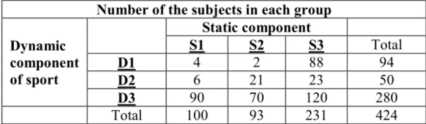
Table 1. – Number of the subjects in each group Number of the subjects in each group Dynamic
component of sport
Static component
S1 S2 S3 Total
D1 4 2 88 94
D2 6 21 23 50
D3 90 70 120 280
Total 100 93 231 424
To examine the seasonal changes of the athletes heart, we observed 15 elite young swimmers (13-15 years of age) and 15 age and gender matched non-athletic sujects. All athletes were members of the same team and members of the national team of their age, swimming in the same team. They had a swimming history of at least four seasons, in average more than 20 hours per week training.
The observation period lasted 1.5 years long, including six measurements according to the macro- and microcyclic periods of training: starting values (S), general enduarnce preparation period 1 (GP1), race specific training period 1 (RP1), detraining (DT), general endurance preparation period 2 (GP2), race specific training period 2 (RP2). On the first and fifth occasion of the study, results were compared to non-athletic controls.
Methods
Study design is presented in Table 2.
4
Table 2. – Study design
Study design
Sport medicine questionnaire: medical, sport, family history Physical examination (AHA 12 point) + blood pressure 12-lead ECG
Echocardiography: Philips HD15
Statistical analysis: Statistica for Windows 12.0
The Echocardiography was performed according to the recommendations of the American Society of Echocardiography.
Data analysis was performed off-line, at least three measurements were averaged. Statistical analysis was carried out with Statistica for Windows 12.0. Normality of the variables was tested with Shapiro- Wilks Test. Normal distribution variables were tested with ANOVA, non-normal distributive parameters with Kruskall-Wallis Test. In the second part of the thesis, Wilcoxon Test, repeated measurement or Friedmann ANOVA were applied. Type I error was set at 5%.
Measured or calculated echocardiographic parameters were the followings: morphological features: wall thickness, ventricular and atrial internal diameters, left ventricular muscle mass (LVMM), hypertrophy index (MQ), ventricular volume (ESV, EDV). Relative (rel) values were related to the body surface area (BSA) by quotients, where the nominator and the denominator has the same power, indices (I) were the values divided with the body surface area.
5
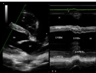
Figure 1. - 2D and M-Mode LV parasternal view
Figure 2. - 2D-Four-chamber view
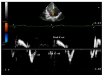
Functional characteristics of the athlete’s heart were determined by pulse-waved and tissue Doppler measurements, velocities and their ratios for the diastolic function are demonstrated in Figure 3 and 4. Systolic function was characterised by TAPSE for the right, and by the EF, VCF, FS and CO for the left ventricle.
Figure 3. - Mitral inflow curve
6
Figure 4. - Mitral annular TDI (septal)
Regulatory characteristics were described by heart rate (HR), stroke volume (SV) and cardiac output (CO) at rest.
4. Results
In the dissertation, all results are published in tabels. Here the most important findings and results are presented.
4.1 Anthropometrical data
No differences were seen in the age of the subjects in each groups. Tallest athletes were found in the S3D3, smallest in the S3D1 group. In the latter group highest weight were observed.
Resting blood pressure didn’t differ among the athletic and control group, unlike resting HR: apart from S2D2 and S1D1, all athletes had lower resting heart rate than the non-athletic subjects.
The non-athletic group had naturally a significantly lower training hour per week than the athletes. Training hours of the athletes didn’t differ in the different categories.
7 4.2. Regulatory features
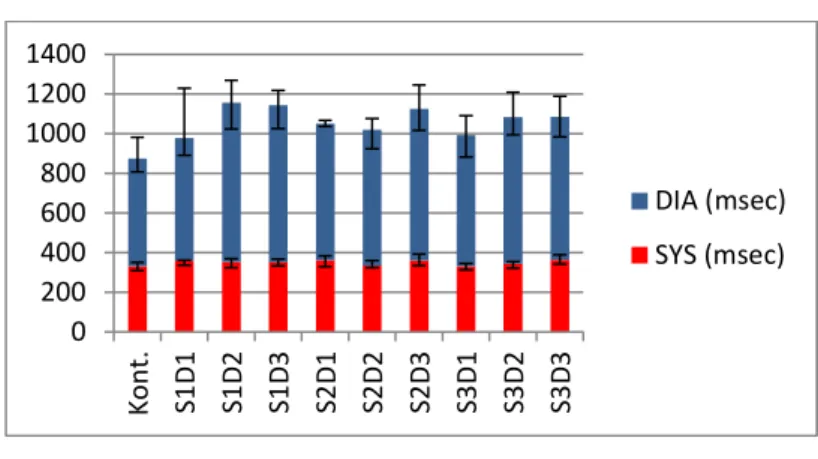
Both the length of the diastole and that of the systole proved to be longer in some athletic groups than in the controls (Figure 5.). Difference is more pronounced in the length of diastole, owing to the diastase alone. Cardiac output at rest was significantly lower in the S1D3 than in the S3D3 category.
Figure 5. – Differences in the length of systole and diastole (median; upper and lower quartiles)
4.3. Left ventricular training adaptation
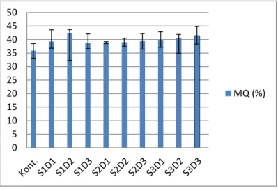
Left ventricular training adaptation was characterized by the wall thickness and internal diameters (both absolute, relative and indexed values). Marked left ventricular hypertrophy was observed in most athletic groups. The highest MQ values were seen in sports with high static components (Figure 6).
2000 400600 1000800 12001400
Kont. S1D1 S1D2 S1D3 S2D1 S2D2 S2D3 S3D1 S3D2 S3D3
DIA (msec) SYS (msec)
8
Figure 6. – Muscular quotient (median;upper and lower quartiles)
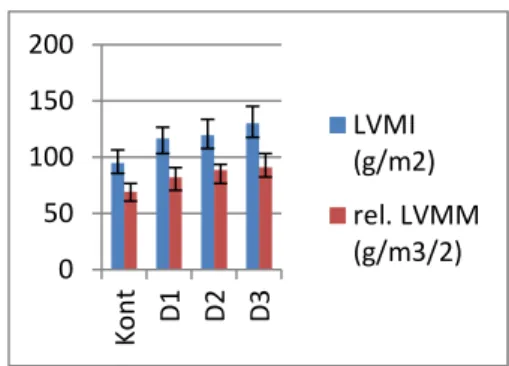
The higher the dinamic component was, the greater left ventricular internal diameters were registered. Impact of the static and dinamic components of sports on LVMM are presented in Figure 7 (blue indexed, red relative values).
05 1015 2025 3035 4045 50
MQ (%)
0 50 100 150 200
Kont S1 S2 S3
9
Figure 7 – Effect of static and dinamic components on LVMM (median; upper and lower quartiles; g/m2 and g/m2/3;
Kont and NA: Non athletes)
Table 3. Differences in the left ventricular muscle mass
LVMM LVM Index Rel. LVMM
Difference Significance Significance Significance NA<S2D3 p<0.00001 p<0.00001 p<0.00001 NA<S3D1 p<0.00001 p<0.00001 p<0.00001 NA<S3D2 p<0.00001 p<0.00001 p<0.00001
NA<S2D2 p<0.01 p<0.01 p<0.01
NA<S3D3 p<0.00001 p<0.00001 p<0.00001 NA<S1D3 p<0.00001 p<0.00001 p<0.00001
S2D3>S3D1 p<0.01 p<0.001 p<0.001
S3D3>S3D1 p<0.00001 p<0.00001 p<0.00001
S3D3>S2D2 p<0.001 p<0.001 p<0.05
S3D3>S1D3 p<0.00001 p<0.00001 p<0.001 Left ventricular diastolic function: the E/A ratio was significantly higher in athletes of sports with high dynamic components than non-athletes, the higher the static component was in the D3 sports, the difference was more pronounced: S1D3 p<0.002, S2D3 p<0.001, S3D3 p<0.000001. Tissue Doppler
0 50 100 150 200
Kont D1 D2 D3
LVMI(g/m2) rel. LVMM (g/m3/2)
10
velocities didn’t differ significantly in the ten groups, such as no difference was observed in the resting systolic function.
4.4. Right ventricular adaptation
A marked morphological adaptation was observed in the sport disciplines with high dynamic component. Internal diameters in diastole showed more pronounced difference: p<0.00001 each diastolic paramerer, p<0.01 each systolic parameter). Where statistical analysis was available, no significant differences were seen in the parameters describing the right ventricular function.
4.5. Training adaptation of the atria and aortic root dilatation
Where statistical analysis was available, the S3D3 category presented greater left ventricular internal diameters and area values than the non-athletic group (LA width p<0.00002, LA length p<0.002, LA area p<0.000001). Differences in the right atrial parameters were even more marked comparing to the control group.
Aortic root dilatation was observed only in the S3D3 (p<0.001) and S3D1 (p<0.00001) athletes when compared to non- athletes.
Bonferroni-Holms correction is advised because of the large number of comparisons. Taking all data into consideration Type I error should be 0.07 %, when only the left ventricle is examined 0.1
%.
11
IV.6. Regression equations for the different groups describing the LV training-adaptation
Non-athletic group: p=0.186
2.542 · + 0.129 · + 6.947 · − 0.399 · + 9.404 ·
= 210.922 : ℎ
: ℎ
:
: : S1D1 category: p=0.009
2.437 · + 0.128 · + 6.630 · − 0.291 · + 9.313 ·
= 204.168 S1D2 category: p=0.013
2.455 · + 0.133 · + 6.824 · − 0.310 · + 10.18 ·
= 213.113 S1D3 category: p=0.200
2.411 · + 0.134 · + 6.854 · − 0.319 · + 9.161 ·
= 208.222
No equations are available for S2D1 group because of the low number of subjects.
S2D2 category: p=0.047
2.446 · + 0.126 · + 6.846 · − 0.327 · + 10.096 ·
= 208.697 S2D3 category: p=0.112
12
2.351 · + 0.124 · + 6.924 · − 0.281 · + 9.821 ·
= 208.327 S3D1 category: p=0.148
2.460 · + 0.126 · + 6.733 · − 0.307 · + 9.958 ·
= 205.837 S3D2 category: p=0.292
2.342 · + 0.119 · + 7.088 · − 0.297 · + 10.489 ·
= 214.154 S3D3 category: p=0,254
2.443 · + 0.130 · + 6.665 · − 0.239 · + 10.066 ·
= 209.086
4.7. Seasonal changes in the athlete’s heart
Height and weight in young athletes followed the normal maturation. No differences between the two genders were observed.
There are no seasonal changes in the resting heart rate and blood pressure in the athletic group. Resting blood pressure values were in normal range for athletes as well.
The non-athletic group was age and gender matched, no differences in the anthropometrical data could be found between athletes and non-athletes in both genders, maturational status was also the same.
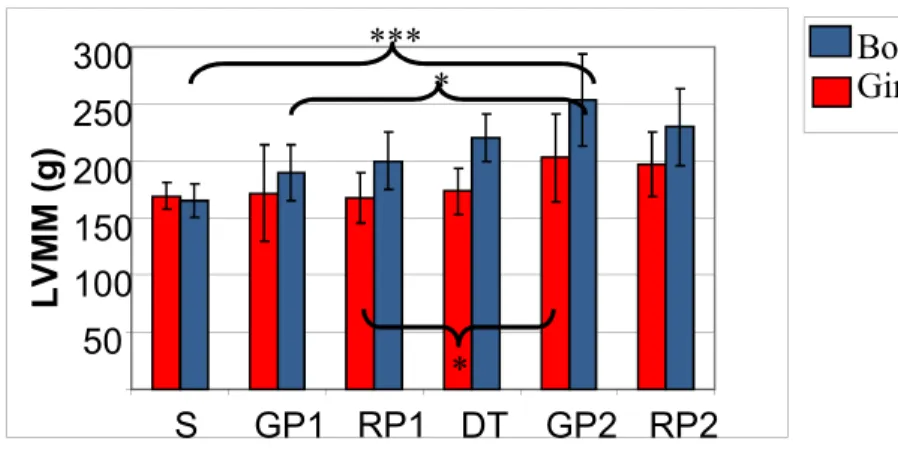
Resting heart rate in young female athletes was significantly lower than that of nin-athletic girls. Seasonal changes bin the LVMM are presented in Figure 8.
13
Figure 8. - Seasonal changes in the left ventricular muscle mass LVMM: left ventricular muscle mass S: starting values, GP1,2:
General Endurance Preparation Period, RP1,2: Race Specific Training Period 1, 2, DT: Detraining. Level of significance: *:
p<0.05, ***: p<0.001. Values are presented with means.
5. Conclusions
Based on our results and findings, the following conclusions can be made:
Body size, quantity and quality of training, type of sport, age (together with gender and ethnicity) are both influencing factors on the athlete’s heart.
According to our present and previous results we suggest to present the data referring to the morphological adaptation of the heart related to the body size. According to our
50 100 150 200 250 300
S GP1 RP1 DT GP2 RP2
LV M M (g )
Girl
*** Boy
*
*
14
suggestions, data indexed to the BSA were the nominator and the denominator have the same power should be used.
Physical activity doesn’t seem to lower resting blood pressure in elite athletes of 18-35 years of age, which also means that excessive physical activity doesn’t influence the blood pressure in an undesired way.
Our findings about the resting heart rate are similar to that described earlier. We measured however not only longer diastole, but longer systole in athletes, however differences in the length of diastole are more pronounced.
No differences could have been observed in the resting systolic function among the groups, which doesn’t mean, that athletes can not have better systolic function during performance.
Findings concerning the left ventricular morphological adaptation are the same as described before. The thickest LV wall and highest internal diameter data were measured in the athletes of sports with high static and high dynamic components. Most of the athletes had bigger LVWT than the control group.
In the S1D1 category (low static and low dynamic components) neither morphological nor regulatory or functional features of the athletic heart were present, that confirms their eligibility in athletes with cardiovascular disorders. Beneficial effects on the heart can not be
15
observed as well (this statement can not refer to the metabolic effects, which we did not examine).
Better diastolic function in athletes were present in sports with high dynamic components. To improve the diastolic function (relaxation) of the heart, only these sports can be recommended.
TDI is essential in differentiating physiological and pathological features of the heart. Our results suggest, that this method doesn’t give any further information on the training adaptation of the heart, but confirms the previous findings with pulsed-wave Doppler.
Similarly to previous results, marked morphological adaptation was observed in the right ventricle, in the athletes with high dynamic components (S1D3, S3D3).
Opposite to the left ventricle no functional adaptation is present in the right one. No differences to the control group mean a good function of the enlarged ventricle, which is an important differentiating factor from ARVD/C.
Morphological adaptation of the atria is only present in the S3D3 group.
Aortic root dilatation doesn’t exceed the normal values in the athletes.
The regression equations help better charachterize physiologic training adaptation of the heart. They also make it possible to compare features of the athlete’s heart of athletes of different origin. Further examinations are needed
16
to objectivise the differences between male and female athletes.
In the future we want to pay attention to the sports of the S1D2 category, to precisely characterise the cardiovasculatory adaptation to this kind of training. We believe, that they can be more extensively recommended for athletes with cardiovascular abnormalities.
New echocardiographic methods are available to characterise better the training adaptation of the heart.
Although we have more precise and modern methods such as MRI to investigate the cardiac adaptation of the heart, echocardiography will be the first method of choice for the near future in athletic heart research
The following conclusions can be made on the seasonal changes in the athlete’s heart:
Seasonal changes can be observed in the morphological adaptation of the athletes heart (LV) in young athletes.
The thickest LV walls and smallest LV internal diameters were found in the second general endurance preparation period, that limits the achievement/performance available in the following race specific period.
On behalf of this we suggest to declare in which training period training adaptation of the heart was examined. This could lead to a precise and reliable comparison of findings of different study groups.
17
Further examinations are needed to judge on the seasonal changes in elite adult athletes.
6. Publications
1. Publications related to the present thesis
Csajági E, Szauder I, Major Z, Pavlik G. Left Ventricular Morphology in Different Periods of the Training Season in Elite Young Swimmers. Pediatr Exerc Sci. 2015;27(2):185-91. IF: 1.452
Pavlik G, Major Zs, Csajági E, Jeserich M, Kneffel Zs: The athlete’s heart. Part II. Influencing factors on the athlete’s heart: kinds of sports, age, gender, performance level. Acta Physiol Hung 2013;100(1):1-27. IF: 0.747
2. Publications not closely related to the present thesis
Szauder I, Csajági E, Major Z, Pavlik G, Ujhelyi G. Treatment of Hypertension: Favourable Effect of the Twice-Daily Compared to the Once-Daily (Evening) Administration of Perindopril and Losartan. Kidney Blood Press Res. 2015;40(4):374-85. IF: 2.123Major Z, Csajági E, Kneffel Z, Kováts T, Szauder I, Sidó Z, Pavlik G. Comparison of left and right ventricular adaptation in endurance- trained male athletes. Acta Physiol Hung. 2015;102(1):23-33. IF:
0.734