DISTRIBUTION OF NOCICEPTIN IN THE PANCREAS AND UTERUS OF NORMAL AND DIABETIC RATS
PhD Thesis SAEED TARIQ
Doctoral School of Pharmaceutical Sciences, Semmelweis University
Supervisor : Professor Kornélia Tekes, Pharm. D., Ph.D., D.Sc
Official reviewers: Professor Erzsébet Fehér, M.D., D. Sc Professor Gábor Pethő, M.D., Ph.D
Head of the Final Examination Committee:
Professor Valéria Kecskeméti M.D., C.Sci
Members of the Final Examination Committee:
Professor Gábor Halmos, Pharm.D., Ph.D Dr.László Kursinszki, Pharm.D., Ph.D
Budapest
2015
1
Contents
Contents 1
List of Abbreviations 5
1. Introduction 7
1.0 Neuropeptides 7
1.1 Nociceptin 7
1.1.1 Structure of nociceptin 8
1.1.2 Nociceptin receptor 11 1.1.3 Distribution and localization of nociceptin and its receptor 11
1.1.4 Physiological roles of nociceptin 11
1.1.5 Nociceptin in pathological conditions 14
1.1.6 Nociceptin receptor (NOP) agonists and antagonists 15 1.1.7 Effects of nociceptin in the pancreas and uterus 15 1.1.8 Methods used to study localization and tissue distribution
of nociception 16
1.1.9 Application of immunohistochemical, Immunofluorescence and 18 electron microscopy methods in the study of nociceptin
1.2 Pancreas 18
1.2.1 Histology of the pancreas 18
1.2.2 Principal cell types of the pancreatic islets 19
1.2.3 Regulatory innervation of the pancreas 19
1.3 Diabetes mellitus 22
1.3.1 Types of diabetes mellitus 22 1.3.2 Role of novel peptides in diabetes mellitus 23
1.3.3 Nociceptin in diabetes mellitus 24
2
1.4 The uterus 24
1.4.1 Histology of the rat uterus 26
1.4.2 The uterine wall 26
2. Hypotheses, Aims and Objectives 28
3. Materials and Methods 29
3.1 Experimental animals 29
3.2 Induction of experimental diabetes mellitus 29
3.3 Experimental design 29
3.4 Body weight 30
3.5 Glucose measurement 30
3.6 Glucose tolerance test on non-diabetic and diabetic rats 30
3.7 Tissue collection 30
3.8 Light microscopy of pancreas and uterus 31
3.9 Immunohistochemical studies of the pancreas 31
3.10 Double- labelling immunofluorescence studies of pancreas 32 3.11 Immunofluorescence microscopy of the uterus 33 3.12 Tissue processing for conventional electron microscopy 33 3.13 Double labelling immuno electron microscopy of pancreas 34
3.14 Immunoelectron microscopy study of uterus 35
3.15 Morphometry 36
3.16 Western blotting of nociceptin in tissues 36
3.17 Statistical analysis 39
4. Results 40
4.1 Pancreas 40
4.1.1 Body and organ weight ratios 40
4.1.2 Glucose measurement 40
4.1.3 Glucose tolerance test in male rats 40
4.1.4 Light microscopy of pancreas 42
3
4.1.5 Immunohistochemistry studies (Avidin Biotin Complex method) 42 4.1.6 Double labelling immunofluorescence study 44
4.1.7 Conventional electron microscopy 46
4.1.7.1 Non-diabetic and diabetic pancreatic β-cells 46 4.1.7.2 Double labelling immunoelectron microscopic study 48
4.1.8 Western blot analysis 51
4.2 Uterus 52
4.2.1 Body weight and organ weight ratios 52
4.2.2 Glucose measurement 52
4.2.3 Glucose tolerance test in female rats 52
4.2.4 Gross morphology of the uterus 54
4.2.5 Light microscopy study 55
4.2.6 Immunofluorescence study 57
4.2.7 Morphometry 57
4.2.8 Western blot analysis 60
4.2.9 Conventional electron microscopy of uterus 61
4.2.10 Immunoelectron microscopy study 64
5. Discussion 66
5.1 Metabolic parameters 67
5.2 Pancreas 67
5.3 Uterus 71
5.3.1 Nociceptinergic innervation 73
5.3.2 Conventional electron microscopy 74
6. Conclusion 75
7. Bibliography 77
8. Publications 101
8.1 Publications related to the PhD thesis 101
4
8.2 Publications not related to the PhD dissertation 102
8.3 Other scientific puplications 104
9. Summary 106
9.1 Összefoglalás 107
10. Acknowledgements 108
11. List of Tables and Figures 109
11.1 List of Tables 109
11.2 List of Figures 109
12. Appendix 112
5
List of Abbreviations
ABC Avidin biotin complex
CMHS College of Medicine and Health Sciences
CNS Central nervous system
CSF Cerebrospinal fluid
DAB 3, 3-diaminobenzidine tetrahydrochloride
dL Deciliter
DM Diabetes mellitus
EM Electron microscopy
FFA Free fatty acid
FITC Fluorescein isothiocyanate
GAPDH Glyceraldehyde 3-phosphate dehydrogenase GDM Gestational diabetes mellitus
GPCRs G-protein coupled receptors
HCS Chorionic somatomammotropin
hPGH Human placental growth hormone
HPLC High performance liquid chromatography
i.p Intraperitoneal
IAPPa Insulin and islet amyloid polypeptide or amylin IDDM Insulin-dependent diabetes mellitus
IEM Immunoelectron microscopy
6
mg/rat milligram per rat
MOP mu opioid receptors
N/OFQ Nociceptin/Orphanin FQ
NC Nociceptin
NE Nuclear envelope
NMDA N-Methyl-D-aspartate
NOP Nociceptin receptor
NOP1 Nociceptin receptor-1
OFQ Orphanin FQ
OP4 Opioid receptor-4
ORL-1 Opioid receptor-like1
PBS Phosphate buffer saline
PNS Peripheral nervous system
PP-cells Pancreatic polypeptide cells
PPNOC Prepronociceptin
RIPA Radioimmunoprecipitation assay buffer
STZ Streptozotocin
TBST Tris Buffered Saline with Tween® 20
TEM Transmission electron microscopy
TNFa Tumor necrosis factor alpha
TRITC Tetramethyl rhodamine isothiocyanate
7
1. Introduction
1.0 Neuropeptides
Neuropeptides are small molecules used by neurons for extracellular signalling. There are a wide variety of neuropeptides depending upon the required brain function and body physiology. For instance, neuropeptides are involved in analgesia, reward, food intake, metabolism, reproduction, social behaviour, learning and memory [1, 2]. Examples of neuropeptides include enkephalin, dynorphin, nociceptin, neuropeptide Y, relaxin, gastrin, cortistatin, somatostatin and calcitonin gene related peptide. All of these neuropeptides have different biological functions. Neuropeptides and neurotransmitters are extracellular signalling molecules involved in a variety of physiological functions. Some peptide hormones, such as somatostatin, also act as neuropeptides. They are secreted from neuroendocrine glands and travel through the blood circulation to distant target tissues and organs. Whereas, neuropeptides are mainly secreted from neuronal cells and send their signals to neighbouring cells. Many neuropeptides may also be co-released with other small-molecule neurotransmitters. For example enkaphalin, neuropeptide-Y and galanin coexist with norepinephrine, a clasical neurotransmitter [3]. Neuropeptides have long been known to play a regulatory role in complex behaviours, such as learning, and pain sensation and memory [1, 2]. Additionally, the metabolism of neuropeptide, such as dynorphin, is impaired in a number of neurological diseases like schizophrenia and addiction [4]. In summary neuropeptides can act as either neurohormones neurotransmitters or neuromodulators where they help to maintain physiological homeostasis and influence important physiological functions [4].
1.1 Nociceptin General information
Nociceptin (NC), also known as orphanin FQ (N/OFQ, is the natural ligand for NC receptor, called opiate receptor-like 1(ORL-1) or non-classical opioid receptor (NOP). NC is an endogenous ligand of G-protein coupled receptor (GPCRs) family. Nociceptin is derivative of prepronociceptin (PPNOC), which is present in a large variety of species [5].
Although, NC is an opioid-related peptide, it binds to its own receptor but does not attach to the classic opioid receptors [6, 7]. This neuropeptide is mainly expressed in neurons of
8
the central (CNS) and peripheral (PNS) nervous systems. The orphan receptor approach regarding the endogenous ligand of the oGPCR ORL-1 was demonstrated successfully with the discovery of N/OFQ, [7, 8].
Meunier et al. [7] used the term nociceptin for the novel peptide that he discovered based on putative pro-nociceptive properties. Almost in th same period, Reinscheid et al. [8]
called this new orphan peptide orphanin FQ, as a ligand of an orphan receptor, whose first and last amino acids are Phe (F) and Gln (Q), respectively.
After 1995, a series of publications have provided detailed descriptions of its pharmacological, physiological and behavioural roles [9–11] and emphasized their biological importance in the body. In addition, the pattern of OFQ/N messenger RNA expression in the CNS has also been reported [11].
1.1.1 Structure of nociceptin
Nociceptin (N/OFQ, NC) is a 17-amino acid peptide (Phe-Gly- Gly- Phe –Thr-Gly-Ala- Arg-Lys-Ser-Ala-Arg-Lys-Leu-Asn-Gln-NH2) (Figure 1) which displays homology in amino acid sequence with opioid peptides, such as dynorphin A, endorphin and enkephalin.
NC and dynorphin A both have 17 amino acids bounded by pairs of basic amino acids which are essential in their assembly from precursors peptides. The other similarity between these two peptides is that they have internal pairs of basic amino acids suggesting the likelihood of further processing. These opioid peptides share an YGGF pattern, where the fifth amino acid is either leucine or methionine (Figure 2). The amino terminal of NC is composed of phenylalanine instead of tyrosine, followed by GGF. As a final point, both peptides contain the same last two amino acids at the carboxyl terminal. NC is generated proteolytically from a larger peptide precursor, preproorphanin in a similar manner to endogenous opioids, which contains additional neuropeptides that may have biological activities [5, 12–16]. The structure of the primary rat and human preproorphanin has been elucidated. Since preproorphanin shares close structural homology to the endogenous opioid peptide precursors like prodynorphin and preproenkephalin, it has been suggested that a synchronized mechanism of evolution may have “alienated” NC from the opioid systems [17–19]. The studies on the regional distribution on preproNC, OP4/NOP receptors and NC mRNA showed that they are highly expressed in various neuronal sites with a
9
pattern wholly distinct from those of classical opioid peptides and share characteristic structural feature mainly with preprodynorphin.
Figure 1: Structural similarities between dynorphin A and nociceptin amino acid sequences [20]. Message: The domain (N-terminal) responsible for the activation of sequences. Address: The segment (C-terminal) involved in the binding of nociceptin and dynorphin A to specific receptors.
Nociceptin: Phe-Gly-Gly-Phe-Thr-Gly-Ala-Arg-Lys-Ser-Ala-Arg-Lys-Leu-Ala-Asn-Gln Dynorphin A: Tyr-Gly-Gly-Phe-Leu-Arg-Arg-lle-Arg-Pro-Lys-Leu-Lys-Trp-Asp-Asn-Gln γ-Endorphin: Tyr-Gly-Gly-Phe-Met-Thr-Ser-Glu-Lys-Ser-Gln-Thr-Pro-Leu-Val-Thr-Leu Met-enkephalin:Tyr-Gly-Gly-Phe-Met
Leu-enkephalin: Tyr-Gly-Gly-Phe-Leu.
Figure 2: Structure of nociceptin and some of the closely related endogenous neuropeptides.
10
Molecular structure of nociceptin
11 1.1.2 Nociceptin receptor
Nociceptin (NC) receptor (NOP) in man consists of seven transmembrane regions with 370 amino acids [21]. The N-terminal of NC receptor polypeptide is a 44-amino acid unit with 3 adjoining sequences for glycosylation (Asn-X-Ser/Thr). In addition, protein kinase A and C can phosphorylate the polypeptide in the second and third intracellular loops, respectively.
Numerous studies have described the structure of NC [11, 22, 23].
1.1.3 Distribution and localization of nociceptin and its receptor
NC and its receptors are widely distributed in the brain region like in thalamus, hippocampus, olfactory bulb, amygdala and cortical areas. [13, 21, 24–33]. NC receptors have been identified in other areas including the dorsal and ventral horns of the spinal cord [29, 34], periaqueductal gray matter of the midbrain and the nucleus raphe magnus.
NOP receptors co-localizes with mu opioid (MOP) receptors [26]. The localization and distribution pattern of NC-NOP system have revealed a role for NOP receptor in the processing of behavioural response to stress and anxiety, motor and balance control, aggression and autonomic control of physiological processes, reinforcement and reward, nociception and sexual behavior, [29, 34].
In addition, NC and its receptors have also been detected outside of the brain in peripheral organ systems such as spleen, vas deferens, intestine and immune system [27, 35, 36].
1.1.4 Physiological role of nociceptin
Nociceptin mediates the modulation of pain via stimulation of NOP in the brain and causes an increase in pain sensation, which suggests that NC may be implicated in the transmission of pain signals. In addition to the regulation of pain signals, NC has been implicated in a variety of physiological functions including, inhibition of locomotive activity [37], reversal of stressed-induced analgesia [37], reduction of stress responses [38, 39], induction of impairment in memory and learning [40, 41], release of neurotransmitters and hormones [42, 43], induction of diuresis and anti-natriuresis [44], neuronal differentiation [45], sexual and reproductive behaviour [46], itching, biting and licking [47], uterine contraction [48] feeding [49] anxiety [50–52], gastrointestinal motility [53]
12
induction of transient hypotension, diuresis-induced bradycardia [54–56], micturition [57]
and antitussive effect in cough [58]. Additionally nociceptin may play a vital role in hypoxic-ischemic brain injury [59–62]. Many other functions regulated by NC include diuresis and sodium balance [44], regulation of temperature [63], vestibular function [64]
modulation of inhibitory neural pathway that inhibit gastrointestinal movement, colonic and mucosal propulsive activity [65]. Nociceptin has been shown to suppress both excitatory [66] and inhibitory [67] synaptic transmission in the murine spinal cord. It also suppresses NMDA receptor-dependent long-term depression in the dentate gyrus of the hippocampus [68]. It also suppresses oxytocin, vasopressin and GnRH release [46] and inhibits tachykinin function [69]. Nociceptin activation of NOP is capable of modulating the activity of neurons in the suprachiasmatic nucleus [70] and lateral amygdale [71]. It has also been shown that nociceptin inhibits enkephalin release [72], mesolimbic dopamine transmission [73] and trigeminal neuronal response to excitatory amino acids [74]. In addition, nociceptin can inhibit endomorphin-1-induced analgesia [75]. In the periphery, N/OFQ inhibits nitric oxide release in the colon of murines [76]. OP4/NOP receptor reduces the activation of adenylyl cyclase and Ca2+ channels while activating K+ channels in a manner similar to opioids.
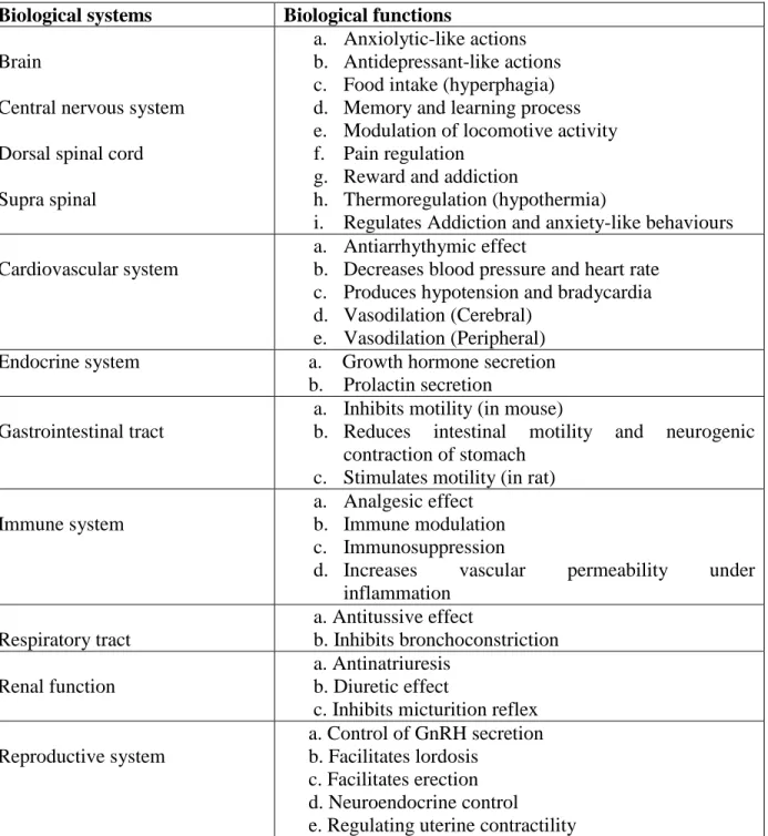
All of these reports show that nociceptin is indeed involved in a large number physiological functions (Table 1).
13
Table 1: Function of nociceptin in different biological systems Biological systems Biological functions
Brain
Central nervous system Dorsal spinal cord Supra spinal
a. Anxiolytic-like actions b. Antidepressant-like actions c. Food intake (hyperphagia) d. Memory and learning process e. Modulation of locomotive activity f. Pain regulation
g. Reward and addiction
h. Thermoregulation (hypothermia)
i. Regulates Addiction and anxiety-like behaviours Cardiovascular system
a. Antiarrhythymic effect
b. Decreases blood pressure and heart rate c. Produces hypotension and bradycardia d. Vasodilation (Cerebral)
e. Vasodilation (Peripheral) Endocrine system a. Growth hormone secretion
b. Prolactin secretion Gastrointestinal tract
a. Inhibits motility (in mouse)
b. Reduces intestinal motility and neurogenic contraction of stomach
c. Stimulates motility (in rat) Immune system
a. Analgesic effect b. Immune modulation c. Immunosuppression
d. Increases vascular permeability under inflammation
Respiratory tract
a. Antitussive effect
b. Inhibits bronchoconstriction Renal function
a. Antinatriuresis b. Diuretic effect
c. Inhibits micturition reflex Reproductive system
a. Control of GnRH secretion b. Facilitates lordosis
c. Facilitates erection d. Neuroendocrine control
e. Regulating uterine contractility Modified from Tariq et al. [77]
14 1.1.5 Nociceptin in pathological conditions
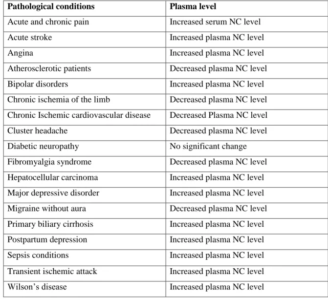
Plasma nociceptin concentration has been found to either increased or decreased in different pathological conditions (Table 2). Albeit, nociceptin is produced in neurons of the CNS and PNS [78] but there is growing evidence that it is also found in body fluids including the plasma, serum, and cerebrospinal fluid (CSF) [79]. The circulating nociceptin may be a good predictor and a biological marker for a variety of pathological conditions.
However, few studies have been done on this topic. [80] noted that the CNS level of NC and prepronociceptin increased significantly in animal models of chronic constriction lesion and diabetic neuropathic pain.
Table 2: Circulating nociceptin (NC) in different pathological conditions Pathological conditions Plasma level
Acute and chronic pain Increased serum NC level
Acute stroke Increased plasma NC level
Angina Increased plasma NC level
Atherosclerotic patients Decreased plasma NC level
Bipolar disorders Increased plasma NC level
Chronic ischemia of the limb Decreased plasma NC level Chronic Ischemic cardiovascular disease Decreased Plasma NC level
Cluster headache Decreased plasma NC level
Diabetic neuropathy No significant change
Fibromyalgia syndrome Decreased plasma NC level Hepatocellular carcinoma Increased plasma NC level Major depressive disorder Increased plasma NC level Migraine without aura Decreased plasma NC level Primary biliary cirrhosis Increased plasma NC level Postpartum depression Increased plasma NC level
Sepsis conditions Increased plasma NC level
Transient ischemic attack Increased plasma NC level
Wilson’s disease Increased plasma NC level
Modified from Tariq et al. [77]
15
1.1.6 Nociceptin receptor (NOP) agonists and antagonists
In addition to the peptide ligands, many classes of non-peptide chemical NOP ligands have been synthetized and identified. They include piperidines, nortropanes, spiropiperidines, 4- amino-quinolines and quinazolines, and many others. A detailed study of NOP agonists and antagonists may be found in the following reviews [10, 77, 81, 82].
1.1.7 Effects of nociceptin in the pancreas and uterus Pancreas
Tekes et al. [79] revealed that long-term diabetes mellitus does not modify the plasma or CSF levels of NC. The pancreas, which regulates a variety of imperative physiological activities, has also been studied in relation to nociceptin and NOP receptors. It is believed that prolonged and extended hyperglycaemia reduces pancreatic β-cell responsiveness to secretagogues [83]. A study was conducted by Linari [84] regarding the regulation of pancreatic exocrine secretion in vitro by NC revealed that the NC-NOP system plays an inhibitory role in the regulation of exocrine pancreatic secretion such as amylase. An experiment was conducted by Matsushita et al. [85] with intracerebroventricular infusion of NC to mice and observed an increased level of plasma insulin. In diabetic rats the role of NC in glucagon secretion still remains to be determined.
Uterus
Klukovits et al. [48] revealed the presence of PNOC in the uterus of rats using radioimmunoassay and radioligand-binding techniques. Deák et al. [86] reported that NC derived from PNOC relaxes uterine muscle in rat. Additionally, NC like any other classical opiates, seems to function as a neuromodulator of the endocrine system of the reproductive system. Bryant et al. [87] reported that NC increased prolactin release in both male and female rats. Using NC knock-out mice and their wild-type littermates as controls, they showed that NC is important in prolactin regulation during lactation. They also showed that offspring survival is decreased when NC is not expressed in post-partum dams. In addition, the role of NC was investigated during the post-partum period by Gu et al. [88]. They suggested that NC may play a role in the pathogenesis of postpartum depression. It is also
16
speculated that the secretion of corticotropin-releasing hormone (CRH) from the epithelium of the endometrium of uterine wall and other hormones secreted from the endometrium may be regulated by NC.
Another important physiological role of the uterine wall is its ability to contract, due to the presence of smooth muscle in the myometrium. Mollereau and Mouledous [89] reported the presence of NC receptors in smooth muscle cells, which is of course an indirect evidence of the presence of NC in smooth muscles. However, no morphological evidence is available for the presence of NC in the muscular layer of the uterus. Physiological studies have shown that NC relaxes the human uterus [86]. However,the exact nature of the role of NC in the uterus and on smooth muscle in general is far from clear, not to mention its pattern of tissue distribution.
1.1.8 Methods used to study localization and tissue distribution of nociceptin.
Several techniques have been employed to study different parameters including, light immunohistochemistry, and ultrastructural localization of NC in tissue and organ systems.
Light and electron microscopy technoiques have been rarely employed to study the pattern of distribution of NC at both the tissue and cellular levels. However, further studies on the location of NC in tissues and cytoplasmic organelles are indeed necessary for understanding its potential clinical application. A basic, reproducible and widely applicable method is needed to achieve this target.
Apart from these research methods and techniques, antisense oligonucleotides targeting NOP receptors or PNOC genes, or applying antibodies against NC directly have been used.
In some cases, the receptors or the peptide precursor genes have been deleted genetically in some animal models [9].
An extensive and robust investigation has been carried out in CNS of murine model to understand the distribution and pattern of NC and its receptors in the brain using in situ hybridization and immunohistochemical techniques [29, 90, 90–92]. There are some other detailed maps that have been compiled by Darland et al. [93] in which the expression of NOP receptors and PPNOC mRNAs where depicted in discrete areas of the rat brain.
17
Due to lack of appropriate techniques it has been difficult to determine the binding sites of NC in organ systems. In order to solve this problem, radioligand technique was used to determine the exact location of this peptide in the tissues. Unfortunately, the method has never been used again because it was not reproducible [77]. The basic cause of failure of this technique to localize the binding site of nociceptin was due to the choice of an inappropriate/insensitive technique or low tissue level of its receptors.
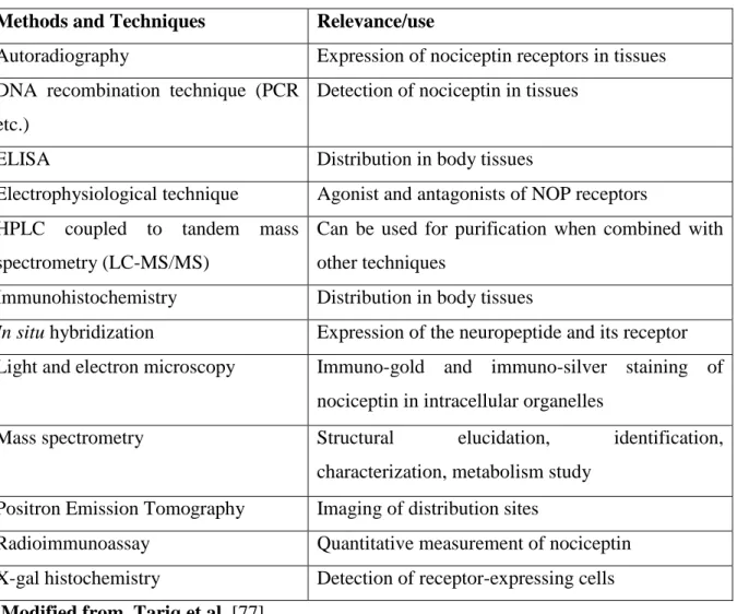
A summary of techniques and methods used to investigate the structure and distribution of NC is shown in Table 3.
Table 3: Methods used to study the structure and distribution of nociceptin in tissues and body systems.
Methods and Techniques Relevance/use Autoradiography Expression of nociceptin receptors in tissues DNA recombination technique (PCR
etc.)
Detection of nociceptin in tissues
ELISA Distribution in body tissues
Electrophysiological technique Agonist and antagonists of NOP receptors HPLC coupled to tandem mass
spectrometry (LC-MS/MS)
Can be used for purification when combined with other techniques
Immunohistochemistry Distribution in body tissues
In situ hybridization Expression of the neuropeptide and its receptor Light and electron microscopy Immuno-gold and immuno-silver staining of
nociceptin in intracellular organelles
Mass spectrometry Structural elucidation, identification,
characterization, metabolism study Positron Emission Tomography Imaging of distribution sites
Radioimmunoassay Quantitative measurement of nociceptin X-gal histochemistry Detection of receptor-expressing cells
Modified from Tariq et al. [77]
18
1.1.9 Application of immunohistochemical, immunofluorescence and electron microscopy methods in the study of nociceptin
Literature search shows that electron microscopy (EM) is rarely used for the pattern of distribution and localization of NC in tissues even though it provides a solid and robust way of determining the cellular and intracellular localization. Electron microscopy comprises conventional transmission electron microscopy (TEM), immuno-EM whereas immunofluorescence microscopy is also a good tool for the morphological studies on nociceptin. In the electron microscopy, double-labelling immunocytochemistry is the most commonly used method for the detection of two or more peptides in cytoplasmic organelles [94].
1.2 Pancreas
1.2.1 Histology of pancreas
The pancreas has two functional components: exocrine part which produces pancreatic juice for digestion and endocrine component that produces insulin and other hormones such as glucagon, somatostatin and pancreatic polypeptide. The exocrine pancreas is formed mostly from acinar cells that secrete digestive enzymes and alkaline buffer to neutralize the acidic chyme formed by the stomach. These acini are surrounded by smaller ducts which drain into the central pancreatic duct. The gross morphology and histology of pancreas are shown in figures 3a and 3b.
The endocrine part of the pancreas is aggregated into small islands of cells called the islet of Langerhans. The islets are more abundant in the tail region of the pancreas. The capillaries of the islets are surrounded by layers of endocrine cells making direct contact with blood vessels. There are four types of cells in pancreatic islets which can be differentiated on the basis of their secretions. These are defined as α-cells which secrete glucagon, β-cells which secrete insulin, δ-cells which secrete somatostatin, and pancreatic polypeptide cells (PP-cells) which secrete pancreatic polypeptide [95].
19 1.2.2 Principal cell types of pancreatic islets
In normal pancreatic islets, the most numerous cell types are β-cells, which form about 70% of the total number of islet cells, and are located in the central portion and secrete insulin. The ultrastructure of pancreatric β-cell shows a homologous electron dense inner core, surrounded by an outer halo. The secretory granules are almost polyhedral in shape and a pale matrix and had a rounded or slightly oval nucleus with irregular contour and narrow perinuclear cisterns. The β-cells contain numerous secretory granules showing a mean diameter of 300 nm [94]. The α-cells constitute about 20% of the total cell population and are generally located peripherally in the islets. These cells are mostly polyhedral in shape and their nuclei are rounded with an undulating contour. The secretory granules of α- cells show homogenous, large rounded moderately dense cored with narrow lucent-halo or well fitted outer membrane. The α-cells secretory granules have a diameter of about 250 nm. The granules are more uniform in size and more densely packed in the cytoplasm than the granules of the β-cells [94] The δ-cells constitute about 5-10% of the total pancreatic endocrine tissue and are also located peripherally in the islets. The δ-cells secrete somatostatin, which is contained in secretory granules that are about 300-350 nm and contain material of low to medium electron density [94].
Polypeptide Pancreatic cells (PP-cells) are located in the peripheral part of the islet. The PP-cells are almost polyhedral in shape and show oval or rounded nuclei with slightly undulating contours. The cytoplasm is moderately granulated, showing immature secretory granules with variable electron dense rounded or oval homogeneous cores. Mature secretory granules having a core with high electron density are separated from the limiting membrane by a narrow electron-lucent halo. The diameter of the PP-secretory granules is 110 nm [94] .
1.2.3 Regulatory innervation of the pancreas
The pancreas receives regulatory stimuli via hormones in the blood and through the autonomic nervous system. These two inputs regulate the secretory activity of the pancreas.
The pancreas is supplied with sympathetic adrenergic, parasympathetic cholinergic
20
neurotransmitters and neuropeptidergic nerves. These types of innervations are implicated in pancreatic secretory activities [96].
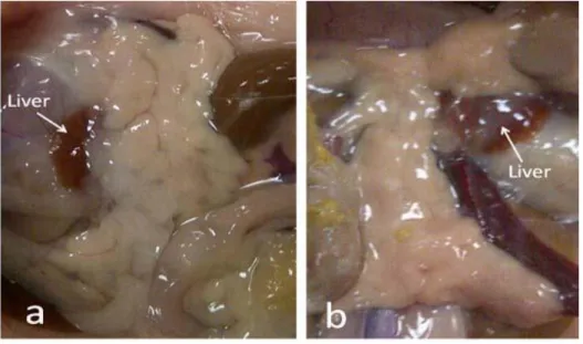
Figure 3a: Gross morphology of non-diabetic (a) and diabetic (b) rat pancreas.
Magnification: X2
21
Figure 3b: Micrographs of the endocrine and exocrine pancreas of non-diabetic (A) and diabetic (B) rats. Magnification: x 400
22 1.3 Diabetes mellitus
Diabetes mellitus (DM) is a complex, chronic metabolic disease, has a heterogeneous group of symptoms and is characterized by disturbances of carbohydrate, fat and protein metabolism, in which the person has high blood glucose levels, either because the insulin production is inadequate, or because the body's cells do not respond properly to insulin, or both. Hyperglycaemia, or elevated blood glucose may lead to various long-term complications like neuropathy, retinopathy and cardiovascular problems [97, 98].
DM is also regarded as a chronic inflammatory disease caused by inflammatory cytokines involving the innate immunity. Modern research indicates that elements of the adaptive immune system may also contribute to this syndrome. Many reports show that inflammatory cytokines do indeed have major role in the pathogenesis of type 2 diabetes.
The etiology of type 1 DM include autoimmune destruction of pancreatic β-cells [99, 100].
1.3.1 Types of diabetes
Type 1 diabetes mellitus (T1DM) results from the body's failure to produce enough insulin. T1DM is characterized by a massive loss or the necrosis of insulin-producing β- cells of the pancreatic islets of Langerhans, leading to insulin deficiency. This form was previously referred to as insulin-dependent diabetes mellitus (IDDM) or juvenile diabetes because β-cells are destroyed in childhood by autoimmune system. Autoimmune destruction of β-cells may be triggered by viruses or chemical toxins [101, 102]. T1DM accounts for about 10-15% of all cases of DM [97] .
Type 2 diabetes mellitus (T2DM) results from insulin resistance, a condition in which cells fail to use insulin properly. This form was previously referred to as non insulin- dependent diabetes mellitus (NIDDM) or adult onset diabetes. The defective responsiveness of body tissues to insulin is believed to involve the insulin receptor.
However, the specific defects are not known. T2DM is the most common type of diabetes and it accounts for almost 85-90% of all DM cases [97, 103, 104].
23
Gestational diabetes: The third main form of DM is gestational diabetes which occurs when pregnant women without a previous diagnosis of diabetes develop high blood glucose level. It may precede the development of type 2 DM. Gestational diabetes mellitus (GDM) is the pathophysiological state of insulin resistance or reduced insulin secretion which is noticed for the first time during mid pregnancy and progresses through the third trimester [105].
1.3.2 Role of novel peptides in diabetes mellitus
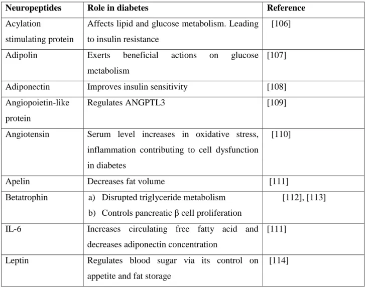
Adipokines, cytokines and chemokines are the primary neuropeptides that play a role in insulin resistance in diabetes mellitus. Some of the neuropeptides and their role in diabetes are shown in Table 4.
Table 4: Novel peptides implicated in the development of type 2 diabetes mellitus
Neuropeptides Role in diabetes Reference
Acylation
stimulating protein
Affects lipid and glucose metabolism. Leading to insulin resistance
[106]
Adipolin Exerts beneficial actions on glucose metabolism
[107]
Adiponectin Improves insulin sensitivity [108]
Angiopoietin-like protein
Regulates ANGPTL3 [109]
Angiotensin Serum level increases in oxidative stress, inflammation contributing to cell dysfunction in diabetes
[110]
Apelin Decreases fat volume [111]
Βetatrophin a) Disrupted triglyceride metabolism b) Controls pancreatic β cell proliferation
[112], [113]
IL-6 Increases circulating free fatty acid and decreases adiponectin concentration
[111]
Leptin Regulates blood sugar via its control on appetite and fat storage
[114]
24
Lipasin Regulates serum triglyceride levels [115]
PAI-1 Modulate insulin signaling [111]
RBP4 Involved in the pathogenesis of type 2 diabetes mellitus
a)To link obesity with its comorbidities
b)Insulin resistance, T2DM, and involved in certain components of the metabolic syndrome
[116]
Resistin Causes resistance to insulin [117]
RIFL Involved in lipid metabolism [118]
TGF-β Increases in GDM [119]
TNF-α Contributes to the development of insulin resistance and diabetes
[111]
Vaspin Insulin-sensitizing with cardioprotective and antiatherosclerotic in diabetes
[120]
Visfatin Have insulin-mimetic actions [111]
1.3.3 Nociceptin in diabetes mellitus
Peripheral neuropathy with significant neuropathic pain is a common complication of diabetes mellitus. It is widely accepted that nociceptin is involved in the pathogenesis of neuropathic pain caused by diabetes [121] and/or in pain regulation systems both at supraspinal and spinal levels. Intracerebroventricular injection of, nociceptin causes hyperalgesia or anti-opioid effects instead of analgesia. However, nociceptin produces allodynia or hyperalgesia when given intrathecally in low doses, but causes anti- nociceptive effects in high doses (Tekes [79, 80]. Liu et al. [80] found that , nociceptin concentrations are raised in the brain, spinal cord and serum of rats with diabetic neuropathy compared to control rats.
1.4 The uterus
The uterus of the rat consists of two horns, and is therefore referred to as a bicornuate uterus. At the tips of the two uterine horns are small lumpy glands called ovaries, which are connected to the horns of the uterus via tiny oviducts. The duplex structure of the uterus
25
enables the rat to have multiple embryos [122]. The gross morphology and histology of uterus are shown in figures 4 and 5.
Figure 4: Gross morphology of the rat uterus (Fig 4A showing the reproductive system of the female rat which has been modified from [123] and in Fig 4B (arrows) shows the ovary and uterus, respectively. Magnification of 4B=X2
26 1.4.1. Histology of the rat uterus
1.4.2 The uterine wall
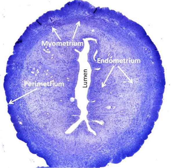
The uterine wall is composed of three layers of tissue. The three layers, from the innermost to the outermost, are as follows endometrium, myometrium and perimetrium [124].
Endometrium: The lining of the uterine cavity is called the endometrium. It consists of functional and basal layers. The functional layer can be further divided into compact and spongious compartments. Blood vessels that supply the functional layer arise from the basal layer. In all placental mammals, the endometrium is shed off periodically or reabsorbed if no pregnancy occurs. Thickness of endometrium of rat varies greatly and ranges from 0.5 mm to 5 mm and depends upon the reproductive cyclic changes [125].
The glandular and vascular tissues of the endometrium support the physiological demands of the growing foetus. Large numbers of uterine glands open onto the endometrial surface and extend deep into the lamina propria, almost reaching the myometrium.
Myometrium: The myometrium is the thickest layer of the uterine wall. The myometrium mostly consists of smooth muscle cells, connective tissue and contains larger blood vessels.
Moreover, it is composed of three indistinctly defined layers of smooth muscle. The middle muscular layer contains numerous large blood and lymphatic vessels. The inner and outer layers of muscles are predominantly oriented parallel to the long axis of the uterus. The myometrium provides much of the contractile force needed to move a large foetus out of the uterus [126].
Perimetrium: The perimetrium is a loose connective tissue around the uterus. This layer is continuous with the lining of the pelvis. The perimetrium surrounds the posterior uterine surface and a significant part of the anterior part. The lower part of the anterior surface is lined by connective tissue [127]
27
Figure 5: General histology of the rat uterus showing, perimetrium, myometrium, endometrium (arrows) and lumen. Magnification: X40
28
2. Hypotheses, Aims and Objectives
Hypotheses
Nociceptin has been implicated in the physiology of pain, including those associated with labour. Nociceptin has also been shown to play a role in endocrine secretion. The aim of the study is to test two hypotheses: Nociceptin is present in endocrine glands e.g. endocrine pancreas. This will allow the nociceptin to play a role in endocrine function.
1. Nociceptin, which has also been localized to smooth muscle, is present in the uterus.
The presence of nociceptin will enable it to participate in uterine pain especially during labour.
2. The tissue content of nociceptin will change after the onset of diabetes mellitus.
Aims of the study
The aims of the study were to:
1. Determine the pattern of distribution of nociceptin in the pancreatic islets cells of rats. Determine the localization of nociception in the uterus tissue of rats.
2. Determine the hypothetic changes of the nociceptin level in the uterus of murine model of streptozotocin-induced diabetes
Objectives of the study
1. Investigate the distribution of nociceptin in the endocrine pancreas of non-diabetic and diabetic rats using immunohistochemical, immunofluorescence, Western blot and immunoelectron microscopy methods.
2. Examine morphology of the uterus after the onset of diabetes.
3. Examine the ultrastructure of the endometrium and myometrium of non-diabetic and diabetic rats using conventional electron microscopy.
4. Investigate the distribution of nociceptin in the uterus of non-diabetic and diabetic rats using immunofluorescence, Western blot and immunoelectron microscopy methods.
29
3. Materials and Methods
3.1 Experimental animals
Twelve adult male or female Wistar rats, weighing 225-250 g, were divided randomly into non-diabetic control group (n = 6) and streptozotocin (STZ)-induced diabetic group (n = 6).
Wistar rats were procured from Harlan Laboratories (Harlan Laboratories, Oxon, England) and bred from the original stock in the Animal Facility of the College of Medicine, UAE University. The rats were housed in plastic cages (six rats/cage) in climate-controlled facilities at 23 ± 1oC and 50 ± 4% humidity. Day and night cycle was maintained at 12 h/12 h. Standard animal chow (Emirates Feed Factory, Abu Dhabi, UAE), and tap water were provided ad libitum. The study was performed with the approval of the CMHS Animal Research Ethics Committee (Approval number: A02/11). During the entire study, the Helsinki guiding principles for the care of and use of laboratory animals were been observed.
3.2 Induction of experimental diabetes mellitus
Diabetes mellitus (DM) was induced either in male or female rats by a single intraperitoneal injection (ip) of streptozotocin, (STZ) (Sigma Chemical Co., St. Louis, MO, USA) at a dose of 60 mg/kg body weight [128]. The STZ was freshly dissolved in citrate buffer (0.5 M, pH 4.5) (see appendix). Following seven days after STZ injection, DM was confirmed by checking blood glucose with test strips using One Touch Ultra 2 glucometer (Life scan Inc., Milpitas, CA, USA). Drops of blood from the tail end of each rat were used for this purpose. The rats were considered diabetic if the random blood glucose levels were
280 mg/dl. The animals were processed for morphological studies two weeks after the onset of DM. Body weight and blood glucose levels were noted before and at the end of the experiment.
3.3 Experimental design
Following the diagnosis of DM, both age-matched healthy non-diabetic controls and STZ- induced diabetic male or female virgin rats were divided into two groups each containing six rats. Female rats were selected on the basis of their oestrous cycle as well [129]. Rats with oestrous cycle on days four were selected for the study. The selection was based on
30
the visual nature of the external genitalia including swollen, moist, and pink vaginal opening with wrinkles and or striations in the posterior and anterior borders.
Groups of experimental animals
Group 1: Non-diabetic healthy controls
Group 2: Diabetic rats
3.4 Body weight
The body and organ (either pancreas or uterus) weights of non-diabetic and diabetic rats were recorded using a 9001 Scale (Sartorius, Hertfordshire, UK) and expressed as mean ± standard error of the mean (SEM) of the body or organ weights.
3.5 Glucose measurement
The blood glucose level was measured for each individual rat of both groups. The blood samples were drawn from the tail end of the rat for blood glucose measurement and expressed as mean ± standard error of the mean (SD).
3.6 Glucose tolerance test on non-diabetic and diabetic rats
Non-diabetic and STZ-induced diabetic rats were subjected to intraperitoneal (i.p) glucose tolerance test, after an overnight fasting for 12 hours. Each rat was given an i.p. glucose load of 2 g/kg body weight according to the method of Kim et al. [130]. The blood glucose measurements were made at fasting zero time (before glucose load), 30, 60, and 120 minutes after the glucose load.
3.7 Tissue collection
Two weeks after the onset of DM, all of the rats from each group were killed humanely under general anesthesia by diethyl ether. A mid-line abdominal incision was made and either the pancreas or uterus was rapidly removed. Representative tissue fragments were taken from the body of the pancreas and uterus and used for immunohistochemical, immunofluorescence, electron microscopy and molecular biology studies.
31 3.8 Light microscopy of pancreas and uterus
Pancreas and uterine tissues from non-diabetic and diabetic female Wistar rats (non- diabetic control n=6) and (diabetic n=6) were dissected out and washed in phosphate buffered saline (PBS) at pH 7.2, blot dried, cut into small pieces and fixed rapidly for 24 hours in freshly prepared Zamboni’s fixative [131]. After washing with running tap water, the samples of pancreatic tissues were dehydrated in ascending series of concentrations of ethanol and cleared in xylene and then embedded in paraffin wax for immunofluorescence and immunohistochemical analysis according to standard procedures [132]. The paraffin blocks were trimmed with razor blade and then sections of 5-6-m thickness were cut with rotary microtome and were placed on slides and stored until further processing.
3.9 Immunohistochemical studies of pancreas
Deparaffinized and rehydrated sections were processed for immunohistochemistry using the avidin-biotin complex (ABC) method [133].
Briefly, the sections were incubated in 0.3% H2O2 solution in methanol for 45 min to block the activity of endogenous peroxidase. Then the sections were rehydrated in descending, gradeed ethanol (100% to 50%) and then transferred into phosphate buffered saline (PBS) three times (5 minutes each). After washing with PBS the tissue sections on the slides were encircled with Dako pen (Dako Cytomation, Glostrup, Denmark) to stop the draining of the solutions from the sections.
The sections were later incubated with blocking buffer for 30 min to start the staining procedure. After draining off the blocking buffer, the sections were allowed to incubate overnight in primary antibodies (anti-rabbit nociceptin antibody; Santa Cruz Biotechnology, Santa Cruz, CA, USA) at a dilution of 1:100 at 4◦C. On the next day the slides were removed from fridge and kept at room temperature for one hour and were later washed 3X in PBS (5 minutes each).
Then the slides were incubated with pre-diluted biotinylated anti-rabbit IgG for 1 hour and again the sections were washed with PBS 3 times (5 minutes each).
32
Then sections were later incubated in streptavidin peroxidase conjugate at a dilution of 1:1000 for one hour and then washed in PBS 3 times (5 minutes each).
The peroxidase activity was observed by incubating the sections in 3,3-diaminobenzidine tetrahydrochloride (DAB) containing 0.03% hydrogen peroxide in PBS for 5 minutes.
Later the slides were then washed under running water for 5 min and then counterstained with haematoxylin for one min. They were later differentiated in acidic ethanol and washed for 2 min with running tap water, then again dehydrated in ascending grades of ethanol for 3 minutes each (50%, 70% & 95% ethanol) and then in 100% ethanol with two changes for 5 min each.
Subsequently the sections were cleared in xylene and mounted on glass slides in Cytoseal 60 (Stephens Scientific, Riverdale, New Jersey, USA). The sections were examined with Zeiss Axiophot microscope (Carl Zeiss, Jena, Germany) and the images of the immunopositive cells were taken.
3.10 Double-labelling immunofluorescence studies of the pancreas
In order to determine whether NC colocalizes with insulin in non-diabetic and diabetic pancreatic tissues, sections were incubated with antibodies against NC and insulin before being immunolabelled with either tetramethylrhodamine isothiocyanate (TRITC; red) or fluorescein isothiocyanate (FITC; Green) according to a previously described method [133].
Deparffinized sections were washed in PBS and circled with a Dako pen to prevent solutions draining away from the tissue section. The sections were incubated first with blocking reagent for 30 min.
Thereafter, the blocking reagent was drained off and the sections were later incubated with the primary antibody (anti rabbit nociceptin polyclonal antibody; 1:100) for 24 hours at 4oC. On the following day, the sections were brought to room temperature for one hour and after washing with PBS, the sections were labelled with secondary antibody (anti rabbit TRITC; 1:100) for one hour at room temperature. The same pancreatic tissue sections were
33
later incubated with the second primary antibody (anti mouse insulin antibody) overnight at 4oC and then labeled with the second secondary antibody (anti mouse FITC; 1:100) (Jackson Laboratory, West Grove, Pennsylvania, USA) for one hour. After washing with PBS three times each, the tissue sections were held in Immunomount® (Shandon, Pittsburgh, PA, USA). Sites of immunoreaction were detected and photographed with Zeiss confocal microscope (Carl Zeiss, LSM 510, confocal microscope Jena, Germany).
3.11 Immunofluorescence microscopy of the uterus
In order to determine the localization of NC in the endometrium or myometrium, deparaffinized sections were incubated with antibodies against NC. Immunolabelling with fluorescein isothiacyanate (FITC) was employed according to a previously described method [133]. Briefly, sections of the uterus of non-diabetic control and diabetic rats were treated with a blocking agent for 30 min at room temperature after rinsing in PBS. The sections were then incubated with anti rabbit nociceptin polyclonal antibody (1:100) for 24 hours at 4oC. Sites of immunoreaction were detected with anti-rabbit FITC (1:100). The sections were held in Immunomount® mounting medium (Shandon, Pittsburgh, PA, USA), examined, and images were taken with a Nikon Confocal Microscope Eclipse 80i, Japan.
3.12 Tissue processing for conventional electron microscopy
Pancreatic and uterine tissues were dissected out from each group of rats (non-diabetic and diabetic) washed in 0.1M phosphate buffer (pH 7.2) and then immersed immediately in Karnovsky’s fixative at pH 7.2 [134] for 24 hours at 4oC. After rinsing with phosphate buffer the tissue samples were post fixed with 1% osmium tetroxide for one hour. After washing with distilled water, the samples were dehydrated in ascending series of graded ethanol from 30% to 95% and 100 % and then finally in propylene oxide. Then tissue were infiltrated and embedded in Agar100 epoxy resin and polymerized at 65oC for 24 hours.
Blocks were trimmed and semithin and ultrathin sections were cut with Reichert Ultracuts, ultramicrotome. Semithin sections (130 nm) on glass slides were stained with 1% aqueous toluidine blue on electro thermal slide drying bench at 55oC and ultrathin sections of golden colour (95 nm) on 200 mesh copper grids then were contrasted with uranyl acetate [135]
followed by lead citrate [136]. Then grids were examined and photographed at different
34
magnifications with Philips CM10 transmission electron microscope (Eindhoven, Netherlands).
3.13 Double-labelling immunoelectron microscopy of the pancreas Fixation of tissue samples
Immunoelectron microscopic studies were carried out by using a previously described method [137]. Pancreatic tissue fragments of six, non-diabetic (n =6) and six diabetic (n = 6) male Wistar rats for each group were used for the immunoelectron microscopy study.
Rat pancreata were quickly excised and cut into pieces (4 x 4 mm3) and fixed in freshly prepared 4% p-formaldehyde + 0.1% glutaraldehyde in 0.1 M sodium phosphate buffer at pH 7.2 [138] fixative for 24 hours at 40C. Dehydration and embedding of the samples into LR white resin (London Resin, Agar Scientific, UK) was performed according to [139]
techniques with some modifications.
Embedding of tissue samples in LR White resin and sectioning
After washing in the fixation buffer, the tissues were dehydrated through a graded series of ethanols from 70%-95%. The dehydrated samples were infiltrated with three mixtures of ethanol and LR white resin (2:1, 1:1, 1:2) staying one hour at each stage. Afterwards, they were transferred into pure LR white resin for 24 hours at 40C. The samples were then placed into fresh embedding medium for 2 hours at room temperature. Then the tissues were later transferred into Agar gelatin capsules of 0 sizes with labels and filled with fresh LR white resin and tightly capped. Polymerization was achieved by irradiation with ultraviolet light (360-365nm) for 24 hours in a TAAB UV chamber at room temperature.
Capsules were removed and blocks were trimmed. Semithin and ultrathin sections were cut with Reichert Ultracuts (Leica Grermany) ultramicrotome. Semithin sections were placed on a drop of water on glass slides and were dried and stained with 1% toluidine blue on a hotplate. Ultrathin sections (golden colour, 95 nm) were cut and collected and mounted onto carbon formvar-coated 200 mesh nickel grids.
35
Postembedding double immunolabelling of ultrathin sections
Nickel grids were then jet-washed with deionized water thoroughly to remove aldehydes from the sections, then the grids were placed in aqueous 10 % H2O2 for 10 minutes. After washing in deionized water the sections were immersed in 0.5 M NH4Cl in 0.01 M PBS (pH 7.2) for 20 min to reduce the staining background. After washing with PBS buffer (pH 7.2) containing, 1% BSA and 0.1% Tween-20 for 5 min, they were then blocked in 20%
NGS diluted in washing buffer for 10 min. The grids were later incubated overnight at 4°C with nociceptin at 1:100 (Santa Cruz Biotechnology, Santa Cruz, CA, USA) and washed in PBS and blocking buffer. They were later incubated with goat anti-rabbit IgG conjugated to 10 nm gold particles at a dilution of 1:20 for two hours at room temperature.
After washing in PBS, the grids were incubated in the second primary antibody (antibodies against insulin, 1:100; Dako, Copenhagen, Denmark) for 24 hours at 4oC. The sections were brought to room temperature for one hour and incubated again in a solution of goat anti-mouse IgG conjugated to 5 nm gold particles at a concentration of 1:20 for 2 hours at room temperature.
After washing with PBS buffer, the sections were fixed in 2.5% aqueous glutaraldehyde and were washed with deionized water then blot dried. The grids were contrasted with 2%
uranyle acetate and lead citrate, for 15 and 7 minutes, respectively. After washing the grids with deionized water and dried on a filter paper, they were examined with Philips CM10, transmission electron microscpe (Eindhoven, Netherlands).
3.14 Immunoelectron microscopy study of uterus
Immunoelectron microscopy study was performed according to the methods described by [94, 140]. Briefly, uterine tissue fragments of non-diabetic control and diabetic Wistar rats were dehydrated in ascending concentrations of ethanol (50% to 70%) and transferred to zero-sized Agar gelatin capsules then embedded in LR white resin as described in section 3.13.
Postembedding single immunolabelling of ultrathin sections
Ultrathin sections were cut and stained as described in section 3.13. Briefly, the uterine tissue sections were incubated overnight with antibodies against nociceptin (1:100; Santa Cruz) at 4°C in humid chamber. The next day the grids were brought to room temperature
36
for one hour then washed and incubated with secondary anti-rabbit IgG conjugated to 10 nm gold particles, at a concentration of 1:20. After washing with PBS the sections were fixed in 2.5% aqueous glutaraldehyde solution, washed with deionized water and then blot dried. The grids were later contrasted with 2% uranyl acetate and lead citrate, for 15 and 7 minutes, respectively. After washing the grids with deionized water, they were dried on a filter paper and stored in petridish. The grids were examined and images were taken with Philips CM10, transmission electron microscope (Eindhoven, Netherlands)
3.15 Morphometry
Six images from each group were randomly selected for quantification. The procedure was carried out on immunohistochemical, immunofluorescent and immunogold electron microscopy images. For the immunohistochemical images, the results were shown as percentages of labeled immunoreactive cell related to the total number of cells (± SEM). In immunofluorescence images, cells containing both nociceptin and insulin were counted.
In transmission electron microscopy images, quantification of nociceptin and insulin peptides were based on secretory granules of β-cells that contain both nociceptin- and insulin-labelled gold particles, 10 nm for nociceptin and 5 nm for insulin. The number of gold particles present on secretory granules was counted and reported to the total number of labelled granules, thus providing a mean density of labeling in each secretory granule.
3.16 Western blotting of nociceptin in tissues
A. Preparation of pancreas and uterine tissues lysates
1. The uteri and the pancreata were retrieved from the animals and kept on ice to prevent tissue lysis.
2. After homogenization, the tissue samples were placed in Eppendorf tubes and stored at -20°C immediately. To a 5 mg piece of tissue, 300 μl lysis buffer (RIPA buffer) was rapidly added before homogenization using electric homogenizer. The blade was rinsed twice with another 2 x 300 μl lysis buffer, then maintained at constant agitation on an orbital shaker for 2 hours at 4°C.
37
3. The homogenate was centrifuged at 10,000 rpm at 4°C in a microcentrifuge for 10 minutes. The supernatants were transfered to a fresh tube kept on ice. The pellet was then discarded.
B. Protein Quantification
1. A small volume (5μl) of supernatant was taken from the total protein extract to perform quantitation using BSA as standard (Bio-Rad Laboratories, Hercules, CA, USA, Cat no. 500-0001). The protein concentration was determined for tissue lysate using Bio-Rad protein Assay kit, (Cat no. 500-0001).
2. To the remaining volume of tissue lysate, an equal volume of 2X Laemmli sample buffer (Loading buffer) was added.
3. Tissue lysate was boiled in sample buffer at 95°C for 10 minutes and aliquot to reduce and denature the protein and stored at -20°C.
4. Prior to electrophoresis, the tubes containing cell lysate were defrosted at 37°C on ice and centrifuged at 16,000 x g in a microcentrifuge for 5 minutes at 4oC.
C. Sample protein loading and separation using gel electrophoresis
The quantity of protein samples loaded into the wells of 10 % SDS-PAGE gel was 40µg along with prestained, molecular weight ladder. 10X diluted running gel buffer was diluted with distilled water up to 1X and filled the tank. The gel was run for 2-3 hours at 90 Volts.
D. Transfer of protein samples from the gel to the membrane
The gel was placed in transfer buffer for 10 minutes and the stack was transferred as follows:
1. The gel was placed on the cathode side and the blot on the anode. The cassette was gently placed in the transfer tank containing transfer buffer and placed on ice block in the tank. The protein was transferred from the gel to nitrocellulose
38
membrane for 2 hours at 4°C in the dark at constant current of 90 volts. The membrane is now ready for antibody staining.
E. Ponceau Staining
The membrane was rinsed in water and incubated in 100% Ponaceau S stain until bands appear to check the transfer quality. Wash off the Ponaceau S stain was washed off with three washes in tris-buffered saline tween-20 (TBST) until proper cleaning.
F. Staining of uterine and pancreatic samples with antibody
The gel membrane was blocked with neutral protein for one hour at 21oC, shaked and mixed continuosly with 5% non-fat milk powder in Tris Buffered Saline with Tween® 20 (TBST).
The membrane was incubated with a primary antibody (anti-nociceptin antibody, ab10277) at a concentration of 1:500 in 5% blocking solution overnight at 4°C.
1. GAPDH (14C10) Rabbit mAb #2118 was used as loading control.
2. Then membrane was washed 3X in TBST for 5 minutes each and then incubated with secondary antibody (goat anti-rabbit IgG-HRP Conjugate #170-6515, Biorad) at 1:5000 dilution in blocking buffer containg 5% milk in TBST at room temperature for one hour.
3. The membrane washed 3X in TBST for 5 minutes each, then rinsed in TBS.
4. The signal was developed by using, Pierce ECL Plus Western Blotting Substrate (Thermo Fisher Scientific Inc.81 Wyman Street Waltham, MA USA 02451) according to manufacturer’s protocol.
5. The images were captured with Typhoon FLA 9500. (GE Healthcare Bio- Sciences AB Björkgatan 30 751 84 Uppsala Sweden )
39 3.17 Statistical analysis
Statistical analysis was done by using SPSS software 21.0. The data obtained were calculated as means ± SEM. Comparisons between data were done and non-parametric Mann-Whitney U test was used to determine the statistical significance. P values of less than 0.05 were taken as significant.
40
4. Results
4.1 Pancreas
4.1.1 Body and organ weight ratios
The average body weight of the non-diabetic male rats was 218±12 g. Pancreas to body weight ratio for non-diabetic rats was 0.0020±0.0004. The average body weight of diabetic rats was 185±10 g and pancreas to body weight ratio was 0.0040±0.0007. Statistical analysis using Mann Whitney test reveals no significance.
4.1.2 Glucose measurement
The random blood glucose level was measured for each individual rat of both groups. The average blood glucose level for non-diabetic controls was 121 ± 12 mg/dL. The average blood glucose level for diabetic rats was 579 ± 10 mg/dL. There was a marked (p <0.05) difference between the glucose level in non-diabetic controls versus diabetic rats.
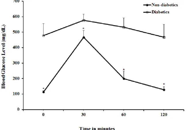
4.1.3 Glucose tolerance test in male rats
Figure 6 depicts changes in the blood glucose concentration during intraperitoneal glucose tolerance test. The glucose load of 2g/kg body weight intraperitoneally generated different profiles in control and STZ-diabetic group. The changes in glucose levels in non-diabetic rats differed from those in the diabetic group. Basal plasma glucose was significantly higher in the diabetic rats and the i.p. glucose load increased glucose levels, in both groups but the level of blood glucose remained significantly (p < 0.05) higher in diabetic rats even 120 minutes after glucose challenge.
41
Figure 6: Glucose tolerance test in male Wistar rats after i.p. load of 2g/kg of glucose.
*P< 0.001 (non-diabetic versus diabetic)
42 4.1.4 Light microscopy for pancreas
Immunohistochemistry by avidin-biotin-complex and double labelled immunofluorescence methods were employed to determine the presence of NC in pancreatic tissues of non- diabetic control and diabetic rats. The results are summarized below.
4.1.5 Immunohistochemistry studies (Avidin Biotin Complex method)
Figure 7 shows nociceptin immunoreactive cells in the central and peripheral portions of the islet of Langerhans of non-diabetic control rats. According to Figure (7a), the number of nociceptin-positive cells in the pancreatic islet of non-diabetic control rats appears to be higher compared to those seen in the islets of diabetic rats ( Figure 7b). The sizes of the islets were found to be reduced in diabetic condition (highlighted with arrows).
43
Figure 7: Light microscopic images of nociceptin-immunopositive cells (arrows) in the endocrine pancreas of non-diabetic (7a) and diabetic (7b) rats. Note that the expression of nociceptin is stronger in the islets of non-diabetic control compared to that of diabetic rats.
Magnification: X400
Figure 8: Percentage distribution of nociceptin-immunoreactive cells in the pancreas of non-diabetic and diabetic rats.Note that the number of nociceptin-positive cells is significantly (p < 0.05) lower in diabetic rats - controls. n=6
44 4.1.6 Double labelling immunofluorescence study
Immunofluorescence technique further corroborates our findings with the ABC method as reported in earlier section (Figure. 7a and b). The result of the immunofluorescence study is shown in Figure 9 which illustrates the co-localization of nociceptin with insulin.
Nociceptin-positive cells are shown in red while that of insulin by green fluorescence.
Yellow colour specifies cells that contain both nociceptin and insulin.
A representative section of non-diabetic rat islets is shown in Figure 9a and diabetic in Figure 9b. The pancreatic islets of non-diabetic control rat pancreas contain significantly (p ≤ 0.05) greater number of NC- and insulin-positive cells compared to diabetic group (Fig. 9). The figures show that insulin and nociceptin immunoreactivity can be observed in β-cells of pancreatic islet. In figure 9c morpohmetric analysis also shows the number of cells containing either insulin, nociceptin or insulin+nociceptins significantly lower (*p<
0.0000003) compared to non diabetic rat pancreas.
45
Figure 9: Immunofluorescence micrographs of nociceptin (red-thick arrows) and insulin (green-thin arrows) in the islets of non-diabetic (9a) and diabetic (9b) rats. Yellow colour indicates cells that contain both nociceptin and insulin. There is a large reduction in the number of nociceptin- and insulin-positive cells in diabetic rat. Magnification: X200
Figure 9c: Number of cells containing either insulin, nociceptin or insulin+nociceptin in pancreas of non-diabetic and diabetic rats.*p< 0.0000003 (Significantly lower compared to non diabetic rat pancreas).
46 4.1.7 Conventional electron microscopy
4.1.7.1 Non-diabetic and diabetic pancreatic β-cells
In non-diabetic pancreatic islets the most numerous cells were β -cells located in the central portion of pancreas that secrete insulin. Most part of the islet was covered with these cells.
Insulin secretory granules of β-cells were observed with homologous electron dense inner core and surrounded by outer peripheral halo with crystalline matrix, which is electron lucent in nature. The secretory granules were almost polyhedral in shape with a pale matrix and have a rounded or slightly oval nucleus with irregular contour and narrow perinuclear cisterns. Non-diabetic β -cells contain numerous secretory granules which showed a mean diameter of 300 nm (Figure 10 A).
In diabetic pancreatic β-cell, the ultra-structural study revealed diverse degrees of injury in the β-cell organelles, such as alterations in the secretory granules with dialated halo space.
Moreover the changes, like damage of chromatin matrix of the nucleus with dilated nuclear envelope, abundant vacoularization in cytoplasm were seen. Broken endoplasmic reticulum and swollen mitochondria with loss of cristae also were observed. Degenerated and sparsely scatted Golgi bodies contribute to explain the appearance of a diabetic syndrome in the rat tissue (Figure 10 B).
![Figure 1: Structural similarities between dynorphin A and nociceptin amino acid sequences [20]](https://thumb-eu.123doks.com/thumbv2/9dokorg/1363178.111157/10.918.137.848.235.395/figure-structural-similarities-dynorphin-nociceptin-amino-acid-sequences.webp)

![Figure 4: Gross morphology of the rat uterus (Fig 4A showing the reproductive system of the female rat which has been modified from [123] and in Fig 4B (arrows) shows the ovary and uterus, respectively](https://thumb-eu.123doks.com/thumbv2/9dokorg/1363178.111157/26.918.132.770.422.774/figure-gross-morphology-uterus-showing-reproductive-modified-respectively.webp)