In vitro microbiological testing of experimental Drug Delivery System
PhD thesis
Dr. Szász Máté Sándor
Doctoral School of Pathology Sciences Semmelweis University
Supervisor:
Dr. Szabó Dóra MD, PhD, DSc
Official reviewers:
Dr. Tekes Kornélia PharmD, PhD, DSc Dr. Kónya József MD, PhD
Head of the Final Examination Committee:
Dr. Cseh Károly MD, PhD, DSc
Members of the Final Examination Committee:
Dr. Ludwig Endre MD, PhD Dr. Kriván Gergely MD, PhD
Budapest, 2015
1
Introduction
Foreign bodies in human body are known as a special problems. Prosthetic joint, prosthetic valve, central venous catheter, peripheral venous canule, nephrostoma or urinary catheter have the ideal surface for the colonisation of microbes. Central venous catheters is a good possibility for the microbes to penetrate continously into the body. Infection of prosthetic joints can cause abscesses at the local tissues. Following the local infection, microbes can be transported to the different organs by the cicrculation forming septic metastates and cause bloodstream infection (BSI).
Latent infections in the human body can cause prosthetic joint infections (PJI) quite frequently. From theese sources of infections, microbes can be also transported by the bloodstream and colonise the surface of the prosthetic. Usually theses microbes come from the nosocomial flore as well. Poly-methyl-metacrilate (PMMA) bone cement widely used for the fixation of prosthetic can cause necrosis in the bone tissue by elevatating the temperature and damaging the capillaries. In the damaged tissues, microbes can cause pyogenic discharge and consequently osteomyelitis can be formed in local bone tissues. These serious event create the ability of recurrent sepsis or prosthetic joint can loose of function in the following years.
Strains causing PJIs can form adhesive factors which can help the bacteria attaching on the foreign surfaces and forming biofilm. PMMA can behave as a receptor of glycocalyx produced by the bacteria. Among Gram-positive bacteria, Staphylococcus epidermidis can form biofilm most frequently on the prosthetic surface. Among Gram-negative ones, Pseudomonas aeruginosa is the most important biofilm forming bacteria.
From the 1950s, coagulase-negative Staphylococci (CNS) strains have been taken an important role in foreign body associated infections. The number of cases caused by CNS strains started to increase, and after 2000, most of the nosocomial infections caused by these bacteria in certain European and American hospitals. In present days, S. epidermidis is one of the most dominant pathogen in nosocimial infections, but the importance of Gram-negative strains is also elevated.
The widely used beta-lactam antibiotics contributed to the spread of methycillin resistant Staphylococcus epidermidis (MSSE) strains. These strains are more invasive then the methycillin susceptible Staphylococcus epidermidis (MSSE) strains, and theese bacteria can be responsible for the deep-prosthetic joint infections (DPJI). MRSE strains usually show reduced
2
susceptible to glycopeptides and numerous studies have been highlighted on the elevated resistance to aminoglycosides in West Europe (more than 60%).
Nowdays, systemic antibiotic therapy can poorly cure the prosthetic joint infection, because the proliferation of microbes under the biofilm can not be influenced in that way.
Therefore, a local therapeutic method is necessary to use to prevent and cure septic discarges of prosthetic joint. The local antibiotic releasing systems called as Drug Delivery Systems (DDSs) are devided in two groups: none-biodegradable and biodegradable systems. From the 1980s, in orthopedic surgery, none-biodegradable systems, as gentamicin containing Septopal- chains and gentamicin-tobramycin impregnated bone cement have been widely used. Nowdays, the elevating resistance to aminoglycosides limitates the usage of theese systems and makes us to develop new, biodegradable systems. The advantage of biodegradable system is, that after releasing the antibiotic, that one can be desintegradet by human body and it is not necessary to remove that one by reoperation. Newly developed systems contains vancomycin, teicoplanin or amikacin, because theese agents can eradicate beta-lactame resistant S. epidermidis strains.
3
Research objectives
My objectives were:
the retrospective epidemiological investigation of pathogens caused prosthetic joint infections between 2001 and 2011 at Orthopedic Clinic of Semmelweis University
to investigate the growing kinetic of Staphylococcus epidermidis in different liquid culture media
to demonstrate the antibacterial effectivity against Staphylococcus epidermidis strain of two different, wax-based drug delivery systems containing different concentrations of vancomycin.
to investigate the drug release profile of two different, wax-based drug delivery systems containing different concentrations of vancomycin.
to ivestigate the biofilm formation of Pseudomonas aeruginosa strains.
to find the certain synergistic antibiotic combinations, which are effective against Pseudomonas aeruginosa strains and inhibit the biofilm formation.
4
Material and methods
Data collection
We collected the clinical data of prostehtic joint infections from Orthopedic Clinic of Semmelweis University between 2001 and 2011. The culture results of clinical speciments were registered at Clinical Microbiology Diagnostic Laboratory of Semmelweis University.
Processing of microbiological speciments
The clinical speciments were transported by transport media (Amies Agar Gel, 108C, Copan Diagnostics Inc, USA) to the laboratory. The storage of the strains were performed in tryptic soy broth culture media (TSB) on -80 ºC. Before the experiments, bacteria were recultivated.
Bacterial strains
For the investigation of drug release and growing kinetic, we used S. epidermidis ATCC 35984 laboratory strain. As control strain, Bacillus subtilis ATCC 6633 (Becton-Dickinson, USA) were used. For the biofilm formation tests, 60 non-related P. aeruginosa strains were collected from the Microbiology Laboratory of Medical Centre, Hungarian Defence Forces. Furthermore, as positive control, Staphylococcus aureus ATCC 12600 and as negative control, S. epidermidis ATCC 12228 strain was used (LGC Standards GmbH, Germany).
Determination of minimal inhibitory concentration
According to the recommendation of Clinical and Laboratory Standards Institute (CLSI) and National Committee Of Clinical Laboratory Standard (NCCLS), determination of minimal inhibitory concentration (MIC) of different antibiotics were presented by microdilution method.
These antibiotics were piperacillin/tazobactam, ceftazidime, cefepime, imipenem, meropenem, gentamicin, amikacin, netilmicin, tobramycin, ciprofloxacin, levofloxacin, chlarithromycin.
Determination of the number of bacteria
We determinated the number of bacteria by dilution method in different broth media. The initial number of colony forming units (CFU) were determinated by McFarland measurement.
Determination of vancomycin concentration in different liquid culture media
For determination vancomycin concentration in different broth media, we used biological, filter paper method. We analysed the correlation between the diameter of the of inhibition zone
5
around the filter paper disc and dilution row of vancomycin concentration (µg/ml). The different liquid culture meda were the following ones: brain-heart infusion (BHI), thyoglycolate (TG), Müller-Hinton broth II (MH broth II) and saline solution.
Investigation of the growing kinetic of Staphylococcus epidermidis
The growing kinetic were analysed by dilution method. The maximal dilution was 105 CFU/ml.
At final dilution, we inoculated 10µl suspension on MH culture media, and after incubation period, we counted the colonies.
Investigation of antibacterial effect of wax discs.
We used Wax1 (Cera alba : Precirol® : Vaselinum album 45 : 45 : 10 composition) and Wax2 (Cera alba : Precirol® 50 : 50 composition) discs with different vancomycin content (0.5, 1, 2 and 4 mg). The antibacterial effect of wax discs were analysed by dilution method BHI media and presented by time-kill curves.
.
Investigation of the drug release of wax discs on solid culture media
The drug releasing kinetics from DDS were demonstrated by Bauer-Kirby method on solid MH culture media.
Investigation of the drug release of wax discs in liquid culture media
The drug releasing kinetics from DDS were demonstrated in BHI culture media by biological method with filter paper discs. We tested theese filterpaper disc on the culture of B. subtilis ATCC 6633 strain.
Investigation of biofilm formation
We used a qualitative method for biofilm screening. The bacteria were cultivated in TSB overnight at 37 ºC in U-wells of polystyrene microtiter plates. The wells were washed three times with phosphate-buffered saline and dried in the inverted position, than we died the plates with safranin. The well-visible film lining of the well walls was considered as positive for biofilm production.
Biofilm susceptibility testing
Bacteria were cultivated in U-wells of polystyrene microtiter plates with the appropriate dilutions of the respective antibiotics in MH broth II and the plates were incubated for 18–20 h
6
at 37 ºC. The wells were washed three times with phosphate-buffered saline and dried in the inverted position. MBICs (the lowest concentration of antibiotic which inhibits biofilm formation; µg/l) were then determined. The strains were tested for susceptibility to certain antibiotics mentioned at MIC determination method.
Biofilm sinergy testing
Serial twofold dilutions of each drug to at least double the MIC were prepared. MH broth II was distributed into each well of the microdilution plates. The first antibiotic of the combination was serially diluted along the ordinate, while the second drug was diluted along the abscissa.
An inoculum equal to a 0.5 McFarland turbidity standard was prepared from each P. aeruginosa isolate in MH broth. After thr inoculation of bacteria, incubation period followed. After washing and dyeing, well-visible film lining of the well walls was considered as positive for biofilm production. The resulting checkerboard contains each combination of two antibiotics, with tubes that contain the highest concentration as control. FBIC of each agent was calculated as follows:
FBICA = MBICA(c) / MBICA(a) FBICB = MBICB(c) / MBICB(a) ƩFBIC = FBICA + FBICB
where subscripts A and B denote antibiotics A and B, subscripts in parentheses c and a denote the activity measurements in combination and alone, respectively.
The summation of both FBICs was used to classify the combination of antimicrobial
agents as synergistic (ƩFBIC ≤ 0.5), partially synergistic (0.5 < ƩFBIC ≤ 1), indifferent (1 <
ƩFBIC ≤ 4), or antagonistic (ƩFBIC > 4).
Statistical analysis
In different liquid media, the growing kinetics were compared with correlation coefficient. For killing curve study the General Linear Model (GLM) was used and to compare each group Fisher LSD test was used, taking in both cases a p value of 0.05 as significant. To compare the initial/early stage of the vancomycin release also the GLM and to compare the CFUs decreasing in each liquid media two tailed t test was used for statistical analysis taking a p value of 0.05 as significant and to compare the peak vancomycin concentrations the two-way ANOVA test was used, taking a p value of 0.05 as significant.
7
Results
Retrospective epidemiological investigation of prosthetic joint infections
We investigated 221 culture results between 2001 and 2011. Gram-positive bacteria caused 64
% of the PJIs. 53 % of the PJIs were caused by Staphylococci. S. epidermidis caused most of theese cases (28%) and S. aureus caused 25 % of theese infections. P. aeruginosa stains were responsible for 8 % of the PJIs.
Deremination of minimal inhibitory concentration of different antibiotics
The average of MIC values of 12 antibiotics tested to 14 P. aeruginos strains are summarised in Table 1.
Table 1: Average of the MIC values (aMIC) of 12 antibiotics.
Determination of vancomycin concentration in different liquid culture media
In BHI and MH broth II media, 2 µg/ml vancomycin concentration caused 18 mm diameter of the inhibition zone. In saline solution this concentration formed 19 mm diameter of the inhibition zone. There was no inhibition zone at 0.125 µg/ml vancomycin concentration in TG culture media. In BHI és MH broth II media, more than 10 mm diameter of the inhibition zone was formed. At 0.25, 0.5 és 1 µg/ml vancomycin concentration, the diameter of the inhibition zone was between 10 and 18 mm in all culture media.
Antibiotics aMIC values (µg/ml)
MIC50 MIC90 Range
Piperacillin/tazobactam 1024 1024 128–1024
Ceftazidime 4 32 2–1024
Cefepime 8 32 2–64
Imipenem 4 16 0.5–128
Meropenem 16 32 2–64
Ciprofloxacin 256 512 8–1024
Levofloxacin 32 62 2–256
Amikacin 4 32 2–1024
Gentamicin 128 256 1–256
Tobramycin 128 512 2–>1024
Netilmicin 4 >256 1–>256
Clarithromycin 256 >1024 32–>1024
8
Investigation of the growing kinetic of Staphylococcus epidermidis
There was a strong, tight, positive correlation between CFU reducing kinetics in MH broth II and TG liquid culture media compared with saline solution. (rsaline, MH = 0,96, rsaline, TG = 0,93).
Strong, postive correlation was observed between BHI media and saline solution (rsaline, BHI = 0,89). There was no initial growing phases of S. epidermidis at 2, 4 és 8 µg/ml vancomycin concentration and after four hours long constant period, there was strong decreasing in MH broth II. In saline solution, the declination phase was close to linear and started eralier. In control BHI containing no vancomycin, we detected the most intensive multiplication. Without vancomycin the count of bacteria reduced below the initial count of CFU after 24 hours in saline solution.
Investigation of antibacterial effect of wax discs.
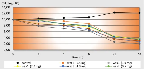
There was a permanent increase of the bacterial count in the group containing no Wax. There was a permanent decrease of the bacterial count in the other groups. Wax2 with 4 mg vancomycin presented the strongest antibacterial effect. After two hours, the initial 9.9 log10 CFU/ml was reduced below 7 log10 CFU/ml, and there was no effect like this at other groups.
None of the groups presented initial growing phase. Wax2 with 4 mg vancomycin showed the strongest decreasing of the initial bacterial count after four hours in BHI comparing to the other liquid media containing no Wax. Wax1 and Wax2 containing 1 and 2 mg vancomycin reduced significant the initial bacterial count only after six hours The same decreasing happened at Wax1 and Wax2 containing 0.5 mg vancomycin after 24 hours (Figure 1).
Figure 1: Decreasing of initial bacterial count at different Wax-based systems (Time-Kill Curves).
0,00 2,00 4,00 6,00 8,00 10,00 12,00 14,00
0 2 4 6 24 48
CFU log (10)
control wax1 (0.5 mg) wax1 (1.0 mg)
wax1 (2.0 mg) wax1 (4.0 mg) wax2 (0.5 mg)
time (h)
9
Investigation of the drug release of wax discs on solid culture media
There was no inhibition zone on the culture of S. epidermidis at Wax1 and Wax2 containing 0.5 mg vancomycin. Wax-based system containing 1 mg or more amount of vancomycin formed more than 18 mm diameter of the inhibition zone after incubation period (Figure 2 and 3).
Figure 2: Inhibition zones of Wax1 containing 0.5, 1, 2 és 4mg vancomycin after overnight incubation.
Figure 3: Inhibition zones of Wax2 containing 0.5, 1, 2 és 4mg vancomycin after overnight incubation.
Wax1 0.5mg Wax1 1mg
Wax2 0.5 mg Wax2 1 mg
Wax2 2 mg Wax2 4 mg
Wax1 2mg Wax1 4mg
10
Investigation of the drug release of wax discs in liquid culture media
In general the vancomycin started to dissolve significantly (p = 0.0134) earlier in the case of the Wax2 than Wax1. Furthermore, the start of vancomycin dissolve depended also on the vancomycin content of the waxes, thus the dissolve started earlier in the presence of the increased vancomycin content. There was a significant correlation (p = 0.000001) between the incubation time and vancomycin concentration. The observed peak vancomycin concentration increased by the used vancomycin content, it is the lowest (0.25 μg/ml) in the case of Wax1 with 0.5 mg vancomycin content and the highest (1 μg/ml) in Wax2 with 4 mg vancomycin.
Differences were observed in the case of vancomycin content comparing the 72 hours peak concentrations but, there was no significant differences (p = 0.0914) between the Wax1 and Wax2 groups. However, there was significant difference (p = 0.0184) in the peak concentrations between the 4 mg vancomycin contaning group and all the other groups. Comparing the groups with 0.5, 1 and 2 mg vancomycin content, no significant differences was observed between the other groups (p = 0.1675). According to the decreasing of initial bacterial count, Wax2 with 4mg vancomycin showed a strong, positive correlation to different liquid culture media containing free vancomycin, and this correlation was very strong to saline solution (Figure 4).
Figure 4: Comparison of Wax2 with 4 mg vancomycin and és 1 µg/ml free vancomycin concentration in liquid culture media according to the decreasing of initial bacterial count.
0 1 2 3 4 5 6 7 8 9 10
2 4 6 24
CFU log (10)
time (h)
wax2 4mg BHI TG BHI MH broth II Saline
11 Investigation of biofilm formation
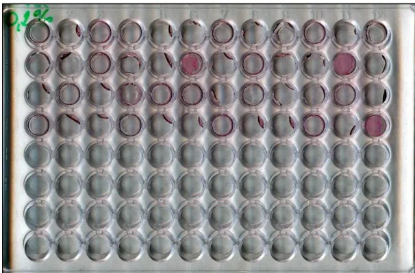
A total of 14 (23.3%) isolates out of 60 isolates of P. aeruginosa were positive for biofilm production. The qualitative method for biofilm screening is presented on Figure 5.
Figure 5: Biofilm screening method. The purple colorisation on the bottom of U-wells shows the biofilm formation.
Biofilm susceptibility testing
Three antibiotics themselves were proved to be efficient biofilm production inhibitors:
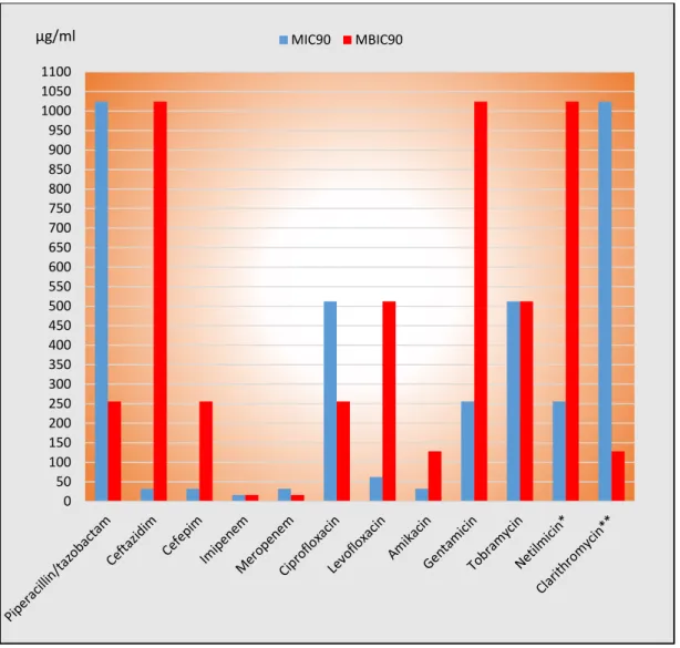
meropenem, piperacillin/tazobactam and clarithromycin, they had the lowest MBIC90 values.
For biofilm-forming P. aeruginosa strains 2-fold to 128-fold higher MBIC values than MIC values were obtained for ceftazidime, cefepime, imipenem, amikacin and netilmicin. The MBIC was 2-fold to 512-fold lower than the MIC values in the case of piperacillin/tazobactam, ciprofloxacin, levofloxacin and clarithromycin. Comparison of MIC and MBIC values are presented on Figure 6.
12
Figure 6: Comparison of the MIC és MBIC values of tested antibiotics to biofilm producing P.
aeruginosa strains.
Biofilm sinergy testing
On eight P. aeruginosa isolates were performed the synergy test (13,3%). Apart from some cases the fluoroquinolones had low FIC-indexes in combination with clarithromycin.
Clarithromycin-levofloxacin combination was proved to be synergistic at 5 of the 8 strains (62.5%). Ciprofloxacin was synergistic with the macrolide at half of the strains,and so the ceftazidime. The aminoglycosides were mainly synergistic with the clarithromycin, and if so, they had very low FIC-index values. The synergy was generally demonstrated for clarithromycin and aminoglycosides, fluoroquinolones and ceftazidime. However, antagonism was detected for clarithromycin and meropenem and cefepime in 62.5% and 75% of the investigated biofilm-forming P. aeruginosa strains. The ceftazidime, the fluoroquinolones and
0 50 100 150 200 250 300 350 400 450 500 550 600 650 700 750 800 850 900 950 1000 1050 1100
µg/ml MIC90 MBIC90
13
aminoglycosides were highly effective against most of the strains in combination with clarithromycin. The carbapenems were mostly ineffective in combination with clarithromycin.
14
Discussion
Certain limitations of the epidemiology investigation are revealed during our study. In European countries, only a few surveillance investigations were presented in the past years, therefore it is very difficult to compare our results to result of European medical centers.
Furthermore, in Hungary, many hospitals use not to take part in annual surveillance programe, so our results can not be compared with them. Anyway, results of our epidemiological investigation shows correlation with European data in general. Our rate of the PJIs caused by S. epidermidis is nearly the same as in foreign medical centers. S. aureus was isolated less than in orher hospitals, but Gram-negtative bacteria caused more sepsis in connection with prosthetic joint.
For the investigation of DDS, it was necessary to determinate the suitable liquid media, which can imitate the physiological environment of human tissue. We tested three enriched liquid culture media (BHI, TG, MH broth II) and saline solution as control. Theese liquid media did not influenced the bactericidal effect of vancomycin. In literature, BHI is the widely used culture media because this one is very rich in nutrients. Saline sollution can keep the starting number of S. epidermidis, but bacteria can not multiply because of the lack of nutrients.
In orthopedic surgery, different kind of antibiotic carrier systems were developed in the past years. These carriers are needed to remove after depletion of antibiotic and cannot be used as vitalisable tissue material. Removing of theese carriers menas the possibility of infection.
Avoiding the reoperation, our team were be able to develop an antibiotic carrier system for preventing the prosthetic joint infections. Our DDS made by wax can be degradable by macrophages after releasing the antibiotic. Cadaver bone tissue, which is widely used in ortopedic surgery can be impregnated with wax, and in that way, theese system can be used as replacement of damaged, infected bone tissue paralelly releasing vancomycin. Vancomycin has a multiple effect againts S. epidermidis, such as the inhibition of cell wall synthesis, which could happen only during the multiplying period.
According to the former studies, 2 µg /ml cocncentration of vancomycin presents the MIC concentration to widely used ATCC 35984 S. epidermidis control strain. In our study, this concentration was caused a rapide bacteria killing effect. Lower concentration of vancomycin was also enough to reduce the number of CFUs for six days. At 1 µg /ml vancomycin concentration, the initial count of bacteria was reduced continously in all kind of liquid media.
15
Wax1 and Wax2 containing 1 mg or more vancomycin could maintain the 18 mm diameter of inhibition zone for six days. Based on our result the Wax 2 with 4 mg vancomycin content had the highest peak concentration in liquid media equal to the minimal inhibitory concentration of S. epidermidis ATCC 35984 and the Wax 2 with 4 mg vancomycin content had the best antibacterial effect, so it could be a potential agent for clinical use in the future.
This type of wax disc can maintain 1 µg /ml vancomycin concentration in physiological environment. According to certain studies, only rifampicin and amikacin has the same strong bactericidal effect as the vancomycin.
Qualitative biofilm screening test is an easy, cheap test to verify biofilm formation avoiding the performance of molecular biological methods. Macrolides may also be beneficial in Pseudomonas infection, even if they have limited direct antibacterial activity against P.
aeruginosa. Macrilides have been shown to reduce the biofilm production in vitro because of inhibiting the quorum sensing mechanism. Various reports on the impact of clarithromycin on biofilm destruction are available, but it was also reported that clarithromycin has no bactericidal action on biofilm synthesis or on pre-formed biofilms. However, some bactericid effects could be demonstrated in this study. Ceftazidime combined with fluoroquinolones has a strong sinergistic effect to inhibit biofilm. Clarithromycin combined with fluoroquinolones and aminoglycosides show also a strong sinergistic effect against biofil formation. Aminoglycoside combined with normaly anti-Pseudomonas agents, such as cefepime, antagonistic effect was performed.
16
Conclusions
1. In our investigation, the rate of PJIs caused by S. epidermidis is nearly the same as in European medical centers. Nowdays, the number of Gram-negtative bacteria caused sepsis in connection with prosthetic joint is elevated.
2. BHI, TG and MH broth II can be used for in vitro testing, because they do not influence the effect of vancomycin. BHI is the widely used culture media because this media is very rich in nutrients. Saline sollution can keep the starting number of S. epidermidis but bacteria can not multiply in that media, so it is not suitable for in vitro tests.
3. Wax 2 with 4 mg vancomycin content has the best antibacterial effect, so it could be a potential agent for clinical use in the future. This type of wax disc can maintain 1 µg /ml vancomycin concentration in physiological environment, which is also enough to reduce bacterial count.
4. Wax 1 and Wax 2 containing 1 mg or more vancomycin can maintain the 18 mm diameter of the inhibition zone for six days. Controled releasing of antibiotic from Wax 2 with 4 mg vancomycin can maintain 1 µg /ml vancomycin concentration in physiologycal conditions for 1 week. With this continously presented concentration of vancomycin, septic discharge of prosthetic joint can be prevented and osteomyelitis als can be treated paralel with parenteral administration of antibiotics.
5. Qualitative biofilm screening test is an easy, cheap test to verify biofilm formation and it is not necessary to do molecular biological test if we are not intrested in the genetic background of biofilm formation.
6. Ceftazidime combined with fluoroquinolon antibiotics can highly inhibit the bacterial biofilm produced by P. aeruginosa. Clarithromycin can be well combined with different antibiotics such as fluroquinolones and aminglycosides to present strong sinergistic effect. Because of the spread of biofilm forming strains, sinergistic antibiotic combinations can be use in DDS to prevent and cure prothetic joint infection.
17
My publication list
1. Szasz M, Lehotkai N, Kristof K, Szabo D, Nagy K: Prevalence and antimicrobial resistance of uropathogens in different inpatient wards. Acta Microbiol Immunol Hung. 2009, 56(4):375- 87. IF. 0,000
2. Kadar B,# Szasz M,# Kristof K, Pesti N, Krizsan G, Szentandrassy J, Rokusz L, Nagy K, Szabo D: In vitro activity of clarithromycin in combination with other antimicrobial agents against biofilm-forming Pseudomonas aeruginosa strains, Acta Microbiol Immunol Hung. 2010, 57(3):235-45. IF: 0,625
# equal contributed
3
.
Kadar B, Kocsis B, Toth A, Damjanova I, Szasz M, Kristof K, Nagy K, Szabo D:Synergistic antibiotic combinations for colistin-resistant Klebsiella pneumoniae. Acta Microbiol Immunol Hung. 2013, 60(2):201-9. IF: 0,780
4. Szasz M, Hajdu M, Pesti N, Domahidy M, Kristof K, Zahar A, Nagy K, Szabo D: In vitro efficiency of vancomycin containing experimental drug delivery systems. Acta Microbiol Immunol Hung. 2013, 60(4):461-8. IF: 0,780


![stroke,andalsoaging[ ].Moreover,inflammatoryresponsesoftheneurovascularunit CerebralPericytesandEndothelialCellsCommunicatethroughInflammasome-DependentSignals](data:image/gif;base64,R0lGODlhAQABAIAAAP///wAAACH5BAEAAAAALAAAAAABAAEAAAICRAEAOw==)