Chapter 4
LICHEN PROPAGULES
F. BRIAN PYATT
ι 17 I. Introduction
II. Vegetative Propagules 1 1 8
A. Isidia and Soredia 1 1 8
B. Other Vegetative Propagules 1 1 9
III. Sexual Propagules 1 1 9
A. Introduction
B. Spore Discharge 1 2 5
C. Spore Germination 137 143
References
I. Introduction
The wide distribution of lichens suggests that these organisms possess efficient means of dispersal. Indeed, quite a wide variety of propagules have been recognized, some characteristic of fungi in general and others unique to lichens. They include both sexual and vegetative types. Ascospores, for example, are produced by the fungal partner of the association and prior to discharge are contained within special chambers: apothecia or perithecia.
In addition, there are two asexual spore types of fungal origin, these being small and large conidia, produced within chambers termed pycnidia (or sper- magonia). The structure and cytological details of these organs are discussed in detail by Letrouit-Galinou in Chapter 2 of this book.
There is a great variation of types of vegetative propagules (or vegetative diaspores) produced by lichens. Such structures are composites containing both fungal and algal contributions. Certain of these propagules, namely, isidia and soredia, are of special interest because they are not produced by nonlichenized fungi. Other types of vegetative propagules include thallus fragments, whole thalli, squamules, and portions or whole lobes of the thal
lus. These structures are heavier than spores and are hence probably gene
rally carried shorter distances.
117
118 F. BRIAN PYATT
Thus, potentially, the lichen thallus often has a variety of propagation
"techniques" open to it, and this has led to a certain amount of controversy over the significance and likely success of the various types of propagules.
But Gregory (1952) believes that the possession, by a fungus, of any organ should be taken as evidence that it does have some function.
II. Vegetative Propagules
A. Isidia and Soredia
The morphology of these unique lichen diaspores is described in Chapter 1 by Jahns. Their importance, or at least potential importance, in lichen dis- persal has been stressed in most texts on lichenology. They have rather high frequency, at least in lichens in temperate regions (Hale, 1967).
Most work has been concerned with soredia. Animals have been recorded as agents of soredia dispersal by Darbishire (1897), Barkman (1958), Hale (1961), and Bailey (1970). Dispersal of soredia by wind has been discussed by des Abbayes (1951), Barkman (1958), Gregory (1961), Smith (1962), Haynes (1964), and Bailey (1966a). Brodie and Gregory (1953) experimen- tally showed soredia being removed by wind from Cladonia cups. Brodie (1951) and Bailey (1966a) demonstrated splash dispersal of soredia, and dis- persal by water trickles has been recorded by Bailey (1968). Soredia are described as being water repellent; they are probably airborne and are likely to be carried farther than isidia but not as far as spores.
There are no known records of active soredia dispersal. Indeed, in all cases, an external source of energy must be applied to the thallus to facili- tate soredia dispersal. From this it does appear that no rhythm of soredia dispersal can exist other than that in some ways rather unpredictable one imposed by the environment itself. The only control that the thallus can exert is with regard to the time of production of these propagules. And so it appears that wind dispersal of soredia could occur at a time when other conditions are unsuitable for further development. However, it can be argued that ascospores can be discharged during a favorable period and car- ried to a region where conditions are unsuitable for germination; but asco- spores are able to survive adverse conditions in a state of dormancy while soredia frequently become infected during periods of enforced dormancy.
Regarding distances of soredia distribution, Bailey (1966a) illustrated that soredia of Lecanora conizaeoides are carried 61.0 cm by 2.2-mm-diameter
"rain" drops falling 366 cm. He noted an increased wind speed was necessary for maximum soredia dispersal as the water content of the thallus increased.
Isidia, being much larger than soredia, probably have a more limited range, and perhaps play a more important role by increasing the surface area
4. L I C H E N P R O P A G U L E S 119
TABLE I
ISIDIA REGENERATION ON THALLI O F Parmelia saxatilis EXPOSED TO SULFUR DIOXIDE"7
Concentration (vpm) 0 5 10 Percentage disks regenerating 15 11 5 Average number of isidia on each regenerating disk 2.8 1.7 1.2
"From Pyatt (1969a).
of the thallus. Barkman (1958) points out that the isidia, which are not water repellent, are likely to be carried down the tree trunk by the stem flow of water. While most isidia are produced diffusely over the upper cortex of the thallus, Kershaw and Millbank (1970) describe the development of squamulose isidia from algal cells underneath the hypothecium of apothe- cial initials in Peltigera aphthosa.
Production and regeneration of isidia can be influenced adversely by air pollution, thereby reducing the reproductive capacity of lichens in polluted areas. Pyatt (1969a), for example, used 1-cm disks of Parmelia saxatilis from which isidia had been removed. The disks were left in a damp petri dish for 6 weeks, and he found that disks removed from material collected near a source of pollution showed less regeneration of isidia than material obtained farther from the pollution source. Disks from material collected from a nonpolluted area were maintained in Perspex boxes subjected to either pure air, 5 vpm million sulfur dioxide, or 10 vpm million sulfur dioxide. After
10 days the material was removed and placed in moist petri dishes for 4 weeks. From Table I, it can be seen that sulfur dioxide limits the ability of lichen disks to regenerate isidia.
B. Other Vegetative Propagules
Various other propagules have been described in lichens and are discussed in detail in Chapters 1 and 3. Most of these, as Barkman (1958) indicates, are the heavier type of vegetative diaspore and, while probably removed by wind, are subsequently carried only short distances. An early example is found in von Schrenk (1898), who recorded dispersal of thallus fragments of
Usnea barbata and pointed out that pendulous forms of Usnea rarely produce apothecia.
III. Sexual Propagules
A. Introduction
Spores occur in a great variety of plant groups. Ascospores are found in both lichenized and nonlichenized fungi, and this type is the most frequently
120 F. BRIAN PYATT
encountered sexual spore in the lichens. The ascocarps of lichens are rela- tively long-lived structures and produce asci, generally at a certain time, each year. Production will continue until, according to Verseghy (1965), "the ascogen hyphae become exhausted."
The division of the ascomycetes into various categories based on hymenial structure (ascohymenial versus ascolocular) and on ascal dehiscence and wall structure (unitunicate versus bitunicate) is elaborated in Chapter 2 by Letrouit-Galinou and Chapter 3 by Poelt.
Dennis (1960), with reference to nonlichenized fungi, indicates that the bitunicate type of ascus generally has a short stalk and contains colored spores which are multiseptate or muriform. However, Richardson and Morgan-Jones ( 1 9 6 4 ) illustrate that in lichens neither of these criteria can be applied. Indeed, they agree with Groenhardt ( 1 9 6 2 ) that one can only be sure the ascus is bitunicate if both endoascus and exoascus can be shown separately and this is rarely possible in lichens.
In the bitunicate ascus the outer wall or exoascus is thin and inextensible while the inner wall or endoascus is thicker and extensible. Richardson and Morgan-Jones ( 1 9 6 4 ) describe how, toward maturity, the exoascus ruptures and the inner wall extends to form a cylindrical sac. Consequently, the asco- spores are carried above the surrounding immature asci. Finally, the asco- spores are discharged through an aperture at the apex of the ascus.
1. ASCOCARPS*
Ascohymenial lichens possess apothecia or perithecia. Apothecia may be stalked or sessile, disk- or cup-shaped. The uppermost region is the fertile hymenium which contains asci in various stages of development and sterile paraphyses. Eventually, with the exception of the mazaedium of members of the Caliciales, the ascospores are actively discharged.
Ascolocular lichens have rather scattered bitunicate asci and no real hymenium. Organized fruiting bodies, which resemble perithecia, are termed pseudothecia.
2. NATURE OF THE SPORE
There are usually eight ascospores present in each ascus; however, one does occasionally find ascospores in lower numbers and in multiples of eight. Erbisch (1964) in a study of two species of Pertusaria found two spores present in each ascus. He points out that the fusion nucleus divides to form four nuclei. Spore walls are subsequently formed and each potential spore
*See also Chapter 2.
4. L I C H E N P R O P A G U L E S 121 captures one to three of the nuclei. The nuclei further divide until at maturity each spore has approximately 100 nuclei.
However, the author has found more variation in the number of ascospores contained within each ascus. In the case of Pertusaria pertusa two ascospores are generally produced within each ascus; asci containing one to three asco
spores have been observed. Furthermore, in the case of asci containing two ascospores, the ascospores occasionally differ greatly in size; probably this results from the unequal cleavage of the cytoplasm of the ascus.
Hale (1967) points out that the smallest spores are probably those of
Acarospora which barely exceed 1 μπι; while the largest are SlO^miSantes- son, 1952) in the tropical lichen Bacidia marginalis. Spores lack sculpturing and may be colorless, brown, or greenish.
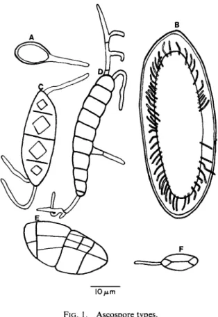
The following classification of spores according to septation is taken from Hale (1967), and some of these types are illustrated in Fig. 1.
I O / i m
FIG. 1. Ascospore types.
122 F. BRIAN PYATT
a. Simple spores: unicellular and unseptate, often small and thin walled
(Lecidea, Lecanora, Parmelia, Usnea), more rarely very large (to 300^m and thick walled (Pertusaria)
b. Transversely septate spores: elongate and multicellular with 1-30 or 40 transverse cross walls (Catillaria, Graphis, Pyrenula)
c. Muriform spores: multicellular with both transverse and longitudinal walls, often large (Phaeographina, Lopadium, Umbilicaria, Diploschistes)
d. Polarilocular spores: two-celled spores with a thick median wall and a thin isthmus, or conversely a single-celled spore with a median constric
tion (Teloschistaceae).
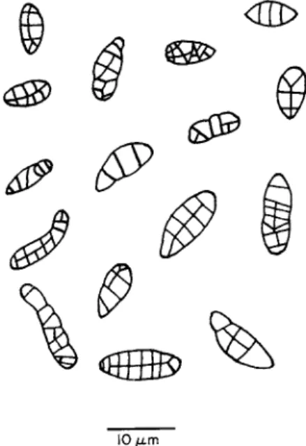
Pyatt (1969a) has found much variation in spore sizes in a given species;
e.g., those of Caloplaca heppiana measure 10.5-13.0 χ 6.3-8.4 μια, while those of Rhizocarpon geographicum measure 25-40 χ 11-18μπι. Septation is also a variable character. Figure 2 shows ascospores of Rhizocarpon geo
graphicum drawn 1 hour after the start of sporulation (Pyatt, 1969a). It appears that the simplest type is that with three transverse septae and that further development occurs to give the more complex types. However, as the ascospores are discharged in various stages of septation, this does reflect some problems in the use of ascospores for species identification.
The multilayered sporangial wall of the fungus Allomyces has been des
cribed by Skucas (1967), and Reeves (1967) has described the fine structure
10 μ. m
FIG. 2. Septation patterns in Rhizocarpon geographicum.
4 . L I C H E N P R O P A G U L E S 123
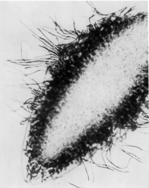
FIG. 3. Ascospore of Pertusaria pertusa.
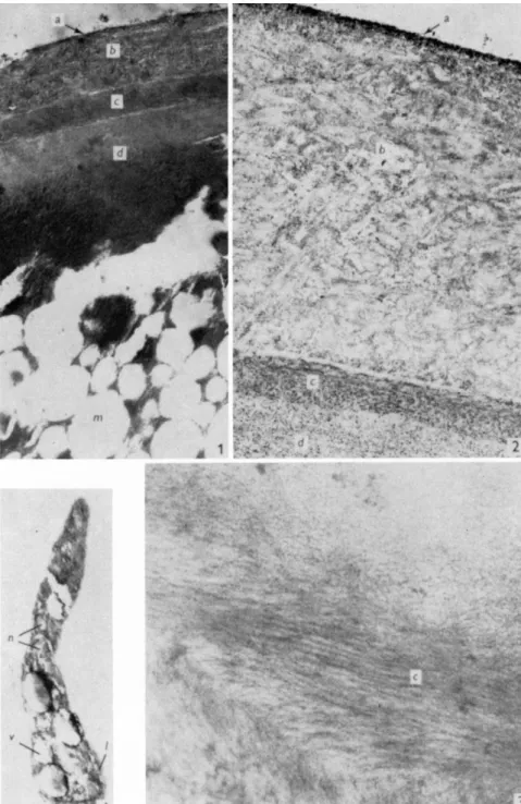
of ascospore formation in Pyronema domesticum. Pyatt (1969b) investigated the ultrastructure of the ascospore wall of Pertusaria pertusa (Figs. 3 and 4).
The ovoid ascospores measure 230 χ 60 μτη and are surrounded by a well- defined wall 9 μπι thick laterally and 11 μτη thick at the poles. The wall is composed of five major layers, one of which is especially rich in regularly
FIG. 4. Pertusaria pertusa. (1) Ascospore wall illustrating various layers, χ 4000. (2) Outer region of ascospore wall, χ 16000. (3) Layer with orientated microfibrils, χ 22000. (4) Germ tube. x 6 0 0 0 . m, medulla; v, vacuoles; 1, limiting membrane; n, nuclei. From Pyatt (1969b).
124
4. LICHEN PROPAGULES 125 orientated, nearly concentrically arranged microfibrils. The germ tubes arise from the inside of the ascospore wall. Initially the germ tube rudiment has a bulbous base in which there is a single nucleus; this nucleus divides into two as the germ tube elongates. One nucleus remains in the base while the other migrates towards the apex of the germ tube where elongation is occurring. Hilitzer (1926) and Kofler (1957) give further accounts of spore discharge and germination.
The medulla of the ascospore is composed of numerous vacuoles as- sociated with cytoplasm containing small and rather ill-defined nuclei.
Although ascomycetes make up the vast bulk of lichens, a few widespread mostly tropical species have a fungal component classified as a basidiomy- cete. They bear basidiospores exogenously on basidia (see Chapter 2 by Letrouit-Galinou for details).
Finally we have asexual conidia which are produced within pycnidia.
Conidia are usually divided into two categories; the very small unicellular microconidia and the much larger, septate, macroconidia.
In all of the above cases the lichen thallus has the potential of producing vast numbers of spores; this spore production and subsequent sporulation must create a high energy demand.
B. Spore Discharge 1. INTRODUCTION
Gregory (1952) recognized two spore types in fungi: (a) sedentary, e.g., chlamydospores, zygospores. These permit the organism to survive periods of adverse conditions. These propagules typically germinate at their site of production; (b) dispersal, e.g., conidia, ascospores, and basidiospores.
This is the category with which we are concerned as these are the spores which allow for colonization of new regions.
Gregory (1952) indicates that a variety of factors help to disperse these spores at random and he recognizes four types of dispersal: (i) active dis- persal; (ii) dispersal by insects and other animals; (iii) rain dispersal; and (iv) wind dispersal.
In these cases he recognizes two stages of dispersal both of which are energy requiring: separation from parent stratum and transport to a dis- tance.
2. TECHNIQUES
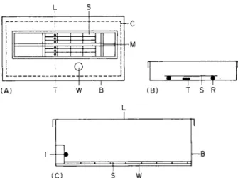
Probably the most useful piece of apparatus which can be used in studies of ascospore discharge is the spore train illustrated in Fig. 5 A. This has been used for work on fungi and has been described by Ingold (1933), Walkey and Harvey (1966), and Pyatt (1969a).
126 F. BRIAN PYATT
FIG. 5. (A) Spore train. (B) "Static" apparatus. (C) Apparatus to measure distance of spore discharge.
A "tram" (T) of microscope slides (S) is drawn, by a cord (M) leading to an electric motor, at a known rate across the lichen thalli (L) which are held on moist cotton wool. Ascospores are caught on the underside of the slides (S). The apparatus is enclosed within a Perspex box (B) and there- fore there is no wind influence; humidity is maintained by the open water surfaces (W) and by moist cotton wool (C) placed around the edge of the box.
The apparatus was kept within a growth chamber at a temperature of 14°±
1°C. A time clock was used to obtain various light regimes. Another method is to make use of an automatic volumetric spore trap as described by Hirst (1952). In this case, spores are "sucked" from the substratum (following discharge) and impact on a microscope slide travelling at 2 mm/hour.
A far simpler "static" set up can occasionally be used (Fig. 5B). A portion of thallus (T) bearing ascocarps is placed into a damp petri dish. By means of a glass rod (R) a microscope slide (S) is suspended above the thallus.
The slide is removed periodically and counts are made of the number of spores present.
3. NATURE OF SPORE DISCHARGE
a. DISTANCE. Brodie and Gregory (1953) made use of Lycopodium
spores to illustrate that funnel or egg-cup-shaped vessels lost spores more readily than rectangular vessels or horizontal slides. They point out that wind passing over the cup creates enough turbulence inside to lift spores into the main airstream.
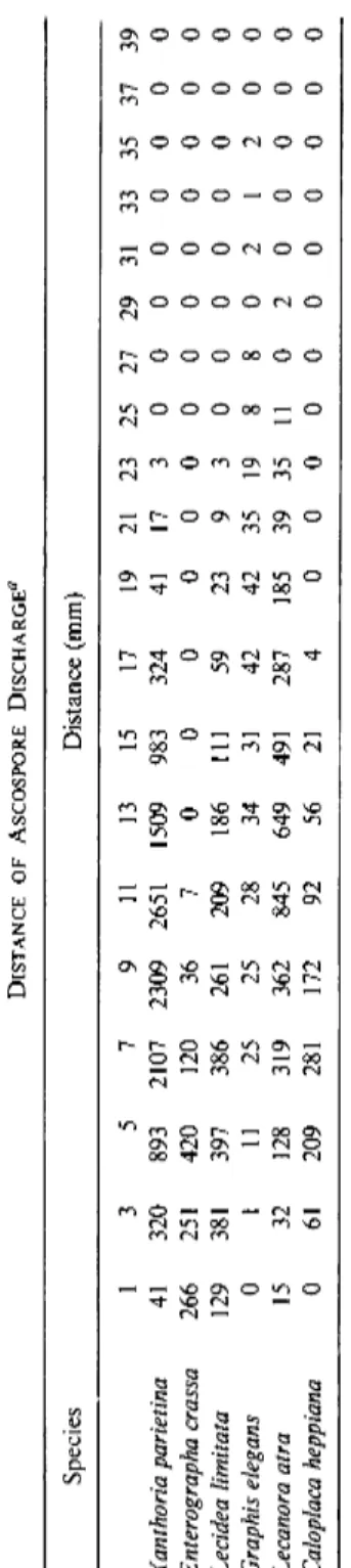
Bailey and Garrett (1968) found a maximum distance of discharge of
TABLE II DISTANCE OF ASCOSPORE DISCHARGE0 Species Distance (mm) 1 3 5 7 9 11 13 15 17 19 21 23 25 27 29 31 33 35 37 39 Xanthoria parietina 41 320 893 2107 2309 2651 1509 983 324 41 17 3 0 0 0 0 0 0 0 0 Enterographa crassa 266 251 420 120 36 7 0 0 0 0 0 0 0 0 0 0 0 0 0 0 Lecidea limitata 129 381 397 386 261 209 186 111 59 23 9 3 0 0 0 0 0 0 0 0 Graphis elegans 0 1 11 25 25 28 34 31 42 42 35 19 8 8 0 2 1 2 0 0 Lecanora atra 15 32 128 319 362 845 649 491 287 185 39 35 11 0 2 0 0 0 0 0 Caloplaca heppiana 0 61 209 281 172 92 56 21 4 0 0 0 0 0 0 0 0 0 0 0 ^Calculated from four experiments.
4. LICHEN PROPAGULES 127
128 F. BRIAN PYATT
45 mm in the case of Rhizocarpon umbilicatum. This figure applies to hori
zontal discharge in a closed system where there is no wind effect.
Pyatt (1969a) investigated the distance of ascospore discharge in six species using the apparatus illustrated in Fig. 5C. From Table II it can be seen that the maximum distance to which a spore was ejected was 35 mm in the case of Graphis elegans. However, maximum spore accumulations were usually found approximately 3-13 mm from the ascocarp. Distances of this order are unlikely to explain the colonization of new areas by lichens but this discharge would be sufficient to carry the ascospores from the substrate on which they were produced into the turbulent air.
Pyatt (1969a) found that the first discharged spores of Lecidea limitata
travel up to lO^am farther than later discharged spores. This is not a reflec
tion of ascospore size but possibly the first discharged spores are expelled more forcibly.
b. RATES OF DISCHARGE. Bailey and Garrett (1968) found the rates of discharge to vary and that the total discharge declined rapidly over a period of 7 days from a peak within the first 24 hours. Pyatt (1969a) also found that ascospore discharge reached a peak 1-2 hours after initiation
T A B L E III
H O U R L Y DISCHARGE RATES ( A V E R A G E OF FOUR REPLICATES)0
Species
Lecanora Pertu Peltigera Lecidea
coni- saria Lecanora Graphis poly- macro- Usnea Xanthoria Time zaeoides pertusa subfusca elegans dactyla car ρ a florida parietina
0900 6030 9 4710 1476 3 0 41 0
1000 7220 86 960 977 1200 47 23 95
1100 3094 28 552 94 6310 23 16 390
1200 1753 11 119 112 4119 620 0 123
1300 1756 9 33 78 972 242 33 107
1400 1142 2 61 80 1058 201 23 81
1500 1101 4 24 63 491 163 11 26
1600 1446 14 11 10 422 81 54 12
1700 1060 10 70 21 312 80 41 4
1800 3826 2 460 435 130 90 46 3 2 06
1900 3843 10 330 294 1600 43 28 3109
2000 2305 12 240 79 389 10 21 294
2100 1364 8 136 54 312 8 11 98
2200 703 3 49 28 109 13 9 61
"From Pyatt (1969a).
^Thallus placed in dry petri dishes.
4. LICHEN PROPAGULES 129 by moistening air-dried thalli. After this the rate of discharge decreased irregularly (Table III). After 9 hours the portions of thalli were transferred to dry petri dishes. In Lecanora conizaeoides, L. subfusca, Graphis elegans, Peltigera polydactyla, and Xanthoria parietina there was a significant increase (Table III) in the degree of spore discharge for 1-2 hours and then values fell; this does lend some support for the observation of Ahmadjian (1961) that ascospores are discharged as the ascus ruptures during drying. How
ever, the observations of Bailey and Garrett (1968) and Pyatt (1968a) that there was massive sporulation following the addition of water to thalli casts much doubt on this.
c. RHYTHMS. Quite a considerable amount of work has been carried out on rhythms and periodicity of spore discharge in nonlichenized fungi by various workers, e.g., Manachere (1968), Carpenter (1949), Ingold and Cox (1955), and Gay et al (1959). Walkey and Harvey (1968a, b) investi
gated the effect of factors such as climate and temperature on the spore discharge rhythms of pyrenomycetes. Fahim (1966) carried out experiments to determine the effect of light and other factors on the sporulation of
Alternaria porri. He found a high degree of sporulation after 2 hours ex
posure to sunlight followed by 48 hours incubation in the dark.
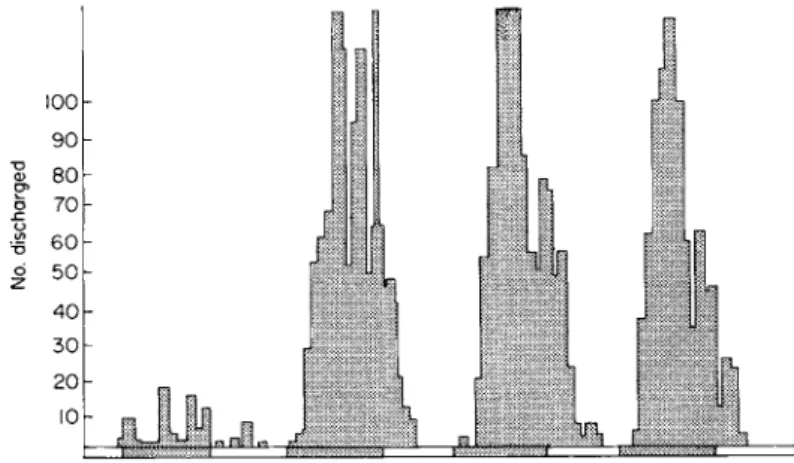
Pyatt (1968a) obtained the following data on rhythms of ascospore dis
charge in lichens.
Lecidea macrocarpa. With a 12/12 (light hours/dark hours) alternation at 14°± 1°C, it exhibited a well-defined nocturnal rhythm (Fig. 6). When transferred to conditions of continuous light, the rhythm persists for a while, but eventually is depressed. From Table IV, it can be seen that
I
6 0ο 50 4 0 σ 70
100 9 0
3 0
20 Pi
10
JbjL
FIG. 6 . Rhythm of sporulation in Lecidea macrocarpa under conditions of 1 2 / 1 2 light/dark alternation at 1 4 ° ± 1 ° C .
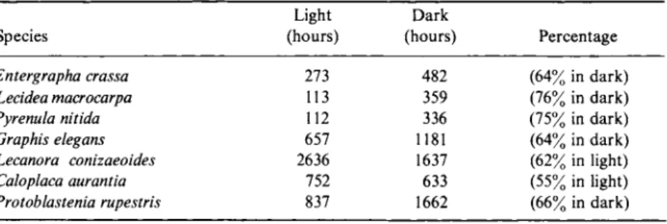
130 F. BRIAN PYATT T A B L E I V
AVERAGE SPORE DISCHARGE VALUES DURING THE LIGHT A N D D A R K PERIODS OF A 12/12 LIGHT H O U R S / D A R K H O U R S A L T E R N A T I O N0
Light Dark
Species (hours) (hours) Percentage
Entergrapha crassa 273 482 (64% in dark)
Lecidea macrocarpa 113 359 (76% in dark)
Pyrenula nitida 112 336 (75% in dark)
Graphis elegans 657 1181 (64% in dark)
Lecanora conizaeoides 2636 1637 (62% in light)
Caloplaca aurantia 752 633 (55% in light)
Protoblastenia rupestris 837 1662 (66% in dark)
"From Pyatt (1969a).
approximately three-quarters of the sporulation occurred during the night period.
Pyrenula nitida. In a 12/12 (light hours/dark hours) regime, no distinct rhythm of sporulation was apparent (Fig. 7).
Enterographa crassa. It shows some degree of nocturnal synchronization, but light was found to be essential for ascospore production and/or dis
charge.
Graphis elegans. No clear rhythm was found; indeed, it shifts from mainly daylight to mainly nocturnal discharge.
Peltigera canina. This tends to be a daylight species but it is greatly in
fluenced by the availability of moisture.
/. Periodicity. Bailey and Garrett (1968) showed that Lecanora coni
zaeoides tends to discharge spores more readily at low temperatures. This agrees with Werner (1927) who indicated that winter and early spring are periods of maximum discharge in Xanthoria parietina.
Scott (1959), however, pointed out that spore discharge is affected by the moistening of apothecia by rain, dew, and mist. In temperate regions the annual distribution of these types of precipitation will, he believes, result
20
10
Ει Ά
FIG. 7. Rhythm of sporulation in Pyrenula nitida under conditions of a 12/12 light/dark alternation at 14° ± 1°C.
4. L I C H E N P R O P A G U L E S 131
T A B L E V
SPORE D I S C H A R G E IN L I C H E N S ^
Species Jan. Feb. Mar. Apr. May June Jul. Aug. Sept. Oct. Nov. Dec.
Lecanora campestris Ρ Ρ L L L L L L Μ Ρ Ρ Ρ
Lecanora conizaeoides R R L L L L Μ Μ Μ Ρ Μ Μ
Lecanora atra Ρ Ρ Μ Μ L L Μ Μ Μ Ρ Ρ Ρ
Lecidea limitata Ρ Ρ L Μ Μ L L L Μ Ρ Ρ Ρ
Lecidea macrocarpa Ρ Ρ Ρ Ρ Ρ Μ Μ Μ Μ Ρ Ρ Ρ
Baeomyces rufus — — Μ Μ — — Μ Μ Μ Μ Ρ Μ
Ochrolechia parella Ρ Ρ L Μ L — — — Μ Ρ Ρ Ρ
Pertusaria pertusa Ρ — — L Μ Μ — L Μ Ρ Ρ Ρ
Xanthoria parietina Ρ Ρ L L Μ Μ Μ Μ Μ Ρ Ρ Ρ
Caloplaca heppiana Ρ Μ L L L Μ Μ Μ Μ Μ Ρ Ρ
Toninia coeruleonigricans Ρ L Μ Μ Ρ Ρ Μ Μ Μ Μ Ρ Ρ
Buellia canescens Ν Ν Ν Ν Ν Ν Ν L Μ Ρ Ρ Ρ
Graphis spp. Ρ Ρ L Μ L Μ Ρ Μ L Μ Ρ Ρ
aF r o m Pyatt (1969c).
^Letters designate sporulation, i.e., R = 0 - 5 0 , L = 50-200, Μ = 200-1000, Ρ = 1000 or more spores discharged. Ν refers to the absence of ascocarps and a dash (—) to the absence of sporulation.
in maximum sporulation during spring and autumn. Verseghy (1965) agrees that the amount of rainfall undoubtedly influences the spore production of lichens and found that in dry years the asci are sterile in both spring and autumn. Also she noted that in rainy years the spore production is intensive in autumn. Des Abbayes (1951) reached the conclusion that in western Europe sporulation is most pronounced in the spring.
Pyatt (1969c), in a monthly investigation over 1 year, found a fairly distinct annual periodicity of spore discharge (Table V), with peaks generally occurring during the period from October to January inclusive.
ii. Propagule Types. Ascospore discharge alone will not lead to the production of new thalli; for this to occur the ascospore must come into contact with a certain specific algal cell. Obviously, it seems most probable that this chance encounter will not always occur. However, in the case of
Pertusaria pertusa and Lecidea limitata (Pyatt, 1969a), and perhaps other species, the fungus-alga association is not always lost even during sporula
tion.
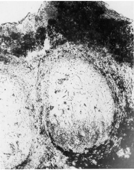
Sections of verrucae (Fig. 8), taken after exposure of ascocarps to various humidities, reveal that the gelatinous matrix or hymenial gelatin within the apothecium cavity tends to shrink as the humidity decreases. From Fig.
8 it can be seen that there are aggregations of algal cells (Protococcus) in the
132 F. BRIAN PYATT
FIG. 8. Section through verruca Pertusaria pertusa.
ostiole region. It appears, then, that at high humidity the gelatinous matrix within the apothecium swells and aids in forcing the large ascospores out
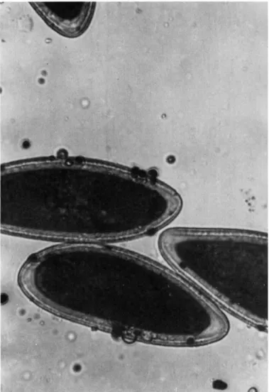
through the ostiole. As the ascospores escape, they frequently pick up algal cells from the ostiole region. An average of 4% of the ascospores examined on slide deposits carried algae (Fig. 9). Of these ascospores approximately 60% carried one algal cell, 30% carried two, and 10% carried three or more.
4. L I C H E N P R O P A G U L E S 133
FIG. 9. Ascospore of P. pertusa with associated algae.
Thus, in the case of Pertusaria pertusa, reestablishment of the composite thallus is likely to occur quite frequently due to the discharge of a joint propagule
F. B. Pyatt (unpublished) has investigated ascospore discharge in Xan-
thoria parietina. A number of spore deposits were collected, in fairly dry petri dishes, and these were investigated to determine the average number of ascospores present in each grouping. From Table VI it is apparent that usually the propagule contains eight ascospores.
134 F. B R I A N P Y A T T T A B L E V I
ASCOSPORE GROUPINGS IN Xanthoria parietina0
Number of ascospores associated
(on slide deposit) Percentage occurrence
1 1.0
2 1.2
3 1.2
4 1.0
5 2.2
6 3.6
7 2.2
8 74.2
12 0.6
16 3.4
22 0.2
24 1.6
50 7.6
°From Pyatt (1969a).
Pyatt and Harvey (1973) have studied the fungus Tichothecium erraticum
which occurs on thalli and apothecia of Caloplaca heppiana and Lecanora
campestris. When infected thalli were enclosed within damp petri dishes, ascospores of both the host and pathogen were discharged within two days.
They used the spore-train technique and observed that the spore discharge of both Caloplaca heppiana and Tichothecium erraticum generally occurred between 10 PM, and 6 AM, i.e., both were nocturnal species.
From an examination of an infected apothecium of Caloplaca heppiana,
it was found that discharged ascospores of the parasite were present on the hymenial surface of the host. Indeed it does seem possible that the smaller spores of Tichothecium erraticum (2-2.5 χ 6-6.5 μίτι) may be picked up by the larger spores of Caloplaca heppiana (10.5-13.0 χ 6.3-8.4 μιτι) at the moment of discharge to yield a composite propagule.
Hi. Animal Dispersal. A somewhat unusual method of ascospore dis
tribution has been described by Pyatt (1968d). Rotifers living on the surfaces of apothecia of Xanthoria parietina actively ingest ascospores. Possibly these rotifers may be splashed off and could then continue to move in a water film. It was observed that there was a 15% germination of excreted asco
spores in those rotifers which had contained 20 or more spores, whereas in those which contained only 3-8 ascospores the voided spores were broken and did not germinate. Gregory (1952) describes how the spores of copro- philous nonlichenized fungi such as Sphaerophorus are deposited on vegeta
tion and are later eaten by animals.
4. LICHEN PROPAGULES 135
4. FACTORS AFFECTING SPORE DISCHARGE
a. LIGHT. Leach (1962) and Leach and Trione (1965) showed the impor- tance of ultraviolet radiation in inducing sporulation in a number of fungi, and have illustrated that the 230 and 290 nm regions of the spectrum are particularly effective. Table IV, from Pyatt (1969a), illustrates the effect of light on lichen sporulation.
b. TEMPERATURE. Ingold (1960) suggested that in most pyrenomycetes, sporulation is probably arrested below 5°C but increases in rate up to 35° C.
He concluded that temperature is probably most important in connection with spore maturation.
However, Hart (1926) found that at any given relative humidity, an in- crease of temperature generally leads to a decrease in the viability of fungus spores, and the lower temperatures, above 0°C, favor longevity. Pyatt (1969a) found that extremes of temperature repressed spore discharge and that in certain species there was more sporulation at 20° C than at 6°C.
c. HUMIDITY AND RAIN. Verseghy (1965) points out that water, which is absorbed over the entire surface of the thallus, is stored in capillaries mostly among the hyphae and so the substratum is less significant in lichens for the absorption of water; the relative humidity of the environment, she notes, is much more important. Her findings are largely in agreement with those of Blum (1964).
Ascospore discharge has seldom been observed when dry thalli are placed in dry petri dishes. Indeed, Pyatt (1969a) showed that spore discharge rarely occurs in Pertusaria pertusa below a relative humidity of 90%.
Pyatt (1968a) showed that when Pyrenula nitida and Peltigera canina were sprayed with fine water droplets there was a massive liberation of spores irrespective of the time or the light regime. Gregory et al. (1959) have shown that fungus spores can be dispersed by splashes.
Fahim (1966), working with Alternaria porri, found the optimum relative humidity for sporulation to be less than 100%. His data agree with those of Cochrane (1958), who indicates that most evidence suggests a generalization that in most fungi reproductive processes are favored by a moderate as dis- tinct from a high atmospheric humidity.
Following the work of Ingold and Marshall (1962) on stimulation of spore discharge by reduced humidity in Sordaria, Austin (1968) investigated the effects of air speed and humidity changes on spore discharge in Sordaria fimicola. She found that a reduction of the relative humidity from 95% to
70% or 35% leads to an immediate but temporary increase in discharge rate (see Table III). A relative humidity of 70% apparently only affected actual discharge, while prolonged exposure to 35% relative humidity interferes as
136 F. BRIAN PYATT TABLE VII
EFFECTS OF PRECIPITATION ON SPORULATION0
Species Effect of dew Effect of flooding
Xanthoria parietina Many spores released Many spores released Pertusaria pertusa Very few released Very few released Lecanora conizaeoides Spores and algae released Spores and algae released Lecanora subfusca Spores released Spores released
Ochrolechia androgyna Spores and algae released Spores and algae released
Usnea florida Spores released Spores released
Lecidea limitata Spores released Spores released
Graphis scripta Spores released Spores released
°From Pyatt (1969a).
well with earlier stages in spore development. She concludes that the im- mediate effect on discharge is physical, not biochemical, and that the perithecial wall seems to play some role in the control of spore discharge.
Pyatt (1969a) carried out additional experiments by enclosing lichen thalli in petri dishes containing water to maintain a humid atmosphere. In some cases droplets of water were pipetted and positioned over ascocarps, i.e., synthetic dew formation. In other cases the thalli were flooded. From Table VII it can be seen that these forms of precipitation do encourage sporulation.
d. WIND. Gregory (1952) points out that in a wind tunnel, at wind speeds in excess of 1 meter/second, the effects of gravity on objects as small as spores are negligible.
Kelly et al. (1951) used an airplane to trap spores and illustrated that convection and turbulence are sufficient to carry spores to great heights in the atmosphere.
The problem of spore removal by wind can be solved in two main ways:
(a) by raising the stalk of the apothecium through the laminar boundary layer into the turbulent air, as in stipitate Calicium; (b) by active discharge into the turbulent air. Once this has occurred, long-distance spore dispersal can occur.
Gregory (1952) found at the same time that under normal turbulence conditions, 99.9% of the spores are deposited within 100 meters of the source.
Eventually the spore is deposited, and Gregory points out that the principal methods are sedimentation, impaction, boundary-layer exchange, turbulent deposition, and rain washing.
e. pH. This has been investigated by Pyatt (1968a, 1969a) in an initial study using both citric acid and phosphate buffer solutions. With Xanthoria
4. LICHEN PROPAGULES 137
parietina a distinct optimum for sporulation occurred at pH 4-5. This is illustrated by the duration of ascospore production and by the total number of ascospores produced. Graphis elegans has a fairly distinct optimum for spore discharge at pH 5-6.
f. SPOROGENIC SUBSTANCES. Trione, et al. (1966) found that certain sub
stances affected sporulation. Whittington and Penn (1967) used camphor vapor as it has been shown to increase the frequency of diploidization in vegetative hyphae (Roper, 1952). They also found that exposure resulted in a definite increase in the number of aberrant asci.
g. MISCELLANEOUS. It has been indicated (Pyatt, 1968a) that ascospore discharge is inhibited in some species when the thallus is maintained upon a nutrient rich substrate. Atmospheric pollutants can also have an effect.
Pyatt (1969a) exposed lichen thalli to 10 vpm of sulfur dioxide for a period of 10 days in sealed jars. From the results in Table VIII it is apparent that sulfur dioxide inhibited the sporulation of the species investigated.
Aggregation of spores may give spores in groups a degree of protection against certain environmental conditions. This can be shown by collecting spore deposits of Lecanora conizaeoides and L. subfusca on microscope slides.
As slides dried the spores moved distances of up to 80μιη until large groups of ascospores were obtained. When these slides were placed in a humid atmosphere the spores separated.
C. Spore Germination
1. INTRODUCTION
Schein and Rotem (1965) have demonstrated that the "germinability" of the spores of nonlichenized fungi is inversely proportional to both temper
ature and atmospheric humidity, and Teitell (1958) indicated that the length of life of spores is affected by environmental conditions.
Fox (1966), working with Ramalina ecklonii, found that the ascospores did not germinate in less than 5 weeks of incubation in a wide variety of
T A B L E V I I I
EFFECT O F SULFUR D I O X I D E ON SPORULATION0
Species Thalli exposed to S0.2 Control
Pertusaria pertusa N o sporulation Sporulation
Xanthoria parietina N o sporulation Prolific sporulation Lecidea limitata Slight sporulation Prolific sporulation
Graphis elegans N o sporulation Prolific sporulation
"From Pyatt (1969a).
138 F. BRIAN PYATT
cultural conditions. He found no difference in germination rates between cultures incubated in light or darkness.
Recent work on spore germination in lichens has been done by Bailey (1966b), who examined discharged spores to see if germination had occurred.
This was also done by Garrett (1968). Pyatt (1968a) showed that ascospore germination in a water film generally commences within 4-5 days after the start of sporulation. He also noted that ascospores of crustose species generally tend to have a higher percentage viability than ascospores of foliose and fruticose types. It is conceivable that in these latter types, the elevation of the thallus into the turbulent boundary layer of the atmosphere favors soredia dispersal and dispersal of thallus fragments. In the case of crustose species, thallus fragmentation is probably less likely to occur and the active discharge of ascospores must be considered an important at
tribute.
2. RHYTHMS AND PERIODICITY
Pyatt (1969c) found that massive sporulation is not invariably associated with high percentage germination values; see Tables V and IX. The following data can be seen in Fig. 10:
Lecanora campestris. Maximum germination values of 30% occurred in October, December, and January; this is also the period of maximum spore discharge.
Lecanora conizaeoides. Spore germination, within the 10-day test period, was only observed in December.
Lecanora atra. Prolific discharge occurs from October to February;
during this period the peak values of germination are 40%. After spore dis-
TABLE IX
ASCOSPORE GERMINATION SHOWING THE A V E R A G E PERCENTAGE VALUES O B T A I N E D E A C H MONTH*7
Percentage germination after 10 days*
Species Jan. Feb. Mar. Apr. May June July Aug.Sept. Oct. Nov. Dec.
Lecanora conizaeoides Pertusaria pertusa Xanthoria parietina Caloplaca heppiana Toninia coeruleonigricans Buellia canescens
0 0 0 0 60 — — 2
0 0 0 0
0 0 1 2
10 0 0 0 Ν Ν Ν Ν 0 5 0 0 0 Ν
0
ο ο ο ο
Ν 0
ο ο
Ν 0 ο
2 0
ο ο
5
0 0 0 1 2 0 26 40 0 6 0 0 9 15 20 15 0 0 0 10 10 30 30 20
°From Pyatt (1969c).
bN refers to the absence of ascocarps and a dash to the lack of spore discharge.
4. L I C H E N P R O P A G U L E S 139
Month
FIG. 10. Percentage of spores germinating after 10 days (monthly figures). The species are Lecanora campestris ( ) , L. atra ( ) , Lecidea limitata ( ) , L. macrocarpa (—), Ochrolechia parella (Ο Ο O), Graphis spp. ( ), and Baeomyces rufus ( ) .
charge has decreased in March, spore germination reaches apeak of45%.
During spring, spore discharge and germination drop and then begin to increase in July.
Lecidea limitata. Sporulates from October to May. During November germination is 100%.
Baeomyces rufus. Prolific spore discharge occurs during November and this is when the highest germination values are found (75%). Discharge did occur but with no germination in March and April. Hence it appears that in this species, ascospores produced during March and April undergo a period of dormancy prior to germination.
Ochrolechia parella. Prolific sporulation occurs during October to February; germination increases from 80% in October to 100% in January and then falls. Pyatt (1968b) noted that some germ tubes tend to swell during their passage through the epispore.
Pertusaria pertusa. The germination of the ascospore of this species has been described by Pyatt (1968c). Prolific spore discharge occurs during October to January and germination increases from zero in October to 60%
in January.
Xanthoria parietina. Germination was only observed in October which is part of the period of maximum discharge.
Caloplaca heppiana. Prolific spore discharge is limited to November, December, and January, and maximum germination (20%) occurs in November.
140 F. BRIAN PYATT Toninia coeruleonigricans. There are peaks of sporulation in November to January and May to June. However, there was only one peak value (10%)
of germination which occurred during December and January.
Buellia canescens. Peak germination values of 30% occurred during the period of prolific discharge.
Graphis spp. Prolific sporulation occurs from November to February and peak germination values (90%) occur in December and January.
Times for germination have been investigated by various workers and the following are some of the results obtained: 10-24 hours, Ahmadjian (1961);
24-144 hours, Kofler and Bouzon (1961); and 10 days or less, Henriksson (1958).
3. TEMPERATURE AND HUMIDITY
Merek and Fergus (1954) found that theendooomaidioiEndoconidiophora fagacearum remained viable longest under dry conditions. Groom and Panni-
seet (1933) noted that lower relative humidities favor retention of viability of specific fungus spores. Cochrane (1958) showed that longevity increases as temperature decreases to 0°C. Cochrane (1945) and Hart (1926) indicate that uredospores of rust fungi have greatest longevity at moderate relative humidities.
4. pH
Pyatt (1968a) found that the pH optimum for ascospore germination was not always the same as that of sporulation and that material collected from different areas showed somewhat different responses.
5. NUTRIENTS AND ALGAE
Scott (1959) found with Peltigera praetextata that an aqueous extract of the phycobiont must be present for successful germination.
Losel (1967), working with the fungus Agaricus bisporus, noted that ger- mination was stimulated by vapor diffusing from dilute solutions of various short-chain fatty acids. Losel suggests that this is not a pH effect, but is due to the entry of the compounds into metabolic pathways.
Pyatt (1968a) investigated ascospore germination on various agars. He found that media with a low nutrient content are more stimulatory to the early stages of ascospore germination than those media with a high nutrient content.
Pyatt (1969a) collected ascopores on agar infused with extracts of the bark on which the lichens were growing. The extracts were prepared by
4 . L I C H E N P R O P A G U L E S 141
T A B L E X
EFFECT OF BARK EXTRACT ON ASCOSPORE G E R M I N A T I O N0
Species
Percent germination Species Agar and bark extract Plain agar
Pertusaria pertusa 15 9
Thelotrema lepadinum 100 100
Lecanora subfusca 92 70
flFrom Pyatt (1969a).
washing a portion of the bark which was then macerated and finally filtered.
The results are shown in Table X.
The condition of sporostasis, where spores fail to germinate on account of inhibitors produced by the parent culture or cultures of another species, has been described by Robinson and Park (1966). This condition is some- times found to depend upon the continuous production of a volatile sub- stance by mycelium.
Another kind of investigation was carried out (Pyatt, 1969a) to determine whether extracts of a given lichen could suppress the germination of an asco- spore of another species (Table XI). Ascospore germination is suppressed in Xanthoria parietina by Parmelia physodes and Ochrolechia parella extracts;
in Pertusaria pertusa by Usnea comosa and Xanthoria parietina extracts. It appears from this initial experiment that extracts of lichens from the habitat of the species being tested are able to inhibit the germination of ascospores from such areas.
T A B L E XI
EFFECT OF O T H E R SPECIES ON ASCOSPORE G E R M I N A T I O N0 Extract^
Species C 1 2 3 4 5 6
Xanthoria parietina 10 10 4 4 10 — —
Pertusaria pertusa 10 — — 10 — 0 0
°From Pyatt (1969c).
^Extracts were prepared by washing portions of thalli and then macerating them in water; the ones used are water ( C ) , Lobaria pulmonaria (1), Parmelia physodes (2), Ochrolechia parella (3), O. androgyna (4), Usnea comosa (5), Xanthoria parietina (6).
cResults are expressed in terms of percentage germination.
TABLE XII PERCENTAGE VIABILITY AFTER DESICCATION (AVERAGE OF FOUR REPLICATES)0 Species Lecanora Lecanora Graphis Lecidea Peltigera Lecidea Usnea Pertusaria Xanthoria Weeks conizaeoides subfusca elegans limitata polydactyla macrocarpa florida pertusa parietina 0 5 20 60 10 0 75 0 16 0 1 0 18 43 10 0 59 0 24 0 2 0 18 39 9 0 29 0 20 0 3 0 14 35 9 0 12 0 19 0 4 0 12 20 8 0 7 0 21 0 5 0 15 18 9 0 7 0 7 0 6 0 13 19 7 0 5 0 8 0 7 0 9 11 4 0 6 0 8 0 8 0 9 4 5 0 5 0 7 0 flFrom Pyatt (1969a).
142 F. BRIAN PYATT
4. LICHEN PROPAGULES 143
6. POLLUTION
Pyatt (1969a) calculated that the average germination of ascospores of
Lecidea macrocarpa, collected from near the pollution source in the steel producing town of Port Talbot, was 62% after 14 days in a humid atmos
phere. The value with material collected from a more distant site was 79%.
This does seem to indicate that air pollution probably adversely affects the viability of lichen ascospores and so possibly those lichen species propagated by ascospores are less likely to colonize heavily polluted regions than species
propagated by other, perhaps more resistant, means.
7. PERCENTAGE VIABILITY OF ASCOSPORES AFTER DESICCATION
Pyatt (1969a) placed ascospore deposits in dry, silicone grease sealed petri dishes for varying amounts of time. They were eventually transferred to a humid atmosphere and the percentage viability was calculated after 8 days of exposure. From Table XII it can be seen that the viability of ascospores falls with extended periods of desiccation.
Acknowledgments
Some of this work was carried out while I was undertaking postgraduate research in the Department of Botany, University College, Cardiff, in association with the Department of Social and Occupational Medicine, Welsh National School of Medicine, Cardiff. I am grate
ful to Professor G. F. Asprey for his encouragement and to my wife who helped in the rather tedious analysis of ascospore data. The work initially was financed by a fellowship from the Nuffield Research Foundation.
References
Ahmadjian, V. (1961). Bryologist 64, 168-179.
Austin, B. (1968). Ann. Bot. (London) [N.S.] 32, 251-260.
Bailey, R. Η. (1966a). J. Linn. Soc. London Bot. 59, 479-490.
Bailey, R. H. (1966b). Rev. Bryol. Lichenol. 37, 852-853.
Bailey, R. H. (1968). Rev. Bryol. Lichenol. 36, 314-315.
Bailey, R. H. (1970). Lichenologist 4, 256.
Bailey, R. H., and Garrett, R. M. (1968). Lichenologist 4, 57-65.
Barkman, J. J. (1958). "Phytosociology and Ecology of Cryptogamic Epiphytes." van Gorcum & Co., Assen.
Blum, Ο. B. (1964). J. Bot. 21, 3 2 - 4 1 . Brodie, H. J. (1951). Can. J. Bot. 29, 224-234.
Brodie, H. J., and Gregory, P. H. (1953). Can. J. Bot. 31, 4 0 2 ^ 1 0 . Carpenter, J. B. (1949). Phytopathology 39, 980-985.
Cochrane, V. W. (1945). CornellUniv.,Agr. Exp. Sta., Mem. 2 6 8 , 1 - 3 9 . Cochrane, V. W. (1958). "Physiology of Fungi." Wiley, N e w York.
Darbishire, Ο. V. (1897). Bot Jahrb. 22, 593-671.