Manuscript Number: MIMET-D-12-00535
Title: A new prototype system for the rapid diagnosis of bacterial and fungal sepsis Article Type: Research Paper
Keywords: Clinically relevant bacteria; Candida; Melting point analysis; Fluorescence resonance energy transfer; Multiplex real-time PCR; bloodstream infections
Corresponding Author: Dr. Ferenc Somogyvári, MSc PhD Corresponding Author's Institution: University of Szeged First Author: Ádám Horváth, MSc
Order of Authors: Ádám Horváth, MSc; Zoltán Pető, MD PhD; Edit Urbán, MD PhD; Csaba Vágvölgyi, MSc PhD; Ferenc Somogyvári, MSc PhD
Abstract: Fungal and bacterial infections can be identified more rapidly and sensitively with
polymerase chain reaction (PCR)-based techniques than with conventional blood culturing techniques.
There was recently been a surge in the number of molecular based infection identifications. There have been numerous reports of new real-time PCR-based pathogen identification, although the clinical practicability of such techniques is not yet clarified.
The present study focuses on the differentiation of the most common bacterial and fungal causative pathogens of bloodstream infections. A multiplex real-time PCR approach is introduced for the detection and differentiation of fungi, Gram-positive (G+) and Gram-negative (G-) bacteria. The Gram classification is performed with the specific fluorescence resonance energy transfer (FRET) probes recommended for LightCycler capillary real-time PCR. The novelty of this system is the use of a non- specific SYBR Green dye instead of labelled anchor probes or primers to excite the acceptor dyes on the FRET probes. In parallel, the use of an intercalary dye allows the detection of fungal amplicons. In this way, fungi, G+ and G- bacteria in the same reaction tube can be differentiated quickly (within an hour after the DNA preparation) via the melting temperatures of the amplicons and probes.
The identification of pathogen species with this modified FRET technique is specific and more rapid than with the gold-standard culture-based methods, and permits the rapid and early evidence-based management of bloodstream infections in clinical practice.
Dear Editor,
I enclose a manuscript entitled “A new prototype system for the rapid diagnosis of bacterial and fungal sepsis” by Ádám Horváth, Zoltán Pető, Edit Urbán, Csaba Vágvölgyi and Ferenc Somogyvári for consideration for publication in "Journal of Microbiological Methods".
By means of real-time PCR and melting point analysis, we have detected and identified the most frequent bacterial and fungal pathogens. Two primer pairs were used for the multiplex amplification of bacterial and fungal DNA, and two hybridization probes to differentiate Gram – and G+ bacteria. The novelty of this prototype system is the use of non-specific SYBR Green dye as a donor molecule instead of a labelled primer or other specific anchor probe.
Thank you in advance.
Yours sincerely,
Ferenc Somogyvari MSc, PhD
Department of Medical Microbiology and Immunobiology Faculty of Medicine, University of Szeged
H-6720 Dom ter 10, Szeged, Hungary.
Tel: +36 303992519 Fax: +36 62545113
e-mail: fsoma99@gmail.com
Dear Mr Volker Gurtler,
I enclose the corrected manuscript entitled “A new prototype system for the rapid diagnosis of bacterial and fungal sepsis” by Ádám Horváth, Zoltán Pető, Edit Urbán, Csaba Vágvölgyi and Ferenc Somogyvári for consideration for publication in "Journal of Microbiological Methods".
The methodological originality has amended in the “Introduction”, and the fungal ITS data have complemented in the “Table 1”. The whole ms have checked again by native English- speaking colleague.
Yours sincerely,
Ferenc Somogyvari MSc, PhD
Department of Medical Microbiology and Immunobiology Faculty of Medicine, University of Szeged
H-6720 Dom ter 10, Szeged, Hungary.
Tel: +36 303992519 Fax: +36 62545113
e-mail: fsoma99@gmail.com
We have detected and identified the most frequent bacterial and fungal pathogens.
Two primer pairs were used for the multiplex amplification of bacterial and fungal DNA, and two hybridization probes to differentiate Gram – and G+ bacteria.
The novelty of this prototype system is the use of non-specific SYBR Green dye as a donor molecule instead of a labelled primer or other specific anchor probe.
1 2 3 4 5 6 7 8 9 10 11 12 13 14 15 16 17 18 19 20 21 22 23 24 25 26 27 28 29 30 31 32 33 34 35 36 37 38 39 40 41 42 43 44 45 46 47 48 49 50 51 52 53 54 55 56 57 58 59
A new prototype system for the rapid diagnosis of bacterial and fungal sepsis
Ádám Horvátha, Zoltán Petőb, Edit Urbánc, Csaba Vágvölgyid, Ferenc Somogyvária *
aDepartment of Microbiology and Immunobiology, Faculty of Medicine, University of Szeged, Dóm tér 10, Szeged, Hungary
bDepartment of Anaesthesiology and Intensive Therapy, Faculty of Medicine, University of Szeged, Semmelweis u. 6, Szeged, Hungary
cInstitute of Clinical Microbiology, Faculty of Medicine, University of Szeged, Semmelweis u. 6, Szeged, Hungary
dDepartment of Microbiology, Faculty of Science and Informatics, University of Szeged, Közép fasor 52, Szeged, Hungary
* Corresponding author: Department of Medical Microbiology and Immunobiology, Faculty of Medicine, University of Szeged, H-6720 Dóm tér 10, Szeged, Hungary. Tel: +36 62545115 Fax:
+3662545113
Email address: fsoma99@gmail.com
4 5 6 7 8 9 10 11 12 13 14 15 16 17 18 19 20 21 22 23 24 25 26 27 28 29 30 31 32 33 34 35 36 37 38 39 40 41 42 43 44 45 46 47 48 49 50 51 52 53 54 55 56 57 58 59 60 61
1. Introduction
Bloodstream infections, which are among the most severe manifestations of bacterial diseases, are life-threatening infections that affect individuals with serious underlying conditions or an impaired immune system (Danai et al., 2006). There were 10,319,418 reported cases of bloodstream infections in the USA between 1979 and 2002, and during this period the number of cases demonstrated an annualized increase of 8.7% (Martin et al., 2003). In patients who require intensive care treatment, the majority of infections are caused by bacteria: 52.1% by Gram- positive (G+) and 37.6% by Gram-negative (G-) species. Fungal sepsis is likewise an important public health problem, but it account for only 4.6% of the overall infections (Martin et al., 2003).
Mixed Candida/bacterial bloodstream infections have been reported to occur in >23% of all episodes of candidaemia (Klotz et al., 2007). Despite its comparatively low frequency, fungal sepsis can progress to severe sepsis and septic shock associated with a drastic rise in mortality.
The early and appropriate treatment of such infections is critical, with the aim of interrupting the progression and improving the outcome (Vallés et al., 2003; Kumar et al., 2009).
There has been recently increasing interest in molecular diagnosis in sepsis cases, because this is more reliable and faster than the classical blood-culturing techniques (Leggieri et al., 2010).
Molecular strategies such as PCR, ligase chain reaction, nucleic acid sequence based amplification, nested PCR, etc. allow nucleic acid-based differentiation (Carroll et al., 2000).
Nevertheless, these molecular approaches are applied only following the positivity of the blood culture, and require a substantial amount of time as compared with the RT PCR-based method described below. The LightCycler PCR assay is relatively easy to perform and may be carried out in small laboratories.
A fluorescence resonance energy transfer (FRET) technique was earlier reported which involves a distance-dependent interaction between the electronic excited states of two dye molecules. The excitation is transferred from a donor (anchor) molecule to an acceptor (quencher) molecule without emission of a photon. This is an appropriate method for discriminating between the most common G+ and G- bacteria that cause sepsis (Klaschik et al., 2002). For better discrimination, subgroups were created within the G+ and G- stains, with differentiation via the melting temperature of the overall PCR product and the melting point of the probes (Klaschik et al., 2004). This system requires less than 4 h, including the time of the DNA preparation and the
4 5 6 7 8 9 10 11 12 13 14 15 16 17 18 19 20 21 22 23 24 25 26 27 28 29 30 31 32 33 34 35 36 37 38 39 40 41 42 43 44 45 46 47 48 49 50 51 52 53 54 55 56 57 58 59
evaluation of the PCR results. The parallel detection of fungal and bacterial infections in a real- time system is still an unresolved problem. Identification of the most common clinically relevant fungal infections is possible through a simple melting point analysis relating to the ITS2 (internal transcribed spacer) region. This non-coding region is a highly variable rRNA region that is adaptable for the identification of clinically relevant fungi in a broad range (Shoch et al., 2012).
Measurments are made at 540 nm and require a non-specific intercalary dye (Somogyvári et al., 2005).
Real-time PCR detection can be performed by using free dyes or labelled sequence-specific probes. One kind of combination of the two techniques was so far being possible that unlabelled probes used for the amplicon detection and Tm determination (Zho et al., 2004). Another, parallel application was the combination of TaqMan chemistry and the very new aspecific dye, BOXTO, as a multiplex PCR (Lind et al., 2006). The novelty of our prototype system that we propose is the use of non-specific SYBR Green dye as a donor molecule instead of a labelled primer or other specific anchor probe. In this way it is possible to examine all three main groups of organisms potentially causing an infection in one tube multiplex PCR reaction.
4 5 6 7 8 9 10 11 12 13 14 15 16 17 18 19 20 21 22 23 24 25 26 27 28 29 30 31 32 33 34 35 36 37 38 39 40 41 42 43 44 45 46 47 48 49 50 51 52 53 54 55 56 57 58 59 60 61
1. Materials and methods
2.1. Reference strains
Strains of 17 clinically relevant bacterial species were collected, as typical of the main causative agents of bloodstream infections (Lucignano et al., 2011). Nine reference strains, Staphylococcus aureus (ATCC 25923), Staphylococcus epidermidis ATCC 12228, Enterococcus faecalis ATCC29212, Listeria monocytogenes ATCC 4701, Bacteroides fragilis ATCC 25285, Pseudomonas aeruginosa ATCC 27853, Haemophilus influenzae ATCC 49247, Escherichia coli ATCC 25922 and Klebsiella pneumoniae ATCC 700603 were from the American Type Culture Collection (ATCC). Streptococcus pyogenes OKI 80002 was from the National Centre for Epidemiology, Hungary (OKI) and Proteus vulgaris HNCMB 60076 was from the Hungarian National Collection of Medical Bacteria (HNCMB). A further 6 clinical strains, Enterococcus faecium, Stenotrophomonas maltophilia, Serratia marcescens, Enterobacter aerogenes,
Enterobacter cloacae and Acinetobacter baumannii were also included. The species identities of the clinical isolates were confirmed by conventional biochemical methods.
Ten fungal strains were examined. Five reference strains, Candida albicans ATCC 10231 and ATCC 14053, C. tropicalis ATCC 750, C. parapsilosis ATCC 22019 and C. glabrata ATCC 39316, were from the ATCC, Cryptococcus neoformans IFM 5844 and IFM 5855 were from IFM Quality Services Pty Ltd (IFM), and Aspergillus fumigatus SzMC 2486, A. flavus SzMC 2536 and A. niger SzMC 2761 were from the Szeged Microbiological Collection (SzMC).
2.2. Bacterial DNA purification
The bacterial strains were grown on Columbia CNA agar base under aerobic circumstances, except that Bacteroides fragilis was grown under anaerobic conditions. The bacterial DNA was extracted with the QIAamp® DNA Blood Mini Kit (QuiaGene Inc, Chatsworth, Calif., USA), following the manufacturer’s instructions in “Protocols for Bacteria”. 1 ml of log-phase culture suspension at a concentration of 107 CFU/mL was used for the preparation. For determination of the sensitivity of the reaction, 100 L of the serially diluted S. aureus reference strain was used
4 5 6 7 8 9 10 11 12 13 14 15 16 17 18 19 20 21 22 23 24 25 26 27 28 29 30 31 32 33 34 35 36 37 38 39 40 41 42 43 44 45 46 47 48 49 50 51 52 53 54 55 56 57 58 59
for DNA extraction. The number of bacterial cells was determined by plating aliquots of serially diluted samples onto Columbia CNA agar base.
For lysis of the rigid multilayered G+ bacterial cell wall, we used a pre-incubation step with 20 mg/mL lysozyme (in 20 mM Tris·HCl, pH 8.0, 2 mM EDTA, 1.2% TritonX100). The spin protocol for “DNA Purification from Tissues” was followed after incubation at 30 oC for 30 min.
The final concentration of DNA was 2.0-13.8 ng/µL with a ratio A260/A280 = 1.6-1.8 after purification.
2.3. Fungal DNA purification
All the clinically important fungi were grown on Sabouraud agar. The fungal DNA was extracted from 1 mL of a log-phase culture suspension contains 9.6 x 10⁷ of fungal cells. For determination of the sensitivity of the reaction, 100 L of the serially diluted C. albicans reference strain was used for DNA extraction. The number of fungal cells was determined by plating aliquots of serially diluted samples onto Sabouraud-glucose agar.
We followed the QIAamp® DNA Mini Kit Protocol for Yeasts. In this case, additional reagents were required for elimination of the complex fungal cell wall structure: sorbitol buffer (1 M sorbitol, 100 mM EDTA, 14 mM -mercaptoethanol) (Lim et al., 2008) was used and the samples were incubated with lyticase for 30 min at 30 oC. Efficient and complete lysis was achieved in 1.5 hour on a shaking water bath. This purification yielded 2.0–25 μg of DNA in 100 μL of water (2.0–13.8 ng/μL), with A260/A280 = 1.6–1.8.
2.4. Bacterial and fungal primer design, FRET probes
Two primer pairs were used for multiplex amplification of bacterial and fungal DNA.
The bacterial primer pair was PLK1 (TAC GGG AGG CAG CAG) forward and PLK2 (TAT TAC CGC GGC TGC T) reverse, which are highly conserved in different groups of eubacteria (Klaschik et al., 2002) and amplify the 16S rRNA sequence. The PLK2 reverse primer was modified and used without the inner fluorescence labelling. Originally the labelled primer excited the Gram specific probes. We applied the non-specific SYBR Green dye for excitation; it also
4 5 6 7 8 9 10 11 12 13 14 15 16 17 18 19 20 21 22 23 24 25 26 27 28 29 30 31 32 33 34 35 36 37 38 39 40 41 42 43 44 45 46 47 48 49 50 51 52 53 54 55 56 57 58 59 60 61
serves for visualization of the fungal amplicons. This primer pair produces a 187 bp fragment in each species.
The previously described hybridization probes were used for the Gram classification (Klaschik et al., 2004). ISN2 (5’-CCG CAG AAT AAG CAC CGG CTA ACT CCG T-3’) labelled with LCRed 640 was specific for G-, and ISP3 (5’-CCT AAC CAG AAA GCC ACG GCT AAC TAC GTG-3’) labelled with Cy5.5 was specific for G+ bacteria. The originally used (Klaschik et al., 2004) ISP2 probe was labelled with LCRed705 at the 5’ end. The producers offered Cy5.5 dye instead of LCred705. This modified probe can serve as a template in further experiments.
The ITS86 (GTG AAT CAT CGA ATC TTT GAA C) forward and ITS 4 (TCC TCC GCT TAT TGA TAG C) reverse primers were used for detection of the medically important yeasts. These primers are located in the ITS2 region, which is a highly variable sequences between the 5.8S and 28S rRNA sequence and amplifies 192-494 bp (Lott et al., 1993).
2.5. Mastermixes/excitation dyes
Different non-specific intercalary dyes are used for real-time PCR investigations. Most of them are accessible in ready-to-use mastermix formulas. Our goal was to choose the best dye for excitation of the labelled probes. The tested dyes were LCGreen “LightCycler® 480 High Resolution Melting Master” (Roche Diagnostic GmbH, Mannheim, Germany); SybrGreen
“LightCycler® 480 DNA Master SYBR Green I”, (Roche); “IQ™ SYBR® Green Supermix”
(Bio-Rad Laboratries, Inc., Hercules, CA, USA) and “Maxime™ SYBR Green qPCR Master Mix no ROX” (Fermentas, Vilnius, Lithuania) and EvaGreen (“LC-FastStart DNA Master
Hybridization Probes” (Roche) combined with EvaGreen dye (Biotium Inc., Hayward, CA, USA) and “Sso Fast™ EvaGreen® Supermix” (BioRad). All mastermixes were used according to the manufacturer’s instructions.
2.6. PCR conditions
PCR was performed on a LightCycler real-time PCR instrument (Roche). The reaction volume of 10 L contained 1 L of DNA, 1 M of each of the primers, 0.7 M of each of the probes, an
4 5 6 7 8 9 10 11 12 13 14 15 16 17 18 19 20 21 22 23 24 25 26 27 28 29 30 31 32 33 34 35 36 37 38 39 40 41 42 43 44 45 46 47 48 49 50 51 52 53 54 55 56 57 58 59
appropiate amount of mastermix and 0.2 mM BSA in the cases of the Fermentas and BioRad mastermixes.
The PCR conditions were as follows: initial denaturation at 95 oC for 600 s, followed by 40 cycles of denaturation (95 oC for 0 s, 20 oC/s), annealing (55 oC for 15 s, 20 oC/s), and extension (72 oC for 20 s, 2 oC/s). The emitted fluorescence was measured after the annealing steps. The melting curve analysis procedure consisted of 1 cycle at 95 °C for 10 s, 40 °C for 120 s, followed by an increase of the temperature to 95 °C at 0.2 °C/s. The fluorescence signal (F) was monitored continuously during the temperature ramp and plotted against temperature (T).
2.7. Data analysis
The melting peaks were evaluated with the LightCycler Software V 3.5. The melting peaks were determined with the manual Tm option on the three detection channels (F1, F2 and F3).
The standard deviation (SD) of the melting points was calculated from five parallel experiments.
The fungal or bacterial infection was verified by gel electrophoresis on 1.5% agarose gel with the help of a low-range DNA ladder.
4 5 6 7 8 9 10 11 12 13 14 15 16 17 18 19 20 21 22 23 24 25 26 27 28 29 30 31 32 33 34 35 36 37 38 39 40 41 42 43 44 45 46 47 48 49 50 51 52 53 54 55 56 57 58 59 60 61
2. Results and discussion
3.1. Discrimination of the fungal, G+ and G- bacterial pathogens
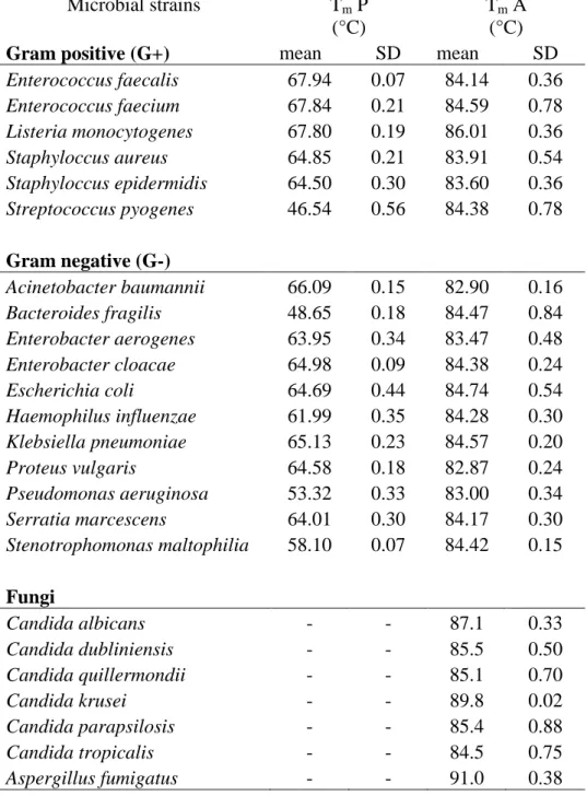
DNA samples from all species studied were prepared and amplified successfully with the SYBR Green dye-based method in the LightCycler instrument. Species-specific Tm-s were obtained by melting point analysis on three detection channel and all pathogens were identified correctly as fungi or G- or G+ bacteria (Table 1). On the F1 channel (540 nm), the melting points of all the amplicons (Tm A) were visible due to the fluorescent signal of the SYBR Green non-specific intercalary dye. On the F2 (640 nm) and F3 (705 nm) channels, the G- and the G+ probes (Tm P), respectively, gave fluorescence signals. After the discrimination of the G- and G+ strains, the fungal pathogens could be screened because the fungal strains gave no signal on the other two channels.
3.2. Determination of the bacterial pathogens
Four G+ and nine G- bacterial subgroups could be distinguished through a joint consideration of the melting points of the probes and the melting point of the overall PCR product (Figure 1). S.
aureus and the S. epidermidis have very close-lying melting temperatures and their species- specific differentiation is not possible via this 16S rRNA sequence (Figure 2). A comparison of the Gene Bank sequences (S. aureus and S. epidermidis NCBI Taxonomy ID: NC_009782.1 and JF_799903.1) of these species revealed a variance of only three base pairs, none of them in the region where the probe is associated with the DNA. Thus, determination of the clinically relevant Staphylococcus species requires other gene sequences whereby the antibiotic resistance can be detected (Kilic and Basustaoglu, 2011). The situation is the same with the two Enterococcus species (Maheux et al., 2011). At the same time, S. pyogenes and L. monocytogenes are clearly differentiable.
Among the G- bacteria, E. coli is one of the main causative agents of bloodstream infections (Bo et al., 2011). Unfortunately, it has almost the same Tm as those of E. cloacae and S. marcescens.
4 5 6 7 8 9 10 11 12 13 14 15 16 17 18 19 20 21 22 23 24 25 26 27 28 29 30 31 32 33 34 35 36 37 38 39 40 41 42 43 44 45 46 47 48 49 50 51 52 53 54 55 56 57 58 59
Other bacterial strains, such as H. influenza, are clearly differentiable through the melting temperature of the probe (Figure 3) or amplicon.
Antibiotic resistance cannot be determined directly with our prototype system. The susceptibility testing of resistant E. coli strains can be performed with a PCR-based technique with other 16S rRNA specific primers (Waldeisen et al., 2011). Unfortunately, these investigations require a PCR after the identification of the bacteria.
3.3. Determination of fungal pathogens
Fourteen frequently encountered fungal pathogens could be distinguished. The highly variable ITS 2 target sequence allowed correct identification of all of the clinically relevant fungal strains through the Tm points on the F1 channel (Gutzmer et al 2004., Somogyvári et al., 2005). There was no signal on the F2 or F3 channel. The sensitivity of the reaction was 5 colony forming units (CFU) per reaction.
The correct differentiation between bacteria and fungi was verified by means of gel electrophoresis with the help of the amplicon length (fungal amplicons 192-494 bp, bacterial 187 bp).
2.4 Calibration of the multiplex PCR
All three non-specific dyes (LCGreen, EvaGreen and SybrGreen) excited all of the labelled probes (LCRed640, LCRed705 and Cy5.5). The best results were obtained with the SybrGreen dye.
The determination of Tm is very sensitive to the composition of the PCR reaction mixture, and primarily to the ionic strength. To avoid Tm bias originating from pipetting errors between PCR runs, the application of mastermixes is advisable. One limitation of the method is that the various forms offered by different suppliers differ in reagent composition, which may influence the Tm values.
Naturally, repeated runs with a given mastermix yield reproducible data. In the event of a change of mastermix, however, calibration is necessary to establish the new Tm data on the fungal strains.
4 5 6 7 8 9 10 11 12 13 14 15 16 17 18 19 20 21 22 23 24 25 26 27 28 29 30 31 32 33 34 35 36 37 38 39 40 41 42 43 44 45 46 47 48 49 50 51 52 53 54 55 56 57 58 59 60 61
The data determined in the present work were obtained with the use of “Fermentas Maxima SybrGreen, no ROX” and five parallel experiments.
4 5 6 7 8 9 10 11 12 13 14 15 16 17 18 19 20 21 22 23 24 25 26 27 28 29 30 31 32 33 34 35 36 37 38 39 40 41 42 43 44 45 46 47 48 49 50 51 52 53 54 55 56 57 58 59
3. Conclusions
Real-time PCR is one of the fastest diagnostic methods currently available. The use of rRNA genes for the detection is based on the conserved 16S rRNA sequences of the bacteria. As regards fungi, the ITS sequence refers to a segment of non-functional RNA situated between 5.8S and 28S rRNAs. To reproduce the results, it is possible to differentiate between fungi and bacteria, or between fungal species by electrophoresis (Ferrer et al., 2001, Turenne et al., 1999) or melting analysis (Khan et al., 2009). The Roche LightCycler PCR was specially constructed to amplify amplicons under 500 bp. The amplicons amplified by PLK1/PLK2 comprised 187 bp, while the fungal amplicons amplified by ITS86/ITS4 primer pair varied between 192 bp (Geotrichum candidum) and 494 bp (Malassezia furfur), values is perfectly suitable for this instrument profile.
We took advantage of the LC system when we used the FRET technique to detect and differentiate the bacterial pathogens. We excited the fluorescent probes with the help of a non- specific intercalary dye, which is an uncommon procedure in real-time investigations. This allows the detection of fungal pathogens in parallel with bacteria. We made use of a multiplex PCR in combination with FRET probes and melting point analysis for the broad-range identification of the most frequent causative agents of bloodstream infections. Among the limitations of the method, neither the G+ S. aureus and S. epidermidis nor the G- E. coli, E.
cloacae and S. marcescens can be distinguished. Additional species-specific probes or primers are needed for the further differentiation of these species, if necessary. In spite of the limitations, the rapidly available information on the bacterial species permits targeted therapy with narrow- spectrum antibiotics instead of empirically administered broad-spectrum antibiotics.
The pathogen load in sepsis is generally below 10 CFU/mL (Loeffler et al., 2000). As the sensitivity of this PCR is 5 CFU per reaction, in combination with a correct preparation method it is suitable for the detection of bloodstream infections.
The incidence of sepsis has continuously increased in recent decades, and the early detection of the pathogens present can have a great impact on the clinical outcome of the infection (von Lilienfeld-Toal et al., 2009, Vince et al., 2008, Wallet et al., 2010, Bauer and Reinhard 2010).
The molecular diagnostic systems allow species identification in less than 24 h, which is a drastic
4 5 6 7 8 9 10 11 12 13 14 15 16 17 18 19 20 21 22 23 24 25 26 27 28 29 30 31 32 33 34 35 36 37 38 39 40 41 42 43 44 45 46 47 48 49 50 51 52 53 54 55 56 57 58 59 60 61
improvement relative to the gold standard culture-based method and Gram staining-based identification methods that yield results in 24 to 72 h (Tissari et al., 2010, Cleven et al., 2010).
In summary, the causative agents of the infection can be detected in 2 h without DNA preparation. This was accomplished by the multiplex PCR with the new combination of aspecific dyes and labelled probes. This system is advantageous to traditional FRET based assays by accurate detecting both the Tm of the probes and amplicons.
4. Acknowledgements
This publication is supported financially by the European Union and cofunded by the European Social Fund. Project title: “Broadening the knowledge base and supporting the long-term professional sustainability of the Research University Centre of Excellence at the University of Szeged by ensuring the rising generation of excellent scientists.” Project number: TÁMOP- 4.2.2/B-10/1-2010-0012.
4 5 6 7 8 9 10 11 12 13 14 15 16 17 18 19 20 21 22 23 24 25 26 27 28 29 30 31 32 33 34 35 36 37 38 39 40 41 42 43 44 45 46 47 48 49 50 51 52 53 54 55 56 57 58 59
References
Bauer, M., Reinhart, K., 2010. Molecular diagnostics of sepsis - Where are we today? Int. J. Med.
Microbiol. 300, 411–413.
Carroll, N.M., Jaeger, E.E., Choudhury, S., Dunlop, A.A., Matheson, M.M., Adamson, P., Okhravi, N., Lightman, S., 2000. Detection of and discrimination between Gram-positive and Gram-negative bacteria in intraocular samples by using nested PCR. J. Clin. Microbiol. 38, 1753- 1757.
Cleven, B.E.E., Palka-Santini, M., Gielen, J., Meembor, S., Krönke, M., Krut, O., 2006.
Identification and characterization of bacterial pathogens causing bloodstream infections by DNA microarray. J. Clin. Microbiol. 44, 2389-2397.
Danai, P.A., Moss, M., Mannino, D. M., Martin G.S., 2006. The epidemiology of sepsis in patients with malignancy. Chest 129, 1432-1440.
Ferrer, C., Colom, F., Frasés, S., Mulet, E., Abad, J.L., Alió, J.L., 2001. Detection and identification of fungal pathogens by PCR and by ITS2 and 5.8S ribosomal DNA typing in ocular infections. J. Clin. Microbiol. 39, 2873-2879.
Gutzmer, R., Mommert, S., Küttler, U., Werfel, T., Kapp, A., 2004. Rapid identification and differentiation of fungal DNA in dermatological specimens by LightCylerPCR. J. Med.
Microbiol. 53, 1207-1214.
Khan, Z., Mustafa, A.S., Alam, F.F., 2009. Real-time LightCycler polymerase chain reaction and melting temperature analysis for identification of clinically important Candida spp. J. Microbiol.
Immunol. Infect. 42, 190-195.
Kilic, A., Basustaoglu, A.C., 2011. Double triplex real-time PCR assay for simultaneous detection of Staphylococcus aureus, Staphylococcus epidermidis, Staphylococcus hominis, and
4 5 6 7 8 9 10 11 12 13 14 15 16 17 18 19 20 21 22 23 24 25 26 27 28 29 30 31 32 33 34 35 36 37 38 39 40 41 42 43 44 45 46 47 48 49 50 51 52 53 54 55 56 57 58 59 60 61
Staphylococcus haemolyticus and determination of their methicillin resistance directly from positive blood culture bottles. Res. Microbiol. 162, 1060-1066.
Klaschik, S., Lehmann, L.E., Raadts, A., Book, M., Hoeft, A., Stuber, F., 2002. Real-time PCR for detection and differentiation of Gram-positive and Gram-negative bacteria. J. Clin. Microbiol.
40, 4304-4307.
Klaschik, S., Lehmann, L.E., Raadts, A., Book, M., Gebel, J., Hoeft, A., Stuber, F., 2004.
Detection and differentiation of in vitro - spiked bacteria by real-time PCR and melting-curve analysis. J. Clin. Microbiol. 42, 512-517.
Klotz, S.A., Chasin, B.S., Powell, B., Gaur, N.K., Lipke, P.N., 2007. Polymicrobial bloodstream infections involving Candida species: analysis of patients and review of the literature. Diagn.
Microbiol. Infect. Dis. 59, 401-406.
Kumar, A., Ellis, P., Arabi, Y., Roberts, D., Light, B., Parrillo, J.E., Dodek, P., Wood, G., Kumar, A., Simon, D., Peters, C., Ahsan, M., Chateau, D., Cooperative Antimicrobial Therapy of Septic Shock Database Researc Group. 2009. Initiation of inappropriate antimicrobial therapy results in a fivefold reduction of survival in human septic shock. Chest 136, 1237-1248.
Leggieri, N., Rida, A., François, P., Schrenzel, J., 2010. Molecular diagnosis of bloodstream infections: planning to (physically) reach the bedside. Curr. Opin. Infect. Dis. 23, 311-319.
Lim, C.S., Tung, C.H., Rosli, R., Chong, P.P., 2008. An alternative Candida spp. cell wall disruption method using a basic sorbitol lysis buffer and glass beads. J. Microbiol. Methods. 75, 576-578.
Lind, K., Ståhlberg, A., Zoric, N., Kubista, M., 2006. Combining sequence-specific probes and DNA binding dyes in real-time PCR for specific nucleic acid quantification and melting curve analysis. Biotechniques. 40, 315-319.
4 5 6 7 8 9 10 11 12 13 14 15 16 17 18 19 20 21 22 23 24 25 26 27 28 29 30 31 32 33 34 35 36 37 38 39 40 41 42 43 44 45 46 47 48 49 50 51 52 53 54 55 56 57 58 59
Loeffler, J., Henke, N., Hebart, H., Schmidt, D., Hagmeyer, L., Schumacher, U., Einsele, H., 2000. Quantification of fungal DNA by using fluorescence resonance energy transfer and the light cycler system. J. Clin. Microbiol. 38, 586-590.
Lott, T.J., Kuykendall, R.J., Reiss, E., 1993. Nucleotide sequence analysis of the 5.8S rDNA and adjacent ITS2 region of Candida albicans and related species. Yeast 9, 1199–1206.
Lucignano, B., Ranno, S., Liesenfeld, O., Pizzorno, B., Putignani, L., Bernaschi, P., Menichella, D., 2011. Multiplex PCR allows rapid and accurate diagnosis of bloodstream infections in newborns and children with suspected sepsis. J. Clin. Microbiol. 49, 2252-2258.
Maheux, A.F., Bissonnette, L., Boissinot, M., Bernier, J.L., Huppé, V., Bérubé, E., Boudreau, D.K., Picard, F.J., Huletsky, A., Bergeron, M.G., 2011. Method for rapid and sensitive detection of Enterococcus sp. and Enterococcus faecalis/faeciumcells in potable water samples. Water Res.
45, 2342-2354.
Martin, G.S., Mannino, D.M., Eaton, S., Moss, M., 2003. The epidemiology of sepsis in the United States from 1979 through 2000. N. Engl. J. Med. 348, 1546-1554.
Schoch, C.L., Seifert, K.A., Huhndorf, S., Robert, V., Spouge, J.L., Levesque, C.A., Chen, W., Fungal Barcoding Consortium; Fungal Barcoding Consortium Author List. 2012. Nuclear ribosomal internal transcribed spacer (ITS) region as a universal DNA barcode marker for Fungi.
Proc. Natl. Acad. Sci. U.S.A. 109, 6241-6246.
Somogyvari, F., Serly, J., Doczi, I., Nagy, E., 2005. Molecular differentiation of most frequent Candida species causing blood-stream infection. Mycoses 2, S198.
Tissari, P., Zumla, A., Tarkka, E., Mero, S., Savolainen, L., Vaara, M., Aittakorpi, A., Laakso, S., Lindfors, M., Piiparinen, H., Maki, M., Carder, C., Huggett, J., Gant, V., 2010. Accurate and rapid identification of bacterial species from positive blood cultures with a DNA based microarray platform: an observational study. Lancet 375, 224–230.
4 5 6 7 8 9 10 11 12 13 14 15 16 17 18 19 20 21 22 23 24 25 26 27 28 29 30 31 32 33 34 35 36 37 38 39 40 41 42 43 44 45 46 47 48 49 50 51 52 53 54 55 56 57 58 59 60 61
Turenne, C., Sanche, S.E., Hoban, D.J., Karlowsky, J.A., Kabani, A.M., 1999. Rapid identification of fungi by using the ITS2 genetic region and an automated fluorescent capillary electrophoresis system. J. Clin. Microbiol. 37, 1846-1851.
Vallés, J., Rello, J., Ochagavía, A., Garnacho, J., Alcalá, M.A., 2003. Community-acquired bloodstream infection in critically ill adult patients: impact of shock and inappropriate antibiotic therapy on survival. Chest 123, 1615-1624.
Vince, A., Lepej, S.Z., Barsic, B., Dusek, D., Mitrovic, Z., Serventi-Seiwerth, R., Labar, B., 2008. LightCycler SeptiFast assay as a tool for the rapid diagnosis of sepsis in patients during antimicrobial therapy. J. Med. Microbiol. 57, 1306–1307.
von Lilienfeld-Toal, M., Lehmann, L.E., Raadts, A.D., Hahn-Ast, C., Orlopp, K.S., 2009. Utility of a commercially available multiplex real-time PCR assay to detect bacterial and fungal pathogens in febrile neutropenia. J. Clin. Microbiol. 47, 2405–2410.
Waldeisen, J.R., Wang, T., Mitra, D., Lee, L.P., 2011. A real-time PCR antibiogram for drug- resistant sepsis. PLoS One. 6, e28528.
Wallet, F., Nseir, S., Baumann, L., Herwegh, S., Sendid, B., 2010. Preliminary clinical study using a multiplex real-time PCR test for the detection of bacterial and fungal DNA directly in blood. Clin. Microbiol. Infect. 16, 774–779.
Zhou, L., Myers, A.N., Vandersteen, J.G., Wang, L., 2004. Wittwer CT. Closed-tube genotyping with unlabeled oligonucleotide probes and a saturating DNA dye. Clin. Chem. 50, 1328-1335.
4 5 6 7 8 9 10 11 12 13 14 15 16 17 18 19 20 21 22 23 24 25 26 27 28 29 30 31 32 33 34 35 36 37 38 39 40 41 42 43 44 45 46 47 48 49 50 51 52 53 54 55 56 57 58 59
6. Glossary
RT PCR real-time PCR
FRET fluorescence resonance energy transfer LC LightCycler
ICU Intensive Care Unit ITS internal transcribed spacer ATCC American Type Cell Culture IFM IFM Quality Services Pty Ltd SzMC Szeged Microbiological Collection Tm melting temperature
SD standard deviation
Tm A melting peak of the amplicon Tm P melting peak of the probe
Table 1. Melting points of bacterial and fungal amplicons and probes. All the amplicons Tm measured at the F1 channel (540 nm). The signal generated by aspecific SybrGreen dye. The G+ specific probes got the signal at the F2 channel (640 nm) the G- probes at the F3 channel (705 nm). The signal induced by the help of special FRET technique.
Microbial strains Tm P (°C)
Tm A (°C)
Gram positive (G+) mean SD mean SD
Enterococcus faecalis 67.94 0.07 84.14 0.36 Enterococcus faecium 67.84 0.21 84.59 0.78 Listeria monocytogenes 67.80 0.19 86.01 0.36 Staphyloccus aureus 64.85 0.21 83.91 0.54 Staphyloccus epidermidis 64.50 0.30 83.60 0.36 Streptococcus pyogenes 46.54 0.56 84.38 0.78
Gram negative (G-)
Acinetobacter baumannii 66.09 0.15 82.90 0.16 Bacteroides fragilis 48.65 0.18 84.47 0.84 Enterobacter aerogenes 63.95 0.34 83.47 0.48 Enterobacter cloacae 64.98 0.09 84.38 0.24
Escherichia coli 64.69 0.44 84.74 0.54
Haemophilus influenzae 61.99 0.35 84.28 0.30 Klebsiella pneumoniae 65.13 0.23 84.57 0.20
Proteus vulgaris 64.58 0.18 82.87 0.24
Pseudomonas aeruginosa 53.32 0.33 83.00 0.34 Serratia marcescens 64.01 0.30 84.17 0.30 Stenotrophomonas maltophilia 58.10 0.07 84.42 0.15
Fungi
Candida albicans - - 87.1 0.33
Candida dubliniensis - - 85.5 0.50
Candida quillermondii - - 85.1 0.70
Candida krusei - - 89.8 0.02
Candida parapsilosis - - 85.4 0.88
Candida tropicalis - - 84.5 0.75
Aspergillus fumigatus - - 91.0 0.38
Figure 1. Differentiation of the bacterial pathogens. The group temperatures indicate the entire Tm of the pathogens. The subgroup temperatures are the melting temperatures of the hybridization probes.
Group I 84 Co Group II 86 Co
64.5 Co S. aureus S. epidermidis
67.8 Co E. faecium E. faecalis
46.5 Co S. pyogenes
67.8 Co L.
monocytogenes
Group II84.5 Co Group I 83 Co
66 Co A. baumannii
64 Co E. aerogenes
48.6 Co B. fragilis
58 Co S. maltophila
62 Co H. influenzae
64 Co S. marcescens E. cloacae E. coli
65 Co K.
pneumoniae
53.3 Co P. aeruginosa
65 Co P. vulgaris
Gram positive
Gram negative
it is impossible to differentiate these Staphylococcus species via the Tm data of the amplicons or probes.
pathogens have very similar Tm-s in the 16S rRNA region, the Tm-s of the probes are clearly different.