Pericentromeric chromatin reorganisation follows the
initiation of recombination and coincides with early events of synapsis in cereals
Andrea Lenyko-Thegze1, Attila Fabian1, Edit Mihok1, Diana Makai1, Andras Cseh2* and Adel Sepsi1,3*
1Department of Biological Resources, E€otvos Lor€ and Research Network, Centre for Agricultural Research, Brunszvik u. 2, Martonvasar 2462, Hungary,
2Department of Molecular Breeding, E€otv€os Lorand Research Network, Centre for Agricultural Research, Brunszvik u. 2, Martonvasar 2462, Hungary, and
3Department of Applied Biotechnology and Food Science (ABET), BME, Budapest University of Technology and Economics, M}uegyetem rkp. 3-9, Budapest 1111, Hungary
Received 16 March 2021; revised 4 June 2021; accepted 14 June 2021.
*For correspondence (e-mail: sepsi.adel@atk.hu; cseh.andras@atk.hu).
[Correction added on 06 August 2021, after first online publication: The email address of Andra´s Cseh has been updated in this version.]
SUMMARY
The reciprocal exchange of genetic information between homologous chromosomes during meiotic recom- bination is essential to secure balanced chromosome segregation and to promote genetic diversity. The chromosomal position and frequency of reciprocal genetic exchange shapes the efficiency of breeding pro- grammes and influences crop improvement under a changing climate. In large genome cereals, such as wheat and barley, crossovers are consistently restricted to subtelomeric chromosomal regions, thus pre- venting favourable allele combinations being formed within a considerable proportion of the genome, including interstitial and pericentromeric chromatin. Understanding the key elements driving crossover des- ignation is therefore essential to broaden the regions available for crossovers. Here, we followed early mei- otic chromatin dynamism in cereals through the visualisation of a homologous barley chromosome arm pair stably transferred into the wheat genetic background. By capturing the dynamics of a single chromo- some arm at the same time as detecting the undergoing events of meiotic recombination and synapsis, we showed that subtelomeric chromatin of homologues synchronously transitions to an open chromatin struc- ture during recombination initiation. By contrast, pericentromeric and interstitial regions preserved their closed chromatin organisation and become unpackaged only later, concomitant with initiation of recombi- natorial repair and the initial assembly of the synaptonemal complex. Our results raise the possibility that the closed pericentromeric chromatin structure in cereals may influence the fate decision during recombina- tion initiation, as well as the spatial development of synapsis, and may also explain the suppression of crossover events in the proximity of the centromeres.
Keywords: Triticum aestivum, Hordeum vulgare, meiosis, meiotic recombination, centromeric crossovers, chromatin dynamism, chromatin packaging, synaptonemal complex.
INTRODUCTION
Chromosomes in the cell nucleus are composed of arrays of chromatin loops irregularly folded into three- dimensional chromosome domains that are framed by prominent structural features called centromeres and telomeres (Heslop-Harrison and Schwarzacher, 2011;
Jerkovic et al., 2020). The spatial organisation of chromo- somes is defined by the extent of this chromatin folding, which has a major impact on key biological processes, such as DNA replication, recombination and transcription
(Bell et al., 2011; Jordan et al., 2020). The capacity to quickly alter chromatin structure during the mitotic and meiotic cell cycle provides a considerable plasticity to chromosomes and allows implementation of a multitude of functions (Ma et al., 2015; Pecinka et al., 2020; Tiang et al., 2012). One fundamental developmental programme requiring abrupt adjustments in chromosome architecture is prophase I of meiosis (Loidl, 2016; Ronceret and Paw- lowski, 2010; Schwarzacher, 2003). At the heart of pro- phase I lies the process of meiotic recombination, which
©2021 The Authors. 1
involves the exchange of genetic material between one of the two chromatids of the homologous chromosomes, resulting in a wide variety of genetic diversity within the gametes. Besides ensuring genetic exchange, recombina- tion provides physical connections between the homolo- gous chromosomes, a fundamental requirement for accurate chromosome segregation. Meiotic recombination begins with the formation of programmed DNA double- strand breaks (DSBs) (Keeney et al., 1997) followed by 50- to 30DNA end resection, homology search and strand inva- sion into an intact, homologous double stranded DNA seg- ment located on one of the chromatids of the homologous partner chromosome (Hunter and Kleckner, 2001; Pradillo et al., 2012). The number of DSBs exceeds 700 per meiosis in hexaploid wheat (Triticum aestivum) and only a minority (typically one per chromosome arm) of them result in crossovers (Gardiner et al., 2019), whereas the majority are processed via repair mechanisms resulting in non- crossovers.
The introduction of genome-wide DSBs coincides with the progressive loading of proteinaceous axial elements along the chromosome axes (Armstrong et al., 2002; Cham- bon et al., 2018). Following homology recognition and strand invasion, the biochemical process of recombination becomes stabilised by the establishment of the synaptone- mal complex (SC) (Higgins et al., 2005; Osman et al., 2006), which builds up between the homologous chromosomes in a meiosis-specific process termed synapsis. The SC is formed progressively by connecting axial elements (now becoming lateral elements) of the homologues via trans- verse filament proteins, which gives rise to the central ele- ment of the SC (Zhang et al., 2014). In many organisms, including higher plants, homologous recombination relies on the SC (Barakate et al., 2014) and crossover maturation occurs when homologues attain perfect synapsis.
These critical DNA- and synaptic events are spatially restricted along the chromosomes, so that mature cross- over distribution (cytologically manifested as chiasmata within metaphase I chromosomes) recombination- and synapsis initiations occur in the euchromatic subtelomeric regions (Higgins et al., 2012; Osman et al., 2021). Chromo- somes in the meiotic nucleus are spatially polarised them- selves by telomere and centromere associations formed at the two extremes of the nucleus (Bass et al., 1997; Murphy et al., 2014; Naranjo and Corredor, 2004; Phillips et al., 2012). In this configuration, each chromosome forms a large loop with the ends (telomeres) attached to the nuclear envelope and folded back at the centromere at the opposite side of the nucleus. Immediately preceding synapsis initiation, a transient nuclear arrangement, the telomere bouquet, is almost universally formed inside the pollen mother cell nuclei (Scherthan, 2001) where telom- eres are gathered together in a single group (Bass, 2003) and associate with the nuclear envelope (Varas et al.,
2015). The telomeres assembled in the bouquet lead mechanical forces from the cytoskeleton to the chromo- somes initiating the rapid movement characteristic of pro- phase I (Sheehan and Pawlowski, 2009; Sepsi and Schwarzacher, 2020). Rapid chromosome movements rep- resent one of the most important elements of homology search because they can facilitate both chromosome encounters and dissociations (Chacon et al., 2016;
Martinez-Garcia et al., 2018). Together with other promi- nent features of chromatin dynamics, prophase move- ments of chromosomes outline a strong mechanical aspect of meiosis that appears to be crucial for the initiation of meiotic recombination, homology recognition and pro- gression of DSB repair pathways (Zickler and Kleckner, 2015). During the period of telomere bouquet formation, a highly dynamic chromatin reorganisation became clear fromin situ hybridisation studies performed on nuclei of wheat and wheat-rye introgression lines (Colas et al., 2008;
Corredor et al., 2007; Maestra et al., 2002; Prieto et al., 2004; Schwarzacher, 1997). An extreme polarisation of the meiotic nuclei was shown by the formation of the large centromere groups close to the nuclear envelope (Martinez-Perez et al., 2003) that led to the massive reor- ganisation of the centromeric chromatin. This implied the transition of the centromeric chromatin from a compact conformation to elongated structures, which coincided with the resolution of the centromeres from the groups and the nuclear periphery (Martinez-Perez et al., 2003). A pre-bouquet conformational change has also been shown for the subtelomeric regions (Prieto et al., 2004) and this phenomenon has subsequently became linked to homol- ogy recognition and pairing (Colas et al., 2008). Recent pro- gress in describing the key modulators of plant meiosis (Mercier et al., 2015) allows the application of protein anti- bodies as meiotic markers to provide the accurate meiotic timing for chromatin dynamics studies supporting an inte- grated understanding of the complex multifaceted process of meiotic prophase I (Colas et al., 2017; Hurel et al., 2018;
Osman et al., 2018; Sepsi et al., 2017; Varas et al., 2015).
For example, centromere clustering and subsequent reso- lution has been confirmed by immunolabelling active cen- tromeres in wheat and was shown to coincide with specific steps of synapsis (Sepsi et al., 2017). However, how chro- matin remodelling at the subtelomeres and chromatin packaging along chromosome arms can be correlated with synapsis progression and the process of DNA recombina- tion remains to be clarified.
Wheat-alien hybrid lines, carrying Robertsonian translo- cations from a related species added to the wheat back- ground, are generally very stable (T€urk€osi et al., 2018) and suitable for following chromosome behaviour during the mitotic or meiotic cell cycle. By combining molecular cyto- genetics with immunohistochemistry, the alien chromo- some segment can be visualised within the accurate
timeline of meiosis and valuable information can be gained about chromosome behaviour during homologous recognition, recombination and synapsis in cereals.
In the present study, we used a 7BS.7HL wheat-barley recombinant chromosome line in which we substituted one pair of wheat chromosome arms to a pair of an entire barley chromosome arms, giving rise to a homologous pair of translocation chromosomes (40 wheat+one pair of wheat-barley translocation chromosome). We followed the chromatin organisation of the two homologous barley chromosome arms as parts of entire chromosomes inside the wheat nucleus from chromosome axis formation to full synapsis. Chromatin organisation of the homologous chro- mosome arms was investigated byin situhybridisation, as detected by optical sectioning with high-resolution laser scanning confocal microscopy, whereas precise meiotic timing including SC formation and recombination initiation was assessed by immunohistochemistry. Our study showed a temporal difference between the meiosis- specific reorganisation of different chromosomal regions, which correlated with recombination initiation and SC for- mation. We showed that, during recombination initiation, homologous subtelomeric regions become synchronously remodelled to an open chromatin structure, coincident with chromosome axis formation. During this period, inter- stitial regions and pericentromeres preserved a closed, highly condensed conformation. Synapsis emerged from the decondensed subtelomeres arranged into the bouquet coincident with the initiation of recombination repair and remodelling of the pericentromeric regions. Late remod- elling of the pericentromeres was in line with a delayed juxtaposition between the homologous pericentromeric regions compared to the subtelomeres. Our study points to a clear temporal correlation between chromatin remod- elling and key meiotic processes such as recombination repair, chromosome juxtaposition and synapsis. We pro- pose that delayed chromatin remodelling within the peri- centromeres has an effect on the fate of recombination repair, such that the closed chromatin structure possibly delays the repair processes, thus lengthening the time- frame for crossover-repair decisions.
RESULTS
Development and cytological characterisation of the wheat-barley translocation line
To investigate the order of chromosome arm pairing dur- ing meiotic prophase I in large genome cereals, we devel- oped a wheat-barley translocation chromosome line that carries 20 pairs of normal wheat chromosomes and one pair of wheat-barley translocation chromosomes. The line was obtained by crossing a wheat/barley 7H addition line (21 wheat chromosome pairs and one added pair of barley 7H chromosome) (Molnar-Lang et al., 2012) with Chinese
Spring wheat carrying theph1bmutation. The wheatph1b mutation (Griffiths et al., 2006) promotes non-homologous recombination at meiosis (see Experimental procedures) and is thus suitable for inducing intergenomic rearrange- ments between the wheat and barley chromosomes. We detected the translocation chromosome in the F2 genera- tion of the cross as a monosome (40 wheat chromosomes and a single translocation chromosome), whereas stable disomics (40 wheat chromosomes and a pair of the translocation chromosomes) (Figure 1a,b) were selected from the F3 generation. Genomic in situ hybridisation showed that the translocation is made up of a wheat chro- mosome arm fused by its centromere to a complete barley (7H) chromosome arm framed by the barley centromere and a telomere (centric fusion) (Figure 1a). The elimination of the ph mutation was confirmed by molecular marker analysis (Xpsr574) showing that the translocation line car- ries the wild-typePh1allele (Figure S1).
Three-colour fluorescence in situ hybridisation (FISH) (with Afa-family, pSc119.2, and pTa71 repetitive DNA probes) allowed identification of all wheat chromosomes present and revealed that the barley chromosome arm translocated to the short arm of wheat chromosome 7B- (7BS) (Figure 1a,b). Barley 7HS and 7HL chromosome arm- specific molecular markers (Bmac0031 SSR and HvCSIF6 STS markers, respectively) (Cseh et al., 2011) identified the barley chromosome arm as 7HL (Figure 1e). These results showed that the translocation occurred between the wheat 7BS and barley 7HL chromosome arms giving rise to the 7BS/7HL translocation. Genomic in situ hybridisation (GISH) combined with in situ hybridisation of the cen- tromeric retrotransposon of wheat (CRW) and the barley centromere-specific G+C repeat revealed that the cen- tromere of the translocation chromosome carries a hybrid centromere, where one half originates from wheat (7BS centromere) and the other half originates from barley (7HL centromere) (Figure 1c,d). The translocation line showed full fertility (Figure 1f) and exhibited stable chromosome inheritance throughout meiosis II (Figure 2). Consequently, 100% of the screened progenies carried the translocation chromosome pair in the background of 40 wheat chromo- somes, indicating that the chromosome set including the barley chromosome arm follows balanced segregation, making the line suitable for studying the behaviour of a chromosome arm during meiotic division of wheat.
SC development and centromere dynamics in the 7BS.7HL translocation line
To study chromosome arm dynamics within an exact mei- otic timing in the 7BS.7HL translocation line, key events of synapsis and recombination were visualised by immunola- belling. In wheat and barley, meiocytes within the three anthers per floret are in synchronised meiotic stages and can therefore be regarded as identical with respect to
meiotic development (Bennett et al., 1971, 1973a; Sch- warzacher, 1997). Meiotic proteins related to the SC axial elements (ASY1) (Armstrong et al., 2002; Boden et al., 2009) and transverse filament proteins (ZYP1) (Higgins et al., 2005) were immunolabelled on preparations made from anthers adjacent to those used to investigate chromo- some morphogenesis viain situhybridisation. In addition to SC proteins, active centromeres were labelled concur- rently by an anti-centromere-specific histone H3 (anti-
CENH3) antibody to confirm the meiotic stages. ASY1 and ZYP1 antibodies revealed the status of the SC progression, whereas CENH3 staining added meiosis-specific cen- tromere dynamics and thus allowed precise measurement of the timing of prophase I. SC formation and centromere dynamics in the 7BS.7HL line proceeded as in wild-type wheat (Desjardins et al., 2020; Osman et al., 2021; Sepsi et al., 2017). Briefly, axial elements formed short stretches along the 40-6-diamino-2-phenylindole (DAPI) stained (a)
Barley genomic DNA G+C repeat TRS repeat
!"
#"
$"
(b)
Afa family Oligo pSc 119.2 Oligo pTa71
2A 6B
7BS.7HL 5B
1B 4A
1D 2A 7A
7BS.7HL 7D 3D
3A 5A 3D 3A 2D 2B
4B 2D 5D
6A 7A 5B
3B 1D 5D
1B7D 1A 5A 4D 3B
4B 6D 2B
6D 4A 6A
4D 6B
1A
(e) L CS 7H 7BS.7HL CS 7H 7BS.7HL 400 bp
300 bp 250 bp 200 bp 150 bp 100 bp
Bmac0031 7HS HvCsIF6 7HL
Barley genomic DNA G+C repeat CRW
(c)
Genotype
Chinese Spring 7BS.7HL
Ave. 0.527476 0.529915
SD 0.268235 0.276070
(f)
Number of grains per spikelet Barley
centromere Wheat
centromere
(d)
Figure 1. Chromosome composition and fertility of the 7BS.7HL wheat-barley translocation line.
(a) Detection of the translocated 7HL barley chromosome arm together with its centromere and telomere by multicolourin situhybridization on mitotic chromo- somes of the 7BS.7HL Robertsonian translocation line. The 7HL barley chromosome arm was labelled with Alexa Fluor 594 (red), the barley centromere-specific G+C repeat sequences (arrow) were detected by Alexa Fluor 488 (green), and all telomeres are shown by the plant telomere-specific repeat sequence (TRS) labelled with Alexa Fluor 647 (far-red and pseudo coloured in grey).
(b) Fluorescencein situhybridization on mitotic chromosomes of the 7BS.7HL Robertsonian translocation line. Repetitive DNA probes Afa family (red), Oligo- pSc119.2 (green) and Oligo-pTa71 (far-red, pseudo coloured in yellow) were used to identify all wheat chromosomes present in the 7BS.7HL translocation line.
The translocation chromosome is marked by an arrow and the chromosomes were counterstained with DAPI (blue). Scale bars=5µm.
(c) Centromere organisation of the translocation chromosome carrying both barley- and wheat specific centromeric sequences (arrow) as shown by multicolour in situhybridization on mitotic chromosomes of the 7BS.7HL Robertsonian translocation line.In situhybridization visualised the 7HL barley chromosome arm (labelled with Alexa Fluor 647, far-red, pseudo coloured in white), the barley centromere-specific G+C repeat sequences (Alexa Fluor 488, green) and the Ty3/gypsy centromeric retrotransposons of wheat (CRWs) (Alexa Fluor 594, red, pseudo coloured in magenta). DAPI staining is shown in blue. Scale bars=5µm.
(d) Close up image of the 7BS.7HL translocation chromosome shown in Figure 1c highlighting the wheat and barley centromeric regions. Scale bars=5µm.
(e) Capillary gel electrophoresis patterns of the Bmac0031 (7HS) and HvCsIF6 (7HL) barley 7H chromosome arm-specific molecular markers on DNA templates
‘Chinese Spring’ (CS) wheat, ‘Asakazekomugi’/‘Manas’ 7H disomic addition line (7H) and 7BS.7HL disomic Robertsonian translocation line (RobT). Chromosome arm-specific bands are indicated by arrows.
(f) Spike morphology and fertility of the 7BS.7HL RobT line compared to the parental Chinese Spring wheat. At-test of independence revealed no difference between the fertility potentials of the two genotypes (MD=0.0024; d.f.=145,P=0.957). The assumption of homogeneity of variances was tested and satisfied via Levene’sF-test (F=0.009,P=0.924).
chromatin and centromeres associated at one half of the nuclear periphery at early leptotene (Figure 3a). Axial ele- ments were fully linear at late leptotene (Figure S2), whereas the associations of telomeres into the bouquet (telomere bouquet) could be distinguished by the prominent concentration of axial elements at the nuclear periphery, opposite the centromeric pole (Figure 3a). Chro- mosome synapsis was detected in early zygotene as short stretches of SC central elements opposite the centromere side (Figure S2). Centromeres begin to disperse from the periphery coincident with the appearance of synapsis and later occupied random locations in the nucleus. To be able to provide a detailed reference for chromosome arm pair- ing as synapsis progressed gradually from early to late zygotene, we subdivided zygotene into four substages according to the extent of the synapsis progression observed within the nucleus. ‘Early zygotene’ was defined by<10% of synapsed chromosome axes; ‘early-mid zygo- tene’ nuclei were distinguished by 10–50% of synapsed axes where significant subtelomeric synapsis was observed with multiple synapsis initiation events located within the chromosome arms; ‘mid zygotene’ carried 50–80% synapsed axes and was characterised by progres- sion of chromosome arm synapsis; and ‘late zygotene’ was defined by 80–95% synapsed axes (Figures 3a and S2).
Chromosomes were fully synapsed by pachytene.
Presynaptic chromatin reorganisation: pericentromeric reorganisation is delayed
Having established the stages of SC development in the 7BS.7HL line, we followed the morphogenesis and pairing of the translocated 7HL barley chromosome arm byin situ hybridisation on anthers adjacent and thus synchronous
(Bennett et al., 1973b) to those accurately staged by ASY1/- ZYP1/CENH3 immunolabelling. To reveal the barley chro- mosome arm and its orientation inside the nucleus, we used a set of different fluorochromes in multicolour FISH experiments and marked barley DNA together with the bar- ley centromere (G+C microsatellite repeat probe) and telomeres (universal telomeric repeat probe). The organi- sation of the barley chromosome arm was visualised from meiotic initiation through SC formation to the appearance of chiasmata showing mature crossovers between the homologous chromosomes.
Multicolour FISH showed that the 7HL chromosome arms lie parallel in separate territories at early leptotene with the centromeres and telomeres anchored at opposite poles of the nucleus (Figure 3a). The detected barley chro- matin was packaged into large globular subdomains at this stage, indicating a closed chromatin structure where the total length of the 7HL barley chromosome arm reached 21.37.47µm (meanSD;n=28) (Figure 3b,c). The dis- tance between the two homologous centromeric or telom- eric loci was >0.2µm (centromeres and telomeres were perceived as two separate spots) in all cases (n=71) (Fig- ure 3d), indicating that no centromere- or telomere pairing occurs during leptotene.
In late leptotene nuclei, the telomere bouquet becomes apparent from the accumulation of the telomeric FISH sig- nals in one narrow group at the nuclear periphery (Fig- ure 3a). Concurrently, one part of the barley chromatin, emerging from the telomere bouquet and progressing into the subtelomeres of the 7HL arm, become elongated (chro- mosome width: 0.22–0.45µm), indicating the decondensa- tion of the 7HL subtelomeric region (Figure 3a–c).
Proximally to this, decondensed subtelomeric
Diakinesis Metaphase I Anaphase I Telophase I
Interphase I! Metaphase II Anaphase II Tetrad
BarleygenomicDNA
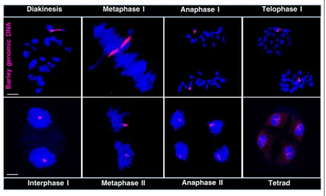
Figure 2. Meiotic segregation of the barley 7HL chromosome arm in the 7BS.7HL wheat/barley disomic translocation line as detected by GISH. The barley geno- mic DNA probe was labelled with Alexa Fluor 594 (red), whereas the DNA was counterstained with DAPI (blue). The meiotic stages shown are: diakinesis, meta- phase I, anaphase I, telophase I, interphase I, metaphase II, anaphase II and tetrad. Scale bar=5lm.
!"
!"
Mid zygoteneLatezygotenePachyteneEarlyleptoteneLateleptoteneEarlyzygoteneEarly-mid zygotene
Lengthof the7HL chromosomearm(µm) 100
80 60 40 20
Early zygotene Late
leptotene Zygotene Pachytene
(b)
Early leptotene Late leptotene Zygotene Pachytene
Packaging and average length of the 7HL arm (ave. 7HL length, µm) with respect to the nuclear diameter (ave. nuclear diameter, µm) during prophase I
(c) Length of the 7HL chromosome arm (µm)
Condensed region Decondensed region Nuclear diameter
Leptotene Early
leptotene
Early-mid zygotene
Mid zygotene
Late
zygotene Pachytene Cellswithpairedloci (%)100
80 60 40 20
(d)
0
Telomere pairing
Centromere pairing
Centromeres Telomeres
Length of the 7HL chr. arm (µm)
Nuclear diameter (µm)
Ave. SD Ave. SD
21.34 7.47 37.56 11.05
37.71 9.14 32.71 9.98
62.03 21.35 46.98 14.74
51.90 22.02 57.55 20.18
EL LL Zy Pa
(a)
C
0 10 20 30 40 50 60 70 T
a
b
c c
Merged Barley
!
Centromeres Telomeres
ASY1 CENH3ZYP1
chromosome segments, the larger proportion of the chro- mosome arm, which also carried the centromere-specific G+C FISH signal, maintained the condensed organisation observed in early leptotene (chromosome width: 0.7– 1.81µm), showing that decondensation of chromatin did not extend to the interstitial- and pericentromeric regions (Figure 3a–c). The decondensed subtelomeric region cov- ered half (53.8%) of the total length of the chromosome arm measured in late leptotene (28.710.1µm;
meanSD; n=27), which corresponded to the length of the diameter of the nucleus (32.710.0µm; meanSD;
n=27) (Figure 3c). This indicated a chromosome arm elongation of 35% compared to early leptotene. The barley chromosome arms become synchronously decondensed along their entire length in early zygotene and reached their maximum prophase I length (62.0221.4µm;
meanSD; n=25) (Figure 3a–c). The quasi-parallel organisation of the homologous chromosome arms was retained up to early zygotene, with the telomeres and the centromeres oriented at the two extremes of the nucleus.
Barley centromeres follow dynamics characteristic of the host genome
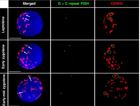
To reveal whether barley centromeres follow the activity of wheat chromosomes during the highly dynamic early stages of prophase I, we performed ImmunoFISH experiments,
where we simultaneously applied barley centromere-specific G+C repeat sequence FISH with CENH3 immunolabelling to meiocytes ranging from leptotene to early-mid zygotene.
CENH3 labelling showed all the active centromeres present, whereas the G+C DNA probe indicated the nuclear localisa- tion of the barley centromere part of the translocation chro- mosome. The barley centromere-specific repeat overlapped with the CENH3 signal in all nuclei analysed (n=147), indi- cating that the barley centromere loads centromere-specific histone protein in the wheat background and thus represents a functional centromere. In leptotene, when large, polarised centromere groups are formed in wheat (ranging from nine to 15 groups), the two homologous barley centromeres were observed to join different groups (Figure 4, top row). In early zygotene (Figure 4, middle row) and early-mid zygotene nuclei (Figure 4, lower row), the barley centromeres were still separated from one another but occupied smaller groups or were perceived individually, most likely as a result of the progressive dispersion of large centromere clusters. We measured the regions marked by the barley centromeric repeat DNA probe from leptotene to early-mid zygotene aim- ing to reveal whether they undergo reorganisation between leptotene and early zygotene, typical of wheat chromosomes (Sepsi et al., 2017). An elongation of the G+C signal was observed between the period of leptotene (number of nuclei=15) and early zygotene (number of nuclei=15)
Figure 3. Chromosome arm morphogenesis and pairing of different chromosomal regions during key events of synapsis at meiotic prophase I.
(a) Left: Examples of 7BS.7HL translocation line nuclei at different substages (leptotene to pachytene) of meiotic prophase I, showing synaptonemal complex progression and centromere dynamics. All active centromeres are visualized by an anti-CENH3 antibody (rabbit, detected by Alexa 594, red). The synaptonemal complex axial elements related protein ASY1 and the synaptonemal complex transverse filament protein visualized by anti-ASY1 (guineapig, detected by Alexa 647, pseudo coloured in grey) and rat anti-ZYP1 antibodies (detected by Alexa 488, green) reveal distinct stages of synaptonemal complex development. Forma- tion of the telomere bouquet is seen by intensive ASY1 staining (arrow on the late leptotene image). For single channel images, see Figure S2. Scale bar=5lm.
Right:In situhybridization showing the orientation and morphogenesis of the homologous barley chromosome arms (red), framed by their centromere (green) and telomere (grey) during substages of meiotic prophase I. Nuclei encompassing meiotic stages from leptotene to pachytene were obtained from adjacent anthers and are thus synchronous to those used for the CENH3, ASY1, ZYP1 immunolabelling (left) (Bennett et al., 1973b). Meiotic nuclei were counterstained with DAPI. Scale bars 5lm. Early leptotene: Telomeres are dispersed within one half of the nucleus and centromeres are located within the other nuclear hemi- sphere. The barley chromosome arms are separated and the chromatin is condensed into globular domains. Late leptotene: The telomere bouquet is formed and the barley telomeres although located in close proximity to one another remain separated. The barley centromeres are separated from one another and located close to the nuclear periphery opposite the telomeres. The pericentromeric and interstitial regions maintain their condensed organisation, whereas the subtelomeric regions are elongated into thin chromatin threads. Early zygotene: Barley telomeres are paired, whereas centromeres (green, highlighted by yellow arrowheads) are at the opposite pole of the nucleus and remain separated from one another. The barley chromosome arms running parallel show an elongated organisation that eventually forms finger-like structures curved out transversally on both sides of the chromosome axis. Early-mid zygotene: Close juxtaposition of the barley 7HL from the subtelomeric region onwards demonstrates synapsis initiation while centromeres and the pericentromeric regions remain separated at a considerable distance from each other. Mid zygotene: Subtelomeres colocalise and interstitial regions show numerous ‘juxtaposed interstitial segments’
with variable length, where the two homologous chromosome arms colocalise. Homologous contacts formed by the ‘juxtaposed interstitial segments’ are inter- spersed with non-colocalised, parallel ‘interstitial chromatin alignments’ of various lengths (for more detail, see Figure 3) and this chromosome configuration extends from the subtelomeres to the pericentromeric regions. Pericentromeres are widely separated from their homologous chromosome segments forming a fork-like structure. Late zygotene: The telomere bouquet disperses; the elongated chromosome arms show multiple ‘juxtaposed interstitial segments’ disrupted by ‘interstitial chromatin alignments’ and this conformation reaches the centromeric regions. Centromeres are juxtaposed on the majority of nuclei. Pachytene:
Synapsis is fully formed between the homologous barley chromosome arms.
(b) Expansion and condensation of the 7HL chromosome arm during substages of meiotic prophase I Statistical analysis revealed a highly significant difference (F=32.614; d.f. 1=3; d.f. 2=96;P=1.24910-14between the chromosome arm length measured at the different stages. Tukey’s Bpost hoctest categorized these differences into three (a, b, c) groups (indicated by lowercase letters): Early Leptotene, Average (ave.): 21.34 (a–shortest); Late Leptotene, Average: 37.58 (b–medium length); Zygotene, Average: 62.03; and Pachytene, Average: 51.90 (both c–longest).
(c) Proportions of the condensed (grey horizontal bar) and decondensed (green horizontal bar) chromosomal regions within the barley chromosome arms (chr.
arm) during prophase I with respect to the nuclear diameter (fine vertical line underneath the bars representing the barley chromosome arm). The positions of the telomere (grey vertical bar) and centromere (red vertical bar) are indicated by uppercase letters (C and T, respectively). Descriptive statistical data is shown on the right side.
(d) Diagram showing the proportion of cells carrying paired 7HL centromeres (green line) and paired 7HL telomeres (grey line) during different stages of pro- phase I. Initiation of pairing is marked by red circles in both cases.
(Figure S3), which did not increase further between early zygotene and early-mid zygotene (number of nuclei=15).
The barley centromere, representing half of the 7BS-7HL cen- tromere, thus followed reorganisation dynamics similar to that of wheat chromosomes during the early stages of mei- otic prophase I.
The progression of pairing between individual chromosome arms during zygotene
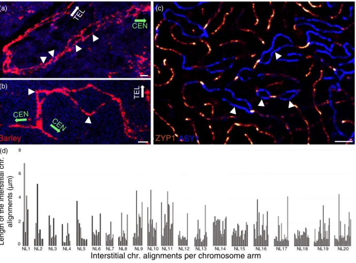
In a fraction of the analysed early zygotene nuclei (34%), homologous 7HL telomeres colocalised, whereas cen- tromeres remained as two distinct foci at a considerable distance from one another (Figure 3a,d). Coincident with the appearance of telomere pairing, GISH showed the juxta- position (the distance between the two chromosome threads was <0.15µm and thus they were perceived as one) of short chromosome segments located in the proxim- ity of telomeres, indicating early events of chromosome synapsis within the subtelomeres (Figure 3a). Subtelom- eres were defined as the chromosomal regions located near the telomeres that extended into the chromosome arms to the length observed to be decondensed at leptotene (17.914.37µm; meanSD) (Figure 3c). This region con- sisted of one-third of the total length of the fully decon- densed chromosome arm as measured at zygotene (Figure 3c). These findings were in agreement with the results obtained from immunolabelling, which showed early traces of linear ZYP1. Progression of subtelomeric jux- taposition was evident in the majority of early-mid zygotene nuclei examined (n=26). In some cases, juxtaposition was measured beyond the subtelomeres indicating early events of homologous interstitial chromosome contacts
(Figure 3a). These colocalised ‘juxtaposed interstitial seg- ments’ did not form a continuum with the juxtaposed sub- telomeric regions; rather, a discontinuous interstitial juxtaposition became evident at a number of foci dispersed throughout the chromosome arms. This implied that homologous chromosome connections were regularly spaced by homologous non-colocalised ‘interstitial chro- matin alignments’ varying in width between 0.2 and 7.3µm (SD 0.7µm;n=26). The occurrence of the parallel align- ments varied from cell to cell ranging from 5 to 23 with alignment lengths extending from 0.2 to 11.9µm (SD 1.4µm) (Figure 5). This was in line with our earlier studies showing multiple synapsis initiation sites within the chro- mosome arms and also with the immunolabelling results observed for the early-mid and mid-zygotene nuclei show- ing multiple synapsis initiation sites within the interstitial chromosome arms (see Figure 3a and Figure S2 and com- pare Figure 5a,b). Furthermore, in situ hybridisation showed that, although subtelomeric and interstitial regions were periodically united, the homologous barley cen- tromeres marked by the G+C probe remained consistently at a considerable distance from one another, exhibiting a fork-like structure (Figure 3a,d). Importantly, centromeres no longer occupied a territory in the proximity of the nuclear periphery but were randomly located in the interior of the nucleus (Figure 3a,d). Their location varied from cell to cell, indicative of dynamic chromosome movement.
Homologous centromere pairing was initially detected within a minority (14%) of the mid-zygotene nuclei (n=70), whereas telomeres colocalised in the 94% of the pollen mother cells (Figure 3d) and juxtaposition of interstitial seg- ments progressed into the pericentromeres (Figure 3a).
LeptoteneEarlyzygotene
Merged G + C repeat FISH CENH3
Early-mid zygotene
Figure 4. ImmunoFISH showing the relative posi- tion of homologous barley centromeres in nuclei of the 7BS.7HL Robertsonian translocation line at dif- ferent substages of meiotic prophase I. Arrows in the merged image (left) indicate the barley cen- tromeres marked by the G+C repeat sequences (FISH, Alexa 594, pseudo coloured in green) over- lapping with the CENH3 (rabbit, detected by Alexa 488, green and pseudo coloured in red) signal, which visualise all active centromeres. The encir- cled (dashed yellow line) signal on the right shows the centromere group including the barley cen- tromere. Meiotic nuclei were counterstained with DAPI. Scale bars=5lm.
Periodical juxtaposition connected a considerable amount of the chromosome arms at late-zygotene and the telomere bouquet began to disintegrate (Figure 3a,d). An increase in the frequency of centromere pairing was also observed (52% of nuclei displayed paired centromeres;
n=58). Full synapsis of the 7HL chromosome arms at pachytene coincided with an apparent arm shortening (52.022.0µm; meanSD; n=20), indicative of chro- matin condensation (Figure 3a,b).
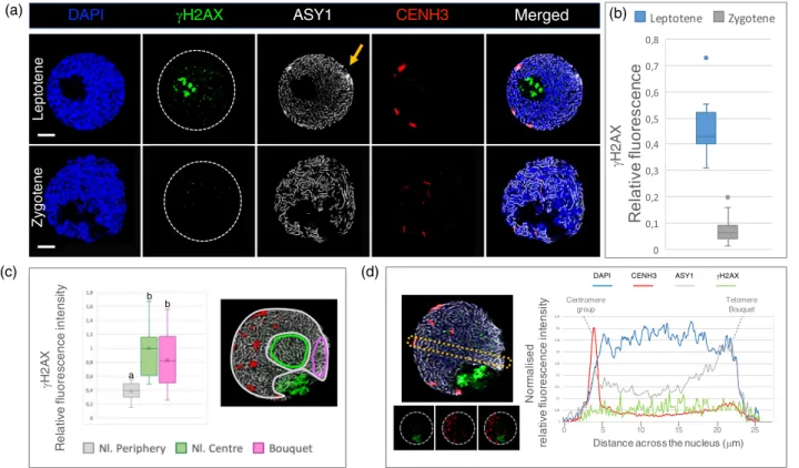
Initiation of meiotic recombination in the 7BS.7HL line Meiotic recombination initiation by SPO11-dependent DSBs can be detected cytologically with antibodies recog- nising the phosphorylation of the histone H2AX at the serine-139 residue, resulting in phospho histone H2AX ser139 (cH2AX) (Ku et al., 2020). To link SC axial element
formation, centromere dynamics and ultimately chromo- some arm morphogenesis with recombination initiation, we immunolabelled cH2AX foci together with ASY1 and CENH3 proteins in the pollen mother cell nuclei of the 7BS.7HL translocation line. Meiotic nuclei between the meiotic leptotene and zygotene stages exhibited numerous chromatin and axis associated cH2AX foci with consider- able variation in fluorescence intensities (Figure 6a). We measured the relative fluorescence intensities of the immunolocalizedcH2AX foci per individual leptotene and zygotene nuclei (n=23) staged according to the ASY1/
CENH3 signal. We observed a high relative fluorescence intensity cH2AX indicative of extensive DSB formation at leptotene (n=12) when the homologous chromosome axes were continuous but still separated (Figure 6a,b). This was in agreement with previous studies reporting
NL1 NL2 NL3 NL4 NL5 NL6 NL7 NL8 NL9 NL10 NL11 NL12 NL13 NL14 NL15 NL16 NL17 NL18 NL19 NL20
Interstitial chr. alignments per chromosome arm Lengthof theinterstitialchr. alignments(m)
(a)
(d)
(c) CEN
CE N CEN
TE L
TEL
Barley (b)
ASY1 ZYP1
6 8
4
2
0
Figure 5. Patterns of homologous juxtaposition within the interstitial chromosome regions and the pericentromeres of the 7HL chromosome arm during zygo- tene.
(a, b)In situhybridisation signal of the barley chromosome arm (in red) shows ‘juxtaposed interstitial segments’ disrupted by numerous non-juxtaposed ‘inter- stitial chromatin alignments’ (arrowheads). The orientation of the centromeres (CEN) and telomeres (TEL) are indicated by green and white arrows, respectively.
(c) ASY1 (blue threads) and ZYP1 (bright orange threads) immunolabelling shows similar patterns of interstitial synapsis interspersed by unsynapsed parallel axis alignments (arrowheads). Bars=2lm.
(d) Examples of the pattern and number of ‘interstitial chromatin alignments’ within the analysed barley chromosome arms, showing the length (y-axis) and number (x-axis) of parallel, non-juxtaposed chromosomal regions per chromosome arms. Groups represent data measured within one nucleus (Nl.) and thus a single pair of barley chromosome arms. Bars within one group from left to right represent measurements onwards the subtelomeres and progressing towards the centromeres.
recombination initiation in late G2-leptotene (Ku et al., 2020; Osman et al., 2021; Storlazzi et al., 2008). Zygotene nuclei (n=11), characterised by partially juxtaposed chro- mosome axes, showed a substantial (85%) drop in the cH2AX relative fluorescence intensity (Figure 6a,b), denot- ing the initiation of DNA repair mechanisms as reported in other studies (Mahadevaiah et al., 2001; Su et al., 2017;
Woglar and Villeneuve, 2018).
DSBs distribution is non-random within the nucleus To understand the spatial distribution of DSBs inside indi- vidual leptotene nuclei, we quantified the relative
fluorescence intensity ofcH2AX foci at the nuclear periph- ery and at the centre. The nuclear periphery carried signifi- cantly lower cH2AX intensity levels (<50%) compared to the nuclear centre (Figure 6c). One specific region of the nuclear periphery distinguished by a concentrated conical ASY1 signal, indicative of the telomere bouquet, carried a cH2AX immunosignal comparable to that observed for the centre of the nucleus (Figure 6c), indicating a higher DSB activity in the vicinity of the telomeres. This is in agree- ment with the subtelomeric initiation of the early DSBs in both wheat and barley (Higgins et al., 2014; Osman et al., 2021).
Zygotene Leptotene
DAPI JH2AX ASY1 CENH3 Merged 3D
JH2AX
b
JH2AX Relative fluorescence intensity
a
b b
Normalised relative fluorescence intensity
Distance across the nucleus (Pm)
0 5 10 15 20 25 DAPI CENH3 ASY1 JH2AX
Telomere Bouquet Centromere
group
Merged
LeptoteneZygoteneZygotene Leptotene
DAPI JH2AX ASY1 CENH3 Merged 3D
JH2AX
JH2AX Relative fluorescence intensity
a
b b
Normalised relative fluorescence intensity
Distance across the nucleus (Pm)
0 5 10 15 20 25 DAPI CENH3 ASY1 JH2AX
Telomere Bouquet Centromere
group
Merged
ZygoteneLeptotene (a)
(c) (d)
(b)
Figure 6. (a) Examples of microscopic images showing variations in thecH2AX levels between the leptotene and zygotene stages of the 7BS.7HL translocation line.cH2AX loci are labelled by an anti-cH2AX antibody (rabbit, detected by Alexa 488, green). For precise stage identification, the synaptonemal complex axial elements related protein ASY1 is shown using anti-ASY1 antibody (guineapig, detected by Alexa 647, pseudo coloured in grey) and CENH3 is sequentially labelled and visualized using anti-CENH3 antibody (rabbit, detected by Alexa 594, red). An orange arrow points to the telomere bouquet apparent by the concen- trated ASY1 signal at leptotene.
(b) Relative fluorescence intensities of thecH2AX signals measured at the leptotene and zygotene stages in the 7BS.7HL translocation line. Significant differ- ences were detected by Student’st-test (P=0.0005).
(c) Spatial distribution of thecH2AX signal within the leptotene nuclei of the 7BS.7HL translocation line. Comparative analysis of relative fluorescence intensities of the nuclear periphery (grey colour, Nl. periphery), the nuclear centre (green colour, Nl. centre) and the nuclear volume including the telomere bouquet (ma- genta, bouquet). Measurements for each region of interest were carried out in three-dimensions (zdirection, right) with LAS X software. Significantly lower rela- tive fluorescence intensities were detected for the nuclear periphery than the nuclear centre. The telomere bouquet, although peripherally located, shows comparable relative fluorescence intensity to the nuclear centre (analysis of variance, d.f.1=2; d.f. 2=30;F=11.07;***P=2.51910-5). Lowercase letters (a, b) indicate significant differences (by Tukey’s honestly significantpost hoctest) between groups.
(d) Comparison between thecH2AX levels within the peripherally located centromeric regions, the chromatin located at the nuclear centre and the subtelomeric regions. Variations in relative fluorescence intensities of thecH2AX signal are shown with respect to DAPI (blue line), CENH3 (red line) and ASY1 (grey line) signals across a single focal point (frame) of a leptotene pollen mother cell nucleus. DAPI demarcates the area of the nucleus. The CENH3 signal shows characteristic periph- eral centromere localisation (indicated by a dotted line, ‘centromere group’), whereas the elevated ASY1 signal intensity at the opposite pole of the centromere indi- cates the location of the telomere bouquet (highlighted by a dotted line). The top image of the microscopic photograph represents an example of the region of interest (yellow dotted line) selected for the measurements. The bottom image aims to represent the decline of thecH2AX signal around the centromeres.
[Correction added on 06 August 2021, after first online publication: Part (d) of Figure 6 has been corrected in this version.]
Although DSBs are formed at leptotene, the centromeres are also peripherally located, opposite the telomeric pole (Carlton and Cande, 2002; Wen et al., 2012). We therefore examined whether the crossover devoid centromeric and pericentromeric regions carry reduced levels of recombina- tion initiations at leptotene, as indicated by the lower DSB signals measured at the nuclear periphery. By selecting single focal planes containing both peripherally located centromeres and a prominent telomere bouquet at the opposite side of the nuclear periphery, we specifically mea- sured fluorescence intensities in two dimensions within the centromeres (n=13), the nuclear centre and the telom- eres. According to our measurements, significantly lower DSB levels were detected within the chromatin marked by CENH3 staining compared to the nuclear centre and the telomeres (Figure 6d), indicating that the core centromere somehow protected but did not completely inhibit DSB for- mation. Centromeres carried 25% lowercH2AX intensities than the nuclear centre and 50% lower intensities than telomeres confirming extensive DSB formation within the subtelomeric regions (Figure S4).
DISCUSSION
Understanding the mechanisms influencing the spatial dis- tribution of early meiotic events is increasingly important in crops where specific allele combinations may determine the productivity of recent varieties (Lambing and Heck- mann, 2018). The present study correlates chromatin dynamics with key steps of recombination and synapsis by assaying a single pair of homologous barley chromosome arms added to the wheat genome (Figure 7) in the form of a translocation (7BS.7HL translocation) at the same time as providing a precise prophase I timing. The stability of the barley chromosome arm in the 7BS.7HL line, leading to stable meiosis II and full fertility, shows that the meiotic morphogenesis of the barley chromosome is relevant to the understanding of chromosome behaviour in wheat.
Additionally, we have demonstrated that the activity of the barley centromeres and their dynamics within the wheat nucleus followed a meiotic program characteristic of the host genome. This is in agreement with previous studies on wheat-rye introgression lines confirming that the pat- tern of chromatin remodelling of the alien (rye) chromo- some at meiosis is directed by the host genetic background in which the chromosome is present (Naranjo, 2018).
Chromatin reorganisation precedes chromosome juxtaposition at the subtelomeres
We established a coherent chronology of chromosome arm pairing by correlating chromosome arm morphogen- esis with the progression of synapsis and recombination.
We showed that, in early leptotene, chromosome arms were condensed and organised into irregular globular
subdomains with telomeres and centromeres pointing to opposite poles of the nucleus. This condensed early chro- mosome conformation and the bipolar orientation of telomeres versus centromeres was in agreement with sev- eral previous studies analysing chromatin dynamics in wheat-rye introgression lines (Corredor and Naranjo, 2007; Maestra et al., 2002; Martınez-Perez et al., 1999;
Mikhailova et al., 2001; Schwarzacher, 1997). We demon- strated that this condensed chromatin structure coincided with recombination initiation by numerous nucleus wide DSBs, whereas homologous chromosome arms were located at considerable distances from one another (Figure 7).
By late leptotene when chromosome axes were fully linear, without any sign of synapsis emerging from the prominent telomere bouquet, the homologous arms become partially and synchronously reorganised. Reor- ganisation explicitly involved the elongation of sub- telomeric chromatin, which emerged from the fully formed telomere bouquet (Figure 7). The elongation of subtelomeric chromatin during the period of telomere bouquet formation has already been demonstrated as one of the earliest events of meiotic prophase I by in situ hybridisation on whole floret vibratome sections of a wheat-rye recombinant chromosome line carrying an interstitial rye chromosome segment. Prior telomere clustering wheat subtelomeric regions elongated and then both rye homologous segments elongated syn- chronously and intimately aligned (Prieto et al., 2004).
The conformational change of the subtelomeric chro- matin was found to be triggered by the mutual recogni- tion of homologous chromosome partners, followed by their intimate alignment, indicating the important role of chromatin unpackaging in the homology recognition process (Colas et al., 2008).
The present study, based on the timeline set up by synaptonemal protein labelling, showed that, although both barley subtelomeres were included in the bouquet in late leptotene, homologous chromosome arms, including the subtelomeres, remained physically separated and loca- lised at a considerable distance from one another. This shows that extensive presynaptic homologous coalign- ments, as observed in Sordaria macrospora (Storlazzi et al., 2010), are absent in large genome cereals. Short regions of chromosome axes coalign in wheat leptotene nuclei as well (Sepsi et al., 2018), although, as a result of the large genome and the multitude of chromosome threads, it cannot be determined whether these alignments involve homologous chromosomes. The present analysis suggests that leptotene axis alignments may occur between non-homologues or represent transient inter- or intra-chromosomal associations as indicated by the tight chromatin packaging within the interstitial and pericen- tromeric chromatin.
SC Recombination Chromosome dynamics
Axial elements loading along
the chromosomes
DSBs formation Non- homologous
centromere associations Dispersed telomeres
Axial elements are continuous
along the chromatin
Single-end invasion
Centromere clustering Telomere bouquet
Subtelomeric SC Initiation and
elogation
DSB repair
Centromere dispersion
Telomere bouquet
Interstitial SC initiation from
multiple points, elongation
Progressive centromere synapsis Telomeres dispersing
Fully-formed SC
Crossover Maturation
Fully synapsed centromere
Telomeres dispersed
Leptotene Zygotene Pachytene
Acce ssi ch ble
rt.
Non-acce ssiblechrt.
Acce ssible
chrt.
Acce ssi ch ble
rt.
Distalregion
Dista l region
Prox imal
region
Proximal region
Barley chrt.
Key Telomere Chromosome arms Centromere
Figure 7. Chromatin dynamics, centromere and telomere associations during prophase I of meiosis. Left: Cartoon diagram of the chromatin morphogenesis and dynamics through the prophase I stages. Central panel: Microscopic images of single focal planes photographed within pollen mother cell nuclei of the 7BS.7HL wheat-barley line labelled within situhybridisation to detect the barley chromatin (Alexa Fluor 594, shown in red). Chrt., chromatin, Scale bars=5µm.
Right: Chronology of synaptonemal complex (SC) development, recombination progression and chromosome dynamics during prophase I of meiosis (present study; Higgins et al., 2012; Osman et al., 2021; Sepsi et al., 2017).
[Correction added on 06 August 2021, after first online publication: The definition for Chromosome arms in the Key of Figure 7 has been corrected in this version.]
DSBs are predominant towards the centre of the nucleus and in the proximity of the telomere bouquet
Here, we showed that, although genome-wide DSBs are introduced at leptotene, the pericentromeric/interstitial chromatin remains initially enclosed in large globular domains, identical to their closed structure detected at the onset of meiotic prophase. Using quantitative image analy- sis, we detected a non-random distribution of DSBs inside the nucleus, where chromatin located at the nuclear periphery showed a lower DSB activity relative to the chro- matin located at the nuclear centre. Subtelomeres, how- ever, assembled into the telomere bouquet, showed an exception to this rule because they demonstrated DSB activity comparable to the nuclear centre. DSBs have been reported to be spatiotemporally confined to the subtelom- eric regions prior to chromosome juxtaposition in wheat (Osman et al., 2021), barley (Higgins et al., 2012), zebrafish (Danio rerio) (Blokhina et al., 2019) and human males (Pratto et al., 2014) and have also been suggested to play a role in homology recognition and pairing.
When specifically assaying the core centromere of lep- totene nuclei, we detected the lowest occurrence of recom- bination initiations, although centromeres were not completely devoid of DSBs. As expected, these events did not lead to crossovers, reflecting the local predominance of non-crossover repair. These results were in agreement with recent molecular work in wheat showing centromeric DSBs to be resolved as gene conversions representing non-reciprocal genetic exchanges (Gardiner et al., 2019).
Examination of maize meiocytes showed that DSB forma- tion at late leptotene coincides with a transient remod- elling of axial elements as well, where a coiled axis morphology transitions to linear structures (Ku et al., 2020), suggesting a possible SPO11-1 dependent mecha- nism to orchestrate changes in axial element structures.
A significant variation in DSB distribution across the lep- totene nucleus showed lower DSB activity for chromatin in proximity to the nuclear periphery, including the con- densed centromeric chromatin. The subtelomeric regions showed an elevated DSB activity that coincided with the presynaptic chromatin remodelling into an open chromatin thread. The present study does not resolve whether the elevated DSB levels trigger chromatin remodelling at the subtelomeres or whether the open chromatin structure renders the subtelomeric region more prone to the DNA damage machinery. Chromatin remodelling is known as a key element of recombination patterning (Szekv€olgyi et al., 2015), although DNA damage itself can also affect chro- matin organisation and mobility (Hauer and Gasser, 2017).
An important question to consider is whether the compact chromatin structure together with the reduced DSB activity observed at the pericentromeres delays the recombination repair pathways and thus promotes non-crossover
outcomes in the proximity of centromeres. This scenario is supported by evidence showing that the crossover/non- crossover decision is taken earlier in meiosis, possibly as early as DSB formation (Allers and Lichten, 2001; Bishop and Zickler, 2004). In line with this, a recent study in mice proposed that the fine scale chromatin structure and tim- ing of DSBs influence the outcome of meiotic recombina- tion. Earlier formed DSBs have been shown to occupy more open chromatin and be more prone to repair as crossovers than those that are formed later, suggesting that the crossover decision may need specific protein com- plexes that are absent at later stages (Chen et al., 2020).
Another element to be considered in terms of restricting crossover events within the centromeric/pericentromeric regions is the formation of early non-homologous centromere-centromere associations. They have been reported in cereal species (present study; Martinez-Perez et al., 2003; Phillips et al., 2012; Sepsi et al., 2017) and the model plant Arabidopsis (da Ines et al., 2012; Pradillo et al., 2014; Ronceret et al., 2009), although they appear in early meiotic prophase I of budding yeast (Saccharomyces cere- visiae) (Tsubouchi and Roeder, 2005) andDrosophila melano- gaster (Kurdzo et al., 2017). Non-homologous centromere associations in wheat are consistent with earlier electron microscopy data on SC formation (Jenkins, 1983), revealing partner switches (homologous alignments interrupted by non-homologous pairing) within the interstitial chromosomal regions during zygotene. Non-homologous centromere inter- actions, thus ensuring spatial separation of the homologous centromeres, may to some extent prevent centromeric DSBs from being repaired via crossover pathways.
The present study suggests that the variation in chro- matin packaging concentrates the whole chromosome recognition machinery to act initially at the subtelomeric regions. Homology testing and recognition promotes SC formation and progression of recombination repair mecha- nisms. Interstitial and pericentromeric regions may be masked by their globular, closed chromatin structure, implying a temporal shift in recognition, juxtaposition and initiation of repair, which results in the preference of the repair processes leading to non-crossovers.
Interstitial chromosome pairing progresses from multiple sites forming periodical juxtapositions interspersed with
‘interstitial chromatin alignments’
The delayed remodelling of interstitial and pericentromeric chromatin compared to the subtelomeric regions (Figure 7) is in line with the temporal shift of the synapsis at the distal and proximal regions (Banerjee and Jones, 1999; Blokhina et al., 2019; Xiang et al., 2014). Initial chromatin juxtapositions occur between the subtelomeres in cereals. This process has been proposed to be mediated by the telomere bouquet and a telomere-led chromosome movement (Martinez-Garcia