ampulla (Ehr.)
ROBERT E . BERREND1
Department of Zoology, University of Wisconsin, Madison, Wisconsin^
Filipodia of Cyphoderia ampulla (Ehr.)
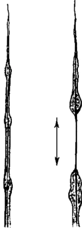
Cyphoderia ampulla (Ehr.) (Fig. 1) is one of the Testaceafilosa De Saedeleer, 1934. Together with the genus Campascus Leidy, it forms the family Cyphoderiidae Deflandre, 1953. T h e test of the animal is colorless to yellowish, and is composed of small round or elliptical scales. It is flask-shaped in outline with a tapering neck curved in such a way that
the aperture is oblique to the main axis of the test. The cytoplasm, densely filled with reserve scales and glycogenic grains does not com- pletely fill the test. T h e large nucleus is located in the posterior portion of the cell body. Slender ectoplasmic filopodia originate from a mass of cytoplasm—the podostyle—which protrudes from the mouth of the test.
It is convenient on the basis of locomotion and the manner of deploy- ment to divide the filopodia of Cyphoderia into three types: directive, nondirective, and secondary. The directive filopodium determines the direction in which the animal moves. It is of interest to note that if two directives should be present, the animal moves along a line which bisects the angle between the two. It is obvious that this situation cannot exist for long unless the two directives are nearly parallel; the larger the angle of divergence the sooner the two directives become opposed to one
1 Editor's note: In the absence of Dr. Berrend, his paper was read at the Sympo- sium by Dr. Lowell Ε. Noland of the University of Wisconsin, under whose supervision the study was carried out.
2 Present address: San Francisco State College, San Francisco, California.
FIG. 1. Cyphoderia ampulla (Ehr.).
433
another and forward movement ceases. Movement begins again when dominance is gained by one of the two, or a new directive originates.
Nondirective pseudopodia vary in number from many to, rarely, none.
They are more abundant when the animal is moving slowly, is on the underside of a surface, or when the surrounding water is in motion.
Secondary pseudopodia spread out laterally from either of the two pri- mary types of filopodia and appear to serve as holdfast devices.
DIRECTIVE FILOPODIA
There is a tendency among nonplanktonic ameboid Protozoa to uti- lize a monopodial or some functional equivalent of a monopodial form of movement. The more pronounced this monopodiality, the greater the distance covered and the greater the area over which the animal may browse. If we postulate an ameboid form with a radial distribution and random dominance of pseudopodia, it is evident that, for the organism to move any distance, some degree of dominance must be maintained for a period of time. The longer the time through which the dominance acts the further the excursion of the organism in that direction. In the ab- sence of trophic responses a random origination of dominant directive pseudopodia will tend to restrain the animal within an area inversely proportional to the rate of replacement of one directive by another.
Actually most ameboid forms established an anterior end by various means (e.g., uroid, bilateral tests). In undisturbed naked amebae the
"limax" pattern of progression is, therefore, the rule. T h e behavioral patterns of the Testacealobosa range from a limax pattern which is par- ticularly well developed in Hyalosphenia minuta to a "crawl" pattern such as is found in Lecquereusia sp. In the latter case along an axis which represents an extension of the median sagittal axis of the test the replacement pseudopodium is formed between the anterior lip of the test and the base of the actively advancing pseudopodium, which in its turn is in a similar relationship, above and in front of its predecessor now re- tracting. The result is the persistence of a relatively permanent anterior end in the animal, which accordingly can be expected to cover a large territory in its movements.
Cyphoderia ampulla, as a representative of the Testaceafilosa also demonstrates monopodiality, but here it is combined with frequent ran- dom changes in direction. The net result is that the range of the animal is small compared with its rate of movement, covering an area of only a few square millimeters in 24 hr; the range is not appreciably larger in 96 hr.
In normal progression there is one directive filopodium along which the animal moves. This directive passes through three stages.
Stage I. The filopodium first originates as a nondirective. It then extends rapidly and has at this time a basal width of 1.5 μ when measured from 10 to 15 μ from the test. At intervals along its length the filopodium widens on contact with the substratum producing a series of thickened areas or nodes connected by narrower internodal sections. These nodes are 1.5 to 2 times the width of the adjacent internodal sections and are slightly elliptical, with the main axis lying in the direction of the direc- tive pseudopodium. Stage I lasts slightly more than a minute.
Stage II. T h e filopodium becomes wider at the base, 3 μ or more, and very long, commonly from 130 to 160 μ in animals with a test length of from 120 to 140 μ. Lengths of 300 μ may occur. During this stage the animal is moving forward, therefore shortening the filopodium proxi- mally. Concurrently the distal portion continues to extend at least as rapidly as the test is moved ahead. Very delicate secondaries originate at the nodes. Stage II lasts from 3 to 4 min.
Stage I I I . When the filopodium no longer extends as rapidly as the test advances, the senile stage commences. T h e base may now be 9 μ or more in width. T h e widening tendency progresses to the tip of the pseudopodium which becomes less sharply pointed. T h e number of secondaries increases at the nodes and they are longer and thicker than in stage II. During the last portion of this stage the new directive may be passing through stage I. Once this new directive is established, the old directive detaches from the substratum and the remnant slowly con- tracts (candles) into the cell body at the podostyle.
NONDIRECTIVE FILOPODIA
Nondirective filopodia are variable in number and length. In the beginning they are not distinguishable from stage I directives. Their principal function appears to be that of adhesion to the substratum.
They originate at any point on the podostyle and in a stationary individ- ual radiate in all directions around the mouth of the test. In a moving animal locomotion produces a constant forward displacement of their origins causing those filopodia that are in contact with the substrate to trail behind the test. These filopodia appear to function as do the strands of mucoid material trailing behind the tests of the Testacealobosa, i.e., they adhere to the substratum and stabilize the test by elastically oppos- ing the directive pseudopodium. T h e nondirective filopodia are com- monly retracted en baïonnette within 2 min. Should the animal be swept free of the substratum, the nondirectives become more rigid. They form angular bends and tend to send out secondary filopodia—a behavioral pattern which would seem to facilitate their snagging in detritus and thus reduce the possibility of the animal being swept away by currents.
SECONDARY FILOPODIA
Secondaries form at the attachment nodes of directives and occasion- ally of nondirective pseudopodia. Their function is apparently that of increasing the contact area of the parent pseudopodium with the sub- stratum. They form an acute angle to the axis of the parent pseudopo- dium, the apex of the angle being toward the test. T h e impression they give is that of guy wires or anchors which resist slippage of the main pseudopodium. This conjecture is strengthened by the observation that secondaries may also be formed along the proximal internode when the animal's direction of movement is such as to pull a nondirective or a codirective in a lateral direction.
Structure and Function of Filopodia
Cyphoderia ampulla is one of the largest of those Rhizopoda that are classed as Filosa. It is a very active animal, and when moving by means of a stage I I directive filopodium, often travels through its own length (140 μ) within a minute. The size of the pseudopodia and their persistence makes C. ampulla particularly useful for observations of the structure of filopodia.
De Saedeleer's (1932) definition of filopodia differs from those of his predecessors in several important details. He notes that the frequency of fusion between separate pseudopodia is common rather than rare or absent as previously stated by many authors. In addition, he stresses the importance of branching and finally the presence of the sheets of proto- plasm which form basally between filopodia or laterally along a filopo- dium (Fig. 2).
This latter observation had been previously made by Bëlaf (1921) on Pamphagus hyalinus. He interpreted this lateral spreading with its re- sulting accentuation of the original central core, as evidence of an axial filament similar to that of the Foraminifera. De Saedeleer, on the other hand, considered that no axial filament was present, either in the small or large pseudopodia. The appearance of a filament being an illusion formed by a transformation of the cortical cytoplasm of a pseudopodium to a more fluid state. The solation of the cortex results in lamellar spread- ing, the untransformed medullary portion then appearing as an axial filament.
Filamentous structures that Bëlaf describes as extending into the tubular portion of the filopodium were considered by De Saedeleer to be illusory. He states, . . pour nous, ces axes, vestiges de pseudopodes en voie de transformation, ne s'observent que dans les lamelles et leur pré- sence ne peut en aucune façon faire interpréter ces pseudopodes comme
des pseudopodes réticulés, car il leur manque le caractère essentiel de ces derniers: les courantes granulaires." The present observations made on C. ampulla tend to support the concept of Bélaf rather than that of De Saedeleer.
Using the phase microscope, bright contrast 45 χ , a dark line 0.5 μ in width can be seen to extend through the length of the pseudopodium except near the distal end where the sides of the pseudopodium are barely wider than this line. T h e dimension of this core does not vary and when seen as it passes through the distal nodes it is the same as in
FIG. 2. Basal confluence of nondirective pseudopodia showing axial core.
the expanded base of the pseudopodium. This core is seen to branch into the secondary filopodium if these become large enough to show it.
In the consolidation of two or more parallel pseudopodia, the cores approach one another as the degree of fusion progresses until they in turn fuse (Fig. 2).
Additional evidence for the presence of a stereoplasmic core is found in the observations of function and misfunction of the pseudopodia. The usual means of retracting the pseudopodium is en baïonnette, that is, the pseudopodium bends in a series of sharp angles and eventually folds into the cytoplasmic mass at the podostyle.
It has been observed that under some conditions, treatment with potassium salts in particular, that the en baïonnette method of pseudopo- dium return is replaced almost completely by candling, a process in
which the rheoplasm runs basipetally to the cell body. Although this type of retraction usually includes the stereoplasmic axis to such a degree that only a small portion of it protrudes distally, occasionally a con- siderable length of the axis may be exposed. In the latter case, the naked stereoplasmic core can again be seen to be of a constant diam- eter. This axis may then be recovered either by folding en baïonnette or by a progressive basal incorporation into the cell body.
FIG. 3. A break in the rheoplasmic layer exposing the apparently naked stereo- plasmic axis.
On several occasions treatment with sodium salts (0.006 N) produced a proximal break in the rheoplasm (Fig. 3). The rheoplasm contracted on either side of the break, the proximal portion moving into the cell body while the distal segments concentrated around the most proximal nodal attachment. This break and withdrawal of the rheoplasm exposed the axis which remained connecting the two masses. The distal proto- plasm was then either recovered by en baïonnette bending of the axial filament or the exposed section of the axis broke and was left behind along with the distal portion of the filopodium.
Rarely in the culture solution and with varying frequency in the
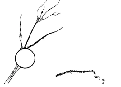
single salt solutions used, the rheoplasm on a filopodium fragmented centripetally and each fragment rounded into beads of cytoplasm strung along the core (Fig. 4). These beads form a graded series, the size of the beads being directly proportional to the width of the pseudopodium at that locus. T h e largest beads are, therefore, proximal and nodal. In a 0.006 Ν CaCl2 solution beading is only a temporary phenomenon, the protoplasm quickly flowing back together and reconstituting the com- plete filopodium. In 0.006 Ν K2H P 04 or ( N H4)2H P 04 solutions beading is always accompanied by loss of the protoplasm involved. There is evi-
FIG. 4 . A drawing to show the effects of sodium ions ( 0 . 0 0 6 N). Note beading of a nondirective pseudopodium. Detail: a single filopodium treated with 0 . 0 0 6 Ν potassium ions showing beaded rheoplasm slipping from the stereoplasmic core.
dently not only a change in state in the rheoplasm in these latter solutions but also a change in the stereoplasm. In potassium and to a lesser extent in sodium and ammonium solutions the beaded segment of the pseudo- podium shortly shows evidence of Brownian bombardment which pro- duces a trembling movement in the internodal portions. Eventually the stereoplasmic core itself fragments, and clusters of beads are moved away by the flow of the solution. In several instances beads were observed to slip one by one from a broken strand of stereoplasm (Fig. 4, detail).
The preceding observations cannot, I believe, be explained by any means other than the presence of an organized core in the pseudopodium.
Moreover, there are indications that the mechanism for the en baïonnette return typical of filopodia is dependent on the presence of such a core.
De Saedeleer (1932) states that filopodia do not contain granular cur- ο
rents of rheoplasm. He asserts that the apparent granules in a drawing by Bëlar (in Hartmann, 1927) of the filopodia of Pseudodifflugia gracilis are in reality of external origin. Observations on C. ampulla made in the present study indicate that these pseudopodia often do contain granules.
The granules in C. ampulla are small and reniform. They are highly refractile under a light microscope, are brighter than the cytoplasm in bright-contrast phase, and darker that the cytoplasm in dark-contrast phase. They appear in both directive and nondirective pseudopodia and are usually confined to the more proximal portions of these structures.
Exceptions to this latter statement occur when treatment by single salt solutions of low pH (Na in particular at pH 4.4) causes the nondirective pseudopodia to be drawn out into extremely long strands. Here the granules are present at considerable distance from the body of the animal.
In small filopodia, such as nondirectives and secondaries, these granules form an outward bulge in the cell membrane as they are moved along and around the pseudopodium. Since this does not occur in the larger basal portion of the directive or in the larger nodes it is probable that this bulging of the membrane is caused by the presence of the more resistant stereoplasmic axis of the pseudopodium. The granules move indepen- dently of each other. It is not uncommon to have two granules moving in opposite directions along the axis of the pseudopodium collide, or nearly collide, then swing around each other as they continue along their original course. T o a lesser extent the granules are independent of the major flow in the filopodium; granules may move proximally in a rapidly elongating pseudopodium or distally in one which is flowing into the cell body. Generally, however, the majority of the granules move with the prevailing direction of the flow in the pseudopodium.
The structure of a filopodium can be deduced from the preceding.
There is present a core or axis of gelled protoplasm of approximately 0.5 μ in diameter. This core is surrounded by a sheath of fluid protoplasm in turn bounded by a thin cell membrane. Initially the cross section of a filopodium is circular and in the smaller portions may remain so but as the structure becomes larger and older there is a lateral spreading.
Whereas the stereoplasmic core seems to be of constant diameter, the amount of rheoplasm increases as the filopodium ages. Some of this in- crease may be apparent, rather than real, caused by a lateral spreading of the rheoplasm. There is a temptation to think of the cortical cytoplasm becoming more solated—De Saedeleer so considers it—but there is no direct evidence that this is the case. A change probably occurs in the membrane at this time as is indicated by the enhancement of adhesive- ness in the more proximal nodal and to a lesser extent in the internodal areas. Probably, however, the major proportion of the increase in width
is caused by protoplasm flowing from the cell body into the filopodium.
Since filopodia are ectoplasmic and the protoplasm is extremely hyaline, this flow is easily overlooked. In C. ampulla the appearance of granules basally and their movement outward along the pseudopodium establishes that such a flow is present and considerable.
Discussion
In many rhizopods, pseudopodium formation and movement should be considered separately. In most laboratory amebae this distinction is normally academic, since such a large volume of the animal is involved in the formation of a pseudopodium that spatial displacement of the organism is automatically involved. However, in C. ampulla the differ- ence between the volume of the pseudopodium and the volume of the cell body is very great, and it is evident that the two (formation and move- ment) were best considered as separate, though related, phenomena.
The formation of a filopodium is rapid. As previously reported in this paper the rate of formation for a directive pseudopodium, exceeds, then equals, and finally falls behind the rate of movement of the organism.
If the filopodium is produced distally, it is evident that the production of axis from cortex requires that the rheoplasm streams at least as rapidly as the animal moves forward. T h e rate of protoplasmic streaming in C.
ampulla has not been satisfactorily determined although it is evident that the velocity meets this requirement. T h e groundplasm of the filo- podium is nongranular. Those large granules that are present are few, proximal, and their positions and movement indicate a considerable degree of autonomy from the principal streaming in the structure; cer- tainly, they are not satisfactory indicators of general protoplasmic flow.
As they do, however, give some insight into possible velocity, their rate of flow was determined. This was found to be about 250 to 300 μ/min.
Rates of streaming indicate that velocities of the required order are easily attained by protoplasm. Measurements of 0.5 μ granules moving along the reticulopodia of Diplogromia showed that 500 μ/min is not uncommon, whereas in the slime mold, Didymium nigripes, the velocity of streaming may reach 1250 μ/min (Vouk, 1913).
As mentioned above the volume of the filopodia of Cyphoderia is small compared with the volume in the body of the animal. Close ob- servation indicates that there is no displacement of the cell body into the pseudopodium in the manner that is characteristic of the naked Lobosa.
In Cyphoderia it appears that the animal streams along the axial fila- ment. The filopodium thus prepares a pathway along which the animal moves.
We may postulate that the axis of the directive filopodium forms a reaction surface. As such it provides the necessary surface for the pseudo- podial sol to stream distally to where, by sol-gel transformation, it ex- tends the reaction surface. The rheoplasm of the base of the pseudopo- dium and probably that of the podostyle streams against the axial fdament. As already noted, at points along its length the filopodium is in close and adhesive contact with the substratum. These nodes in addi- tion to adhering to the substratum are centers of secondary filopodium formation, thus increasing the contact area. Each nodal locus functions as an anchor for the axial filament. Streaming along such a reaction surface which in its turn is fixed in its position external to the cell body must result in movement of the organism. Conversely, if the reaction surface were not fixed and were of smaller mass than the organism, it would be ingested by the organism, cf. the feeding of Amoeba vespertilio on filamentous algae or Histomonas on bacteria. In Cyphoderia the re- trieving of a pseudopodium by the candling process suggests just this sort of process.
Several observations support the contention that the axial filament must serve as the locomotor reaction surface, just as the cytoplasm of the cortex or of the podostyle must serve as the actual locomotor force. Both the Frey-Wyssling (1947) and Loewy (1949) theories require that the reaction surface be a comparatively rigid structure. This condition is only satisfied by the axial gel. The cortex, although slightly elastic, does not have enough rigidity to serve in itself as a reaction surface. If the filopodium were in contact with the substratum throughout its length, (his would not be a valid objection since the substratum itself could serve as a traction surface, as it apparently does in some slime molds (Loewy, 1949). It has been observed, however, that only rarely do the internodal sections of the filopodium adhere to the surface. T h e most proximal internode is often lifted at a considerable angle above the surface. The animal occasionally moves along its pseudopodium at a rate faster than the streaming of the peripheral rheoplasm. When this occurs the rheoplasm is seen to wrinkle in advance of the oncoming mass of the organism. If the cortex were serving as the reaction surface, such wavelike folding could not occur as it would be pulled taut in opposition to the movement. Since the axis is the only portion of the pseudopodium with sufficient rigidity to be free from the substratum, except at anchor points, and to serve as a reaction surface, this function must reside in the gelled axis.
T o summarize, a filopodium is an organelle consisting of a delicate cell membrane over an actively streaming cortex, the rheoplasm, which in turn surrounds a gelled core. A filopodium is formed by the streaming of the rheoplasm along a reaction surface, which in this case, is the stereo-
plasmic axis. Stereoplasm is produced by a distal sol-gel transformation of the rheoplasm. If a filopodium functions as a directive simultaneously with its distal elongation, it is found that the cell body advances along the proximal section. It is suggested that this advance of the cell body is what would be expected from a streaming action against a reaction surface firmly fixed to the substratum, as is the axial rod of the filopo- dium.
REFERENCES Bëlaf, K. (1921). Arch. Protistenk. 94, 93-160.
De Saedeleer, H. (1932). Arch. Zool. Exptl. Gen. 74, 597-626.
De Saedeleer, H. (1934). Mem. Museé R. Hist. Nat. Belg. 60, 112 pp.
Frey-Wyssling, A. (1947). Exptl. Cell Res. Suppl. 1, 33-42.
Hartmann, M. (1927). "Allgemeine Biologie," 756 pp. Jena, Germany.
Loewy, A. G. (1949). Proc. Am. Phil. Soc. 93, 326-329.
Vouk, V. (1913). Denkschr. Akad. Wiss. Wien. Math-Nalurw. Kl. 88, 652-672.
DISCUSSION
DR. BOVEE: I shall show a film which will demonstrate many of the points in Dr. Berrend's paper. However, I would like to disagree on one point: I don't think the axial rod is formed at the time the pseudopod is extended from the body. These pseudopods almost always extend upward into the water and then down. T h e head forms a node of attachment. There seems to be a gelation of this strip between the point of origin at the shell and the point of attachment. T h e next part of the pseudo- pod then may drop down and form a second point of attachment and the rod seems to appear in that area.
DR. NOLAND: T h e first ones that fall down are quite narrow, and it would be very hard to see the rod. However, they are extremely straight, suggesting they must have some stiffening structure.
DR. BOVEE: Where they have formed a node, you can see the gelation between the node and body. T h e pseudopod may go forward in sections and there may be one, two, and possibly a third node by the time it is extended. As each node makes contact, there is apparently a local change sufficient to show contrast and suggest that this axial rod is in formation.
DR. ALLEN: I would be a little bit doubtful about the existence of this rod on the basis of the fact that a number of excellent older investigators, such as W . J . Schmidt, F. Doflein, and others, described such rods and drew them in pictures of Foraminifera, but in later studies with phase contrast and polarized light, no one has succeeded in demonstrating these rods. They certainly may occur in some organisms, but I think that the burden of proof of their existence should lie with the observer.
It seems to be assumed that such a rod must exist to explain the rigidity. Actually, all that is required to explain the shape and rigidity of these pseudopodia is the presence of gel structure.
DR. NOLAND: It is hard to interpret the straight, rigid strand between the droplets of solating cytoplasm in any other way.
DR. BOVEE: Also the bending and folding in segments.
CHAIRMAN MARSLAND: Did Dr. Berrend use phase contrast?
DR. NOLAND: Yes.
DR. KITCHING: T h e axopods of Heliozoans can be made to show similar bending effects and they certainly do have an axial rod.