1
Renal complications and ways of prevention in a model of vascular surgery of the lower limb arteries
Conditioning methods affecting ATP-dependent K- channels
Doctoral theses
dr. Péter Arányi
Semmelweis University
Doctoral School of Clinical Medicine
Supervisor: Attila Szijártó, MD, D.Sc.
Official reviewers: György Deák, MD, Ph.D.
Levente Kiss, MD, Ph.D.
Head of the final examination committee: László Entz, MD, D.Sc.
Members of the final examination committee: Zoltán Járai, MD, D.Sc.
Edit Dósa, MD, Ph.D.
Budapest
2018
2 1. Introduction
Acute arterial occlusion of the lower limbs and vascular surgeries performed in arterial occlusion lead unavoidably to an ischaemia-reperfusion injury to the tissues distal from the occlusion, mainly a vast skeletal muscle mass. Ischaemic rhabdomyolysis occurs, sometimes even with compartment syndrome, and often with large circulatory redistribution, local and systemic haemodynamic changes, as well as inflammatory response. An acute renal dysfunction, or even failure is one of the most common postoperative complication. The mechanism of the pathophysiology of this renal impairment is multifactorial: it may be a consequence of the circulatory alterations, the nephrotoxic agents released from the injured muscle fibres, neuroendocrine responses or the inflammatory reactions. The most determining event is the release of myoglobin into the circulation during ischaemic rhabdomyolysis, which is harmful for the tubular cells of the kidneys leading to acute tubular necrosis.
Ischaemic postconditioning is a method when performing several brief episodes of additional ischaemia and reperfusion induced immediately after the relief of a longer occlusion. Different mediators in the ischaemic tissues trigger an intracellular phosphorylation cascade leading eventually to the activation and opening of the mitochondrial ATP-dependent potassium (mitoKATP) channels. As an effect of these channels, the opening of the mitochondrial permeability transition pore channels are inhibited that would be the final effector channels in cell necrosis after ischaemia.
Levosimendan is a positive inotrope and a vasodilator that is used in the treatment of severe acute and advanced chronic heart failure. It is also described about this agent that as an agonist it opens mitoKATP channels and thus has a protective role against ischaemia-reperfusion injuries.
2. Objectives
An experimental model was created on rats that resulted in bilateral lower extremity ischaemia-reperfusion injuries with distal organ and systemic consequences representing the most important complications of the arterial reconstructive vascular surgery. We investigated in the model two conditioning method and their effects on the postoperative renal complications. The two methods have in common that they involve in their
3 mechanisms of action the opening of the mitoKATP channels (at least in the ischaemia- reperfusion injuries of cardiomyocytes). We sought answer for the following questions:
1. Does postconditioning (performed on the infrarenal abdominal aorta) have any effect on muscle fibre injuries in the rat model of 3 hours long bilateral lower limb ischaemia?
2. What degree and sort of kidney injury appears in this model, what timely characteristics does it have and how can this be affected by the application of postconditioning?
3. What mechanisms does the pathophysiology of the postoperative renal failure have and what effect does postconditioning have on these mechanisms?
4. How does mean arterial pressure and heart rate change in time in this model and can we alter the haemodynamic changes happening at the onset of reperfusion with the application of the method postconditioning?
5. What changes in the microcirculatory flow on surface of the limb muscles and in the kidney cortex measured with laser Doppler flowmetry? Does postconditioning have any effect on the microcirculation?
6. Can any inflammatory reactions be detected by measuring serum TNF-α levels and what happens when postconditioning is applied at the onset of reperfusion?
7. Does anything change when levosimendan is applied in the same model in terms of the lower limb muscle viability?
8. Does levosimendan have any protective effect for the postoperative renal dysfunction?
9. How does levosimendan affect systemic haemodynamics?
10. Does this drug have any impact on the microcirculatory flow of the limb muscle tissues and the renal cortex during the first 4 hours of reperfusion?
11. What happens with the release of the cytokine TNF-α when levosimendan is infused during the operation?
4 2. Methods
2.1. Ethical background
The experiments were performed with allowance of the Semmelweis University Animal Care Committee (license Nr.: 22.1/2409/3/2011.), with respect to the pertinent laws and regulations (Nr. XXVIII. 1998. and 243/1998.XII.31.) also in accordance with the internationally set requirements (US National Institute of Health guidelines, Publication No. 85–23, revised in 1996; MD, USA).
2.2. Animals
250-350 g, spf (specific pathogen free), inbred, male, Wistar rats (Charles River Hungary Ltd, Isaszeg, Hungary) were given standard rat food and water ad libitum, in the last 12 hours before operation only water. They were kept under 22-24 °C, with artificial light representing circadian lighting. Operations were performed at the same time of the day.
2.3. Anaesthesia
Rats were anaesthetized by intraperitoneal injection of 75 mg/bwkg ketamine (Calypsol®, Richter Gedeon, Budapest), and 7,5 mg/bwkg xylasin (Xylasin®, Produlab Pharma, Raamsdonksveer, Netherlands). The right jugular vein was cannulated (Harvard Apparatus, Holliston, MA, USA) for intraoperative saline infusion (3 ml/bwkg/h), maintaining anaesthesia (25 mg/bwkg/h ketamine, 2,5 mg/bwkg/h xylasin), and injection of heparin (60 IU/bwkg before the aortic clamping).
2.4. Experimental settings, surgical procedure
In the 4 h reperfusion subgroups, a 22-gauge catheter was inserted into the right carotid artery to monitor arterial blood pressure. Through a median laparotomy the retroperitoneal space was opened. Except for the sham-operated animals, all rats underwent 180 min of bilateral lower limb ischaemia by infrarenal crossclamping of the abdominal aorta using an atraumatic microvascular clip (Aesculap FT260T; B.Braun AG, Melsungen, Germany). During ischaemia the wound of laparotomy was covered with a wet sterile towel.
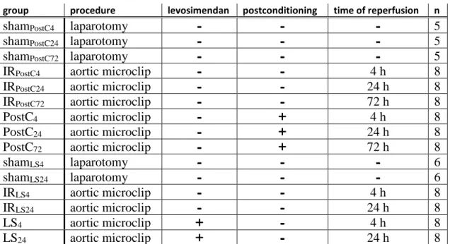
5 2.4.1. Groups of animals – 1st and 2nd experiment (Table 1)
Table 1: Group of animals.
Group categories, name of the groups and basic caracteristics in the 1st and 2 nd experiment.
group procedure levosimendan postconditioning time of reperfusion n
shamPostC4 laparotomy 5
shamPostC24 laparotomy 5
shamPostC72 laparotomy 5
IRPostC4 aortic microclip 4 h 8
IRPostC24 aortic microclip 24 h 8
IRPostC72 aortic microclip 72 h 8
PostC4 aortic microclip 4 h 8
PostC24 aortic microclip 24 h 8
PostC72 aortic microclip 72 h 8
shamLS4 laparotomy 6
shamLS24 laparotomy 6
IRLS4 aortic microclip 4 h 8
IRLS24 aortic microclip 24 h 8
LS4 aortic microclip 4 h 8
LS24 aortic microclip 24 h 8
2.4.2. 1st experiment: Postconditioning in the model of lower limb IR-injury After 180 minutes of ischaemia in one group of animals, with repeated opening and closure of the microvascular clip postconditioning was applied (2 minutes, with 10 s reperfusion, 10 s reocclusion in 6 cycles). Microclip was removed and the wound of laparotomy was sutured. In one group of animals narcosis was maintained for 4 hours and haemodynamic monitoring was performed. In these animals samples were taken after 4 hours. Urine samples were collected during the last 3 hours of the monitored reperfusion period and completed with puncture of the urinary vesicle. In other groups of animals narcosis was interrupted, the venous cannula was removed, the vein was knotted, the wounds were sutured. 24 h and 72 h reperfusion time was allowed with conventional holding of the animals. Samples were taken under anaesthesia. In the sham-operated group, rats were not subjected to aortic clamping, other interventions and measurements were performed in the same manner as in the animals subjected to limb ischaemia.
2.4.3. 2nd experiment: Levosimendan in the model of lower limb IR-injury First steps of the experiment were the same as described above until aortic occlusion.
Levosimendan (Simdax® 2,5 mg/ml, OrionPharma Ltd, Hungary), dissolved in 5 g/100
6 ml glucose solution, was administered as a continuous intravenous infusion (0,2 μg/bwkg/min, 0,48 ml/bwkg/h with the dilution of 25 μg/ml, through the left jugular vein), initiated at the onset of infrarenal aortic clamping and throughout the whole course of ischaemia and the first 3 hours of reperfusion (6 hours in total). In the first 10 minutes of administration an increased (bolus) dose of 12 μg/bwkg levosimendan was given to mimic the clinically recommended administration protocol. IR and sham-operated groups received only the vehicle (5 g/100 ml glucose solution). Samples were taken after 4 and 24 hours of reperfusion time.
2.5. Methods
2.5.1. Haemodynamic monitoring
By invasive blood pressure monitoring system (Kent Scientific Corporation, Torrington, CT, USA), blood pressure was recorded by DasyLab V9.00.02. (National Instruments Corporation, Austin, TX, USA), calculating and registering heart rate (HR), systolic (SBP) and diastolic blood pressure (DBP) values every 5 seconds. Parameters underwent mathematical transformations using c++ (ISO/IEC14882 standard) code edited in Code::Blocks (10.05 rev6283m Code::Blocks Team) and compiled by MinGW (2012.
mingw.org). For eliminating the incidental measurement errors and failures due to external influences, some criteria were defined and enforced by deleting those data that failed these preset conditions. The invalidated data were replaced by the last known accepted values. As criteria, upper and lower bounds for SBP, DBP and HR values were adjusted for each individual animal. Sudden changes in these parameters were also considered as results of external factors. Thus data were cancelled and replaced when exceeding a preset percentage of difference compared to the last accepted value. When replacing multiple consecutive faulty values, the percentage interval of admissible parameters was increased exponentially with respect to the duration of the measurement error. The resulting sequences of data underwent further mathematical transformations (Gaussian smoothing) for the purpose of calculations and better comparability of the individual animals. For each parameter a mean of nearby values weighted with Gaussian function was taken. The following calculations were performed in each animal:
7 (a) At each time point the quotient of HR and SBP was registered and the mean was taken for the period of ischaemia and for reperfusion. Dividing this mean of reperfusion by the mean of ischaemia, the quotient is characteristic for the haemodynamic changes occurring after revascularisation and the efficacy of the given animal’s compensative mechanisms.
(b) To quantify the observed drop of blood pressure at revascularisation, the lowest mean arterial pressure (MAP) was identified within the first 20 min of the reperfusion period.
The time intervals were determined between this time point and the beginning of reperfusion, as well as the degree of this drop expressed as percentage of the last registered MAP of the ischaemic period.
(c) After the drop of blood pressure, a gradual rise was apparent in each animal reaching again a plateau state. With help of ’IF function’ of Microsoft Excel (Microsoft Corporation, Redmond, WA, USA), the point in time was determined when mean arterial blood pressure reaches the beginning of the plateau phase.
2.5.2. Histopathology
Right kidney and right biceps femoris muscle were removed and fixed for 24 h in 4 % formalin, then embedded in paraffin. 3-5 µm slices were cut and analysed after haematoxilin-eosin (HE) staining with light microscopy.
2.5.3. Muscle viability
Rectus femoris muscle samples were rapidly frozen in liquid nitrogen and stored at -70
°C until processed. Slices (3 μm) were incubated in dark for 30 min in 0,025 % nitroblue tetrazolium chloride (NBT, Sigma-Aldrich Inc, St. Louis, MO, USA), dissolved in pH 7,6 TRIS buffer containing 0,05 mmol/l NADH. Unbound NBT was eliminated from the sections with 30, 60 and 90 % acetone solutions in increasing and decreasing concentrations. Under standardised conditions photographs were taken, under light microscope (5 randomly selected areas per each animal). Staining density was measured using a morphometric software (Leica Qwin Pro, Leica Microsystems GmbH, Weltzer, Germany). The relative area of a preset wavelength-range (blue: 175/66, green: 105/0, red: 150/0 on the RGB scale) was determined as a quotient of the occupied area and the total area of the picture. In all operated animals, viability was expressed as a percentage of untreated animals’ staining density.
8 2.5.4. Laboratory measurements
Blood samples were harvested from the right ventricle of the heart by direct puncture.
Samples were snap-frozen in liquid nitrogen and stored at -70 ºC. Analysis was performed by an automated clinical chemistry analyzer (Beckman Coulter AU480/2011, Beckman Coulter Inc, Brea, CA, USA). Serum creatine kinase (CK), aspartate aminotransferase (AST), lactate dehydrogenase (LDH) activities, serum Na+, creatinine és carbamide concentrations were given and also urinary sodium and creatinine concentrations. Serum carbamide/creatinine ratio, Renal Failure Index (RFI = UNa * Pcreat / Ucreat), and fractional sodium excretion (FENa = ClearanceNa / Clearancecreat * 100 = UNa * Pcreat * 100 / (Ucreat
* PNa)). (UNa: urinary Na+; PNa: serum Na+, Ucreat: urinary creatinine; Pcreat: serum creatinine concentrations).
2.5.5. TNF-α measurements
TNF-α levels were assessed by sandwich ELISA kits (R&D Systems, Minneapolis, MN, USA). Mouse TNF-α standard (200 mL, in serially decreasing dilutions: 750–23,4 pg/mL), and 200 mL of TNF-α conjugate (polyclonal antibody to TNF-α conjugated with horseradish peroxidase) was added to each well in the ELISA microtiter plate, and incubated. A color reaction developed with a substrate containing equal volumes of stabilised hydrogen peroxide and tetramethyl benzidine (stopped after 20 min by addition of hydrochloric acid). Absorbance was measured at 450 nm. Each optical absorbance value was calculated as a percentage of the average optical absorbance.
2.5.6. Lower limb and kidney cortex microcirculation measurements
Kidney and lower limb muscle microcirculation were measured by a laser Doppler flowmeter (LDF; Moor DRT4, 2 mW laser power at λ = 632,8 nm; DP1T surface probe, Moor Instruments Ltd, London, UK). Data were registered (at a sampling rate of 10/min) with the manufacturer’s own software (MoorSoft for Windows v1.2, Moor Instruments Ltd, London, UK). All measured fluxes were normalised against and expressed as a percentage of mean of baseline flux. Two parameters were calculated: reperfusion area (RA, integral of the reperfusion segment of the graphs, proportional to the average blood- flow during reperfusion), and the plateau maximum (PM, mean of the last, plateau-shaped 10 min of the reperfusion-slope).
9 2.5.7. Acid-base status
In the 1st experiment arterial blood samples were analysed using Radiometer ABL80 (Radiometer Medical ApS Åkandevej 21 DK-2700, Brønshøj, Denmark). For further calculations, base excess (BE) was chosen as an indicator of an acidic load of the systemic circulation. Relative base excess (RBE) parameter was defined: a quotient of each measured BE and the BE registered before the onset of reperfusion.
2.5.8. Anti-myoglobin immunohistochemistry
In the 1st experiment kidney samples were fixed in 4 % neutral-buffered formalin, washed with phosphate-buffered saline. Endogenous peroxidase activity was blocked with 3 % hydrogen-peroxide. Antigen retrieval was performed in 10 mmol/l citrate buffer, in a pH 6,0 medium. For inhibition of nonspecific binding, tissue was blocked with bovine serum albumin. Samples were incubated (12 h, 4 °C) with polyclonal anti-human anti- myoglobin antibodies raised in rabbits (1:50, Diagnostic BioSystems Inc, Pleasanton, CA, USA) and hybridized for 1 h with peroxidase-conjugated secondary antibody (EnVision®, Dako Denmark A/S, Glostrup, Denmark). For visualization 3- diaminobenzidine was applied.
2.5.9. Heat shock protein 72 (HSP72) analysis
In the 1st experiment kidney samples in the non-surviving animal groups were homogenised in a buffer medium (10 mmol/l HEPES pH=7,9, 1,5 mmol/l MgCl2, 10 mmol/l KCl, 1 mmol/l DTT, 1 mmol/l PMSF, 4 mmol/l benzamidine, 100 U/ml aprotinin, Gedeon Richter PLC, Budapest) with a sonicator (Cole-Parmer Instrument Co., Chicago, IL, USA). HSP72 determination was performed as described previously with Western blot analysis, using rabbit anti-HSP72 antibodies (1:10.000) incubated for 1 h at room temperature and hybridized with horseradish peroxidase-coupled goat anti-rabbit secondary antibody (1:10.000, 1 h; Dako A/S, Glostrup, Denmark). Bands were visualised by chemiluminescence, scanned and quantified by ImageJ software (NIH, Bethesda, MD, USA). To control for equal loading of protein, the expression of the housekeeping protein glyceraldehyde 3-phosphate dehydrogenase (GAPDH) was also checked on the same membrane by subsequent hybridisation with mouse anti-GAPDH (1:10.000 dilution, 1 h; Biodesign International, Saco, ME, USA) and horseradish
10 peroxidase-coupled goat anti-mouse (1:10.000 dilution, 1 h; Dako A/S, Glostrup, Denmark) antibodies.
2.5.10. Lipidperoxidation
After 4 h of reperfusion, right kidney samples were homogenised, diene conjugate contents were extracted by adding isooctane (5 ml isooctane pro 1 g wet tissue sample).
After storing under hypoxic conditions (room temperature, 20 h), the conjugated diene concentrations were detected using a Lumat LB 9051 spectrophotometer (Lumat, Berthold, Windbad, Germany) on 232 nm wavelength. Formation of diene conjugates represents the first step of lipid peroxidation thus indicates oxidative injury in kidneys.
2.5.11. Statistical analysis
Data were analysed using IBM SPSS Statistics Version 20 (IBM Corporation, Armonk, NY, USA). Values are presented as means ± standard deviation. For every group of animals the normality of each measured parameter’s distribution was tested using Kolmogorov-Smirnov statistic. Levene’s tests for homogeneity of variances were performed. In cases when homogeneity of variances was assumed, one way ANOVA (analysis of variance) tests were calculated with Scheffe’s post hoc test for between groups comparisons. When heterogenity of variances was assumed, Brown-Forsythe test was performed with Games-Howell post hoc multiple comparisons. RBE measurements were tested using one-way repeated measures ANOVA. GraphPad (GraphPad Software Inc, La Jolla, CA, USA) was used to generate flow graphs. Any differences were considered statistically significant with p values less than 0,05.
3. Results
3.1. 1st experiment
3.1.1. Haemodynamic monitoring
(a) In the IRPostC4 group a higher „shock-index” occurred for the period of reperfusion as for ischaemia (quotient > 1), whereas in the PostC4 group the quotient was <1, showing a non-elevated shock-index after revascularisation (1,040 ± 0,22 vs. 0,814 ± 0,19, p=0,044).
(b) The lowest systolic blood pressure within the first 20 min of reperfusion occurred at 9,41±7,05 min in the PostC4, and at 7,87±5,06 min after revascularisation in the IRPostC4
11 group. This drop of blood pressure proved significantly lower in degree in the PostC group (IR: 17,11±3,59 % vs. PostC: 11,58±6,04 %, p=0,038).
(c) After the drop of the blood pressure, the pressure curve reached the plateau phase sooner in the PostC4 than in the IRPostC4 group (IRPostC4: 22,92±20,42 min vs. PostC4: 13,90±12,93 min after revascularisation). Because of the high standard devitation, the difference was found non-significant (p=0,565).
3.1.2. M. rectus femoris histopathology
On HE stained sections only minor differences were observed between the groups. In the 4 hours reperfusion group fibre striation and staining of the nucleus were preserved, no oedema was seen, only minimal vaculoisation was detected in both IRPostC4 and PostC4
groups. Some fibres in the IRPostC4 animals showed early signs of rhabdomyolysis with undulate sarcolemma, and intracellular desorganisation. After 24 hours, in samples of the IRPostC24 groups, leukocyte infiltration showed inflammatory changes. In some fibres striation was lost with otherwise intact morphology. Irreversible changes were not seen.
In the PostC24 group only minor oedema was detected. After 72 hours a regression was present in all groups.
3.1.3. Kidney histopathology
On sections of the IRPostC4 animals some degree of acute tubular injury was present (hyaline cylinders in the lumina, the cells are swollen with vacuolisation, hydropic degeneration). In the PostC4 groups milder alterations were seen (hyaline cylinders but not obstructing the lumen, and less cells are degenerated). After 24 and 72 hours reperfusion signs of injury were milder, without any obvious difference between the postconditioned and non-conditioned (not sham) animals (qualitative analysis).
3.1.4. Muscle fibre viability
4 hours after revascularisation a marked muscle injury was detected in both ischaemic subgroups, without any significant difference (IRPostC4: 43,27 ± 30,14 % vs. PostC4: 42,93
± 19,20 %). Till the end of the first postoperative day, a regeneration seemed to be in process in the PostC group, while in the IR group viability remained on a similar level as it was 4 hours after reperfusion (significant difference, IRPostC24: 41,15 ± 16,95 %;
PostC24: 62,65 ± 27,13 %). 72 hours after the revascularisation, a further improvement could be detected in both subgroups, with no significant difference between the
12 postconditioned and the non-conditioned animals (IRPostC72: 77,64 ± 30,64; PostC72: 79,82
± 45,11).
3.1.5. Laboratory measurements – muscle injury
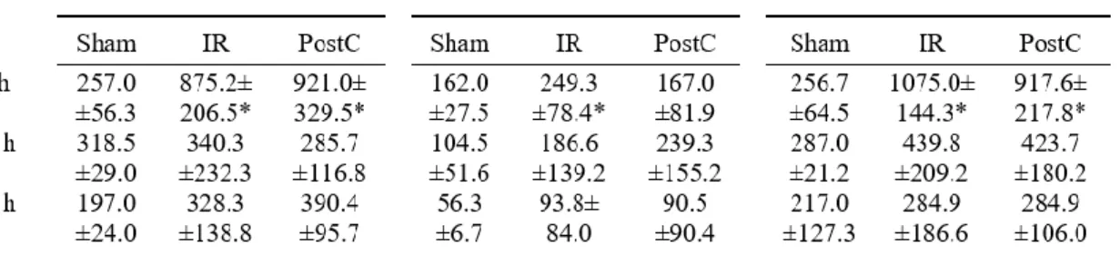
Elevated creatine kinase, aspartate aminotransferase and lactate dehydrogenase serum activities confirmed rhabdomyolysis in the 4th postreperfusion hour in both IRPostC4 and PostC4 groups. Till the end of the 3rd postoperative day these enzyme activities return to normal range. No significant difference were seen between PostC and IRPostC groups.
Table 2: 1st experiment, serum creatine kinase, aspartate aminotransferase, lactate dehydrogenase activities. No significant difference between IRPostC and the corresponding PostC subgroups. In the 4 hours’ reperfusion groups, significantly elevated levels compared to the sham operated animals in the IRPostC4 and (except for ASAT) in the PostC4 subgroups.
3.1.6. Laboratory measurements – renal failure
All animals subjected to limb IR developed acute kidney injury of some extent. PostC4
group developed significantly lower serum creatinine elevation than the IRPostC4 group.
For characterisation of the evolving renal dysfunction, clinical renal functional parameters were calculated. At the end of the fourth hour of reperfusion, both serum carbamide/creatinine ratio and the fractional sodium excretion (ClearanceNa/Clearancecreatinine*100) indicated a tubular type of kidney failure, but only in the IR group, whereas the values in the PostC group were not characteristic for any type of kidney failure, according to usual definitions. Renal failure index (RFI=[Na]urine*[creatinine]serum/[creatinine]urine) indicated persistent kidney damage with significantly lower values in the PostC subgroups.
13 Table 3: 1st experiment, laboratory renal functional measurements: †: p < 0,05 vs.
the corresponding IRPostC group; ‡: p < 0,001 vs. the corresponding IRPostC group.
3.1.7. TNF-α measurements
Compared to the sham operated 4 hours group (6,615 ± 1,423 ng/ml) significantly elevated TNF-α levels were measured in both IRPostC4 (44,904 ± 8,693 ng/ml) and PostC4
(22,906 ± 4,909 ng/ml) groups. TNF-α levels were significantly less elevated in the PostC4 group than in the IRPostC4 group (p<0,05).
3.1.8. Lower limb microcirculation
In IRPostC4 animals hypoperfusion was detected during reperfusion (compared to the measured fluxes before aortic occlusion). In the PostC4 group, after release of aortic clamping and postconditioning a transient hyperaemia happens and afterwards microcirculation settles at a level comparable to the one before aortic clamping. So in PostC4 animals significantly higher RA and PM values were calculated, compared to the IRPostC4 group. (RA: PostC4: 96,15 ± 17,48 % vs. IRPostC4: 63,99 ± 15,73 %, p = 0,038, PM: PostC4: 82,87 ± 23,19 % vs. IRPostC4: 60,85 ± 15,33 %, p = 0,194).
5.1.9. Kidney cortex microcirculation
Kidney microcirculation remained at the baseline level after clamping of the infrarenal aorta. At revascularisation flux dropped and after a lower, stable level, microcirculation improved in the PostC group nearly reaching the normal baseline level, while it further deteriorated in the IR group. The perfusion of the kidney cortex was significantly better
14 in the PostC group than in the IR group (RA: PostC4: 99,01±2,76% vs. IRPostC4: 82,31±12,23%, p=0,024, PM: PostC4: 96,81±6,14% vs. IRPostC4: 77,21±14,81%, p=0,037).
5.1.10. Acid-base status
Relative base excess (RBE) is an arbitrary parameter, formed as a quotient of each registered base excess values (BE, measured at 4 points of time at the beginning of the reperfusion) and the base excess value just before the onset of the reperfusion in the same animal. As a quotient of two negative numbers, a higher positive RBE represents a higher acid-load. PostC animals developed significantly less increased RBE values, than the IR animals (1st min: PostC4: 1,80±0,66 vs. IRPostC4: 3,19±0,27, 2nd min: PostC4: 2,44±1,14 vs. IRPostC4: 3,38±0,17, 3rd min: PostC4: 2,07±0,82 vs. IRPostC4: 5,32±2,91, 4th min:
PostC4: 2,05±0,56 vs. IRPostC4: 3,28±0,32, p=0,037).
5.1.11. Anti-myoglobin immunohistochemistry
4 hours after reperfusion presence of myoglobin epitopes was detected in the lumen of the kidney tubules and also in the apical lysosome vacuoli of the proximal tubular cells.
Sections demonstrated a milder myoglobin-load in the PostC animals with less tubular myoglobin casts and sparse presence in the intracellular lysosomes. 24 and 72 hours after revascularisation, all kidney sections proved myoglobin-negative (no intraluminal stained casts or intracellular signs, due probably to the dissolved or transformed myoglobin- epitopes).
5.1.12. Heat shock protein 72 (HSP72)
All HSP72 measurements were normalised and standardised by registering as a quotient of the housekeeping gene glyceraldehyde 3-phosphate dehydrogenase (GAPDH) expression in the same sample. Although there was a tendency for kidney HSP72 expression to be upregulated in both the IRPostC4 (1,78±0,45), and PostC4 (1,41±0,13) groups vs. the sham-operated group (0,98±0,23), we did not find any significant differences between the groups.
15 5.1.13. Lipid peroxidation
Conjugated diene contents detected by spectrophotometer from homogenised kidney samples were elevated in both IR and PostC animals in comparison with the sham- operated group, showing an early evolving lipid peroxidation in the post-revascularisation 4th hour. Conjugated diene concentrations (expressed in absorption units/ml) were significantly higher in the IRPostC4 than in the PostC4 group (ShamPostC4: 0,12 ± 0,01 U/ml, IRPostC4: 0,17 ± 0,04 U/ml, PostC4: 0,14 ± 0,01 U/ml, p=0,032).
5.2. 2nd experiment
5.2.1. Haemodynamic monitoring
(a) Calculated ’shock-index’ was higher for the period of reperfusion as for ischaemia in the IR group (the quotient is >1). The quotient was also >1 in the Levosimendan group, with no statistically significant difference between the two groups (1,09±0,25 vs.
1,07±0,30, p>0,05).
(b) The lowest systolic blood pressure within the first 20 min of reperfusion occurred at 6,00±2,86 min in the LS4, and at 7,37±4,97 min after revascularisation in the IRLS4 group (p>0,05).
(c) This drop of blood pressure was greater in the IRLS4 group (16,61±3,61 % of the last registered systolic blood pressure of the ischaemic time) than in the LS4 group (8,56±8,22
%), though because of the high standard deviation, the difference was found non- significant.
(d) After the drop of the blood pressure, the pressure curves reached the plateau phase within a similar time interval in both the LS4 and the IRLS4 group, with high standard deviation (IRLS4: 17,53±11,23 min vs. LS4: 17,42±19,83 min after revascularisation, p>0,05).
5.2.2. Histopathology
Light microscopy and HE staining was not able to detect any definitive histopathological lesions in the samples taken from the tibial anterior muscle in any groups at any measured time points.
Kidney samples showed signs of acute tubular injury in the IR group with swollen, vacuolised tubular cells and precipitated hyaline-cylinders within the tubular lumina after
16 4 hours of reperfusion. 24 hours after aortic declamping slightly milder histological damage could be observed. Levosimendan administration resulted in lesser degree of histological injury at both examined reperfusion intervals compared to the corresponding IR group (qualitative analysis).
5.2.3. Muscle viability
4 hours after revascularisation, a marked decrease in viability was detected in the IR group, as well as results showed no regeneration even after 24 hours of reperfusion.
Levosimendan administration resulted in significantly preserved viability compared to the IR group at both measured time points (4 and 24 hours), however the values of the LS24 group failed to reach the levels of the sham operated animals. (4 h: shamLS4 99,2±2,6
%, IRLS4: 36,6±6,5 %, LS4: 60,7±3,9 %, p<0,05; 24 h: shamLS24: 99,1±2,7 %, IRLS24: 39,7±4,1 %, LS24: 66,6±4,7 %, p<0,05).
5.2.4. Laboratory measurements
At the end of the fourth hour of reperfusion, kidney injury was evident in the IRLS4 group according to markedly elevated serum creatinine levels (p<0,001). Meanwhile in the 4 h levosimendan treated group, significantly (p=0,006) less elevated serum creatinine levels, less expressive kidney injury could be observed.
Serum carbamide/creatinine ratio and the fractional sodium excretion indicated a tubular type of kidney failure in the IRLS4 group, whereas the values in the LS4 group were not characteristic for any type of kidney failure, according to usual definitions. Differences were significant (p<0,001). Renal failure index indicated persistent kidney damage at the 24th hour after reperfusion with significantly lower values in the levosimendan treated group (p<0,001).
5.2.5. Serum TNF-α measurements
TNF-α concentrations were significantly increased in the IRLS4 group compared to the sham operated group (p<0,001) after 4 hours of reperfusion. Levosimendan administration was able to significantly reduce serum TNF-α levels compared to the IRLS4
group (ShamLS4: 4,96±0,72 ng/mL, IRLS4: 36,79±7,78 ng/mL, LS4: 14,51±2,30 ng/mL, p<0,001 between IRLS4 and LS4).
17 Table 4: 2nd experiment, laboratory renal functional measurements: †: p < 0,05 vs.
the corresponding IRLS group; ‡: p < 0,001 vs. the corresponding IRLS group.
5.2.6. Microcirculation of lower limb muscle and kidney cortex
Sham operation resulted in no alterations in either kidney or skeletal muscle flow throughout the experiment. Kidney microcirculation remained at the baseline level after clamping of the infrarenal aorta in both IRLS4 and LS4 groups. After revascularisation, flux deteriorated gradually in the IRLS4 group while levosimendan administration was able to maintain renal cortical microcirculation at a significantly higher level, close ot the values measured in the ShamLS4 animals (PM: IRLS: 78,02±12,63 % vs. LS4: 96,90±4,67
%, p=0,009; RA: IRLS4: 82,91±9,75 % vs. LS4: 98,84±1,85 %, p=0,004).
Muscle microcirculation dropped at the onset of aortic occlusion in both the IRLS4 and LS4 groups. At the onset of reperfusion, microcirculation increased to about 100 % of baseline flux in the LS4, in contrast to IRLS4 group where it reached only 80 % of baseline values. (PM: IRLS4: 62,87±14,58 % vs. LS4: 89,25±6,70 %, p=0,004; RA: IRLS4: 66,01±14,92 % vs. LS4: 90,26±6,55 %, p=0,006).
18 6. Conclusions
1. In our model of 3 hours’ bilateral limb ischaemia postconditioning did not have any positive effect on the skeletal muscle fibre ischaemia-reperfusion injuries, based on conventional HE stained histopathology and serum CK, LDH and ASAT enzyme activities. After 24 hours of reperfusion time, a significantly less decreased (more preserved) muscle viability could be detected.
2. After reperfusion laser Doppler flowmetry revealed a microcirculatory failure in the lower limb muscle. With application of the method postconditioning a significantly less decreased flow was measured. At the same time the inflammatory marker TNF-α levels were less elevated in the postconditioned animals.
3. In the model an acute kidney dysfunction and failure developed. In the background of the pathophysiology we demonstrated the nephrotoxic myoglobin released from the injured muscle fibres, lipid peroxidation, systemic acidosis, inflammatory response, failure of the kidney cortex perfusion and partly systemic haemodynamic changes. Postconditioning resulted in a less expressed acute tubular injury. This is mostly independant of the lower limb muscle rhabdomyolysis and parhaps is due to the mechanical characteristics of the method. Release of myoglobin and other acidic and nephrotoxic metabolites from the injured limb tissues into the circulation is fractionated, delayed with the use of the method.
4. It is likely that also due to the mechanical aspects of the method, the circulatory disturbances at the onset of reperfusion are less expressed using postconditioning, and the lower shock-index implies a better circulatory autoregulation and adaptation.
5. Levosimendan applied in the same model significantly protected muscle viability measured at both 4 and 24 hours after reperfusion, and also based on microcirculatory flowmetry. These local effects on IR injuries may be explained by the opening of mitoKATP channels and mitochondrial protection.
6. Levosimendan resulted in a less decreased microcirculation of the lower limb muscle tissues. This is accompanied by a less expressed release of the inflammatory cytokine TNF-α.
19 7. With infusion of levosimendan a lesser extent of acute tubular kidney injury was detected. In background of the pathophysiology, the potential protective effect of levosimendan on muscle mitochodrial integrity is implied, also an amelioration of the inflammatory processes and this is completed by the favourable cardiovascular effects of the drug.
7. Bibliography of the candidate’s publications Publications the dissertation is based upon:
1. Aranyi P, Turoczi Zs, Garbaisz D, Lotz G, Geleji J, Hegedus V, Rakonczay Z, Balla Z, Harsanyi L, Szijarto A. (2015) Postconditioning in major vascular surgery: prevention of renal failure. J Transl Med, 13:(1)21.
2. Onody P, Aranyi P, Turoczi Z, Stangl R, Fulop A, Dudas E, Lotz G, Szijarto A.
(2016) Levosimendan Administration in Limb Ischemia: Multicomponent Signaling Serving Kidney Protection. PLoS One, 11(9):e0163675.
Other publications:
1. Szijártó A, Gyurkovics E, Arányi P, Ónody P, Stangl R, Tátrai M, Lotz G, Mihály Z, Hegedüs V, Blázovics A, Kupcsulik P. (2009) Postconditionálás kísérletes alkalmazása aortakirekesztés kapcsán. Magy Seb, 62(4):180-187.
2. E. Gyurkovics, P. Aranyi, Zs. Turóczi, D. Garbaisz, M. Varga, V. Hegedüs, G.
Lotz, P. Kupcsolik, A. Szijarto. (2010) Postconditioning attenuates remote organ injury after lower limb arterial occlusion. Interventional Medicine & Applied Science, 2(4):169-177.
3. Szijártó A, Arányi P, Turóczi Zs, Kupcsulik P, Gyurkovics E. (2010) Ischaemiás- reperfúziós károsodás csökkentésének elvi és gyakorlati lehetőségei az érsebészetben. Irodalmi áttekintés. Érbetegségek 4:113-120.
4. Szijártó A, Arányi P, Turóczi Zs, Kupcsulik P, Gyurkovics E. (2010) A reperfúziós szindróma és a postconditionálás sejtszintű mechanizmusa. Irodalmi áttekintés. Érbetegségek 17(1):11-19.
5. Szijártó A, Turóczi Zs, Arányi P, Nagy Z, Gyurkovics E. (2010) Akut kritikus ischaemia az alsó végtagon. Irodalmi összefoglaló. Orv Hetil, 2;151(50):2057-66.
20 6. Szijártó A, Turóczi Z, Arányi P, Garbaisz D, Varga M, Stangl R, Lotz G, Kupcsulik P. (2010) Hosszú idejű végtagi verőér-elzáródás és izomszövet- életképesség vizsgálata kísérletes állatmodellben. Magy Seb, 63(6):374-9.
7. E Gyurkovics, P Aranyi, R Stangl, P Onody, G Ferreira, G Lotz, P Kupcsulik, A Szijarto. (2011) Postconditioning of the lower limb - Preconditioning against the reperfusion syndrome. J Surg Res, 169(1):139-47.
8. Arányi P, Turóczi Zs, Garbaisz D, Varga M, Lotz G, Kupcsulik P, Szijártó A.
(2011) Postconditionálás: érsebészeti technika posztoperatív veseelégtelenség megelőzésére – Állatkísérletes eredmények. Hypertonia és Nephrologia, 15(3):117-124.
9. Szijarto A, Turoczi Z, Szabo J, Kaliszky P, Gyurkovics E, Aranyi P, Regali L, Harsanyi L, Lotz G. (2013) Rapidly progressing fatal reperfusion syndrome caused by acute critical ischemia of the lower limb. Cardiovasc Pathol, 22:(6)493- 500.
10. Garbaisz D, Turoczi Z, Fulop A, Rosero O, Aranyi P, Onody P, Lotz G, Rakonczay Z, Balla Z, Harsanyi L, Szijarto A. (2013) Therapeutic option for managing lung injury induced by infrarenal aortic cross-clamping. J Surg Res, 185(1):469-76.
11. Garbaisz D, Turóczi Z, Fülöp A, Rosero O, Arányi P, Onody P, Lotz G, Rakonczay Z, Balla Z, Harsányi L, Szijártó A. (2013) Postconditioning can reduce long-term lung injury after lower limb ischemia-reperfusion. Magy Seb, 66(3):146-54.
12. Turóczi Zs, Arányi P, Lukáts Á, Garbaisz D, Lotz G, Harsányi L, Szijártó A.
(2014) Muscle fiber viability, a novel method for the fast detection of ischemic muscle injury in rats. PLoS One, 9:(1)e84783.
13. Garbaisz D, Turoczi Z, Aranyi P, Fulop A, Rosero O, Hermesz E, Ferencz A, Lotz G, Harsanyi L, Szijarto A. (2014) Attenuation of skeletal muscle and renal injury to the lower limb following ischemia-reperfusion using mPTP inhibitor NIM-811.
PLoS One, 9:(6) e101067.
14. Arányi P, Tomcsányi J. (2016) Differential diagnostic dilemmas after use of an out-of-date antihypertensive medication. Case report. Orv Hetil, 158(11):426- 431.