MORPHOLOGICAL EVALUATION OF EXPERIMENTAL AUTOLOGOUS RECTUS FASCIA SHEATH VASCULAR
GRAFTS USED FOR ARTERIAL REPLACEMENT IN A DOG MODEL
Péter CSÉBI1*, Csaba JAKAB2, Attila PATONAI3, Attila ARANY-TÓTH1, László KÓBORI3
and Tibor NÉMETH1
1Department and Clinic of Surgery and Ophthalmology and 2Department of Pathology and Forensic Veterinary Medicine, Faculty of Veterinary Science, Szent István University,
István utca 2, H-1078 Budapest, Hungary; 3Transplantation and Surgical Clinic, Semmelweis University, Budapest, Hungary
(Received 9 April 2014; accepted 25 June 2014)
Although experimental autologous patch or tubular conduit vascular grafts made from the internal rectus fascia sheath (IRFS) have been reported in the lit- erature, thorough morphological evaluation and verification of the histological ar- terialisation of such grafts are lacking. Four purpose-bred Beagle dogs were util- ised to create eight arterial internal rectus fascia sheath (ARFS) grafts implanted between bisected ends of the external iliac arteries. Four out of the eight ARFS grafts were patent after three months. Haematoxylin-eosin and Azan staining veri- fied that the grafts gained a vessel-like layered structure with the presence of large amounts of collagen fibres. Although the inner surface of the intact IRFS was originally covered with claudin-5-negative and pancytokeratin-positive mesothe- lial cells in control samples, the internal cells of the ARFS grafts became claudin- 5 positive and pancytokeratin negative like in intact arteries. Spindle-shaped cells of the wall of ARFS grafts were α-smooth muscle actin (α-SMA) positive just like the smooth muscle cells of intact arteries, but α-SMA immunoreactivity was negative in the intact IRFS. According to these findings, the fibroblast cells of the ARFS graft have changed into myofibroblast cells. The study has proved that ARFS grafts may be used as an alternative in arterial replacement, since the graft becomes morphologically and functionally similar to the host vessel via arterialis- ation.
Key words: Rectus fascia sheath, autologous vascular graft, immunohisto- chemistry, claudin-5, alpha-SMA, desmin, pancytokeratin, veterinary field
Several different types of vascular substitutes are used in vascular surgery including synthetic vascular prostheses, autologous vessels, heterologous donor
*Corresponding author; E-mail: pcsebi@gmail.com; Phone: 0036 (1) 478-4197;
Fax: 0036 (1) 478-4196
vessels, xenografts of bovine or ovine origin and cryopreserved vessels (Bíró et al., 2001). The greater saphenous vein (Akimaru et al., 2000) and the internal jugular vein (Takayama et al., 1995) are often used as autologous vascular im- plants in the human clinical practice. The use of the autologous internal rectus fascia sheath (IRFS) as patch or tubular vascular conduit grafts for replacing ar- terial or venous defects has been reported in the human and veterinary literature (Kóbori et al., 2000, 2003, 2005, 2008; Hammond et al., 2004; Németh et al., 2005; Csébi et al., 2011). The vascular use of the IRFS was based on its previous application for aortic patch repair in dogs (Cousar and Lam, 1952), as an autolo- gous graft in cardiac surgery (Bilfinger et al., 1983) and for numerous other indi- cations such as choledochoplasty (Pacholewicz et al., 1998) and reconstruction of abdominal wounds (Gondolesi et al., 2009). The viability and the haemody- namic characteristics of the IRFS were proven to be clinically adequate and, due to the early appearance of the neoendothelial layer, its thrombogenicity was also found to be low (Coltharp, 1989). Despite the numerous applications, the thor- ough and detailed morphological evaluation and verification of the histological arterialisation of the IRFS graft are lacking. The goal of this study was to exam- ine arterial autologous internal rectus fascia sheath (ARFS) grafts, control arter- ies and control intact IRFS by haematoxylin-eosin and Azan staining, by claudin- 5, alpha-SMA, desmin and pancytokeratin immunohistochemistry and by elec- tron microscopy. The hypothesis of the study was that the ARFS graft can take over the function of an artery owing to changes in graft morphology through ar- terialisation.
Materials and methods
Implantation, monitoring and removal of the rectus fascia sheath vascular grafts This study was approved by the competent authorities acting upon the rec- ommendation received from the Scientific Ethics Committee on Animal Experi- mentation (authorisation number: 73/2008). The animals were treated according to the international guidelines for the care and use of laboratory animals (Decla- ration of Helsinki). Four purpose-bred Beagle dogs were utilised to create eight arterial internal rectus fascia sheath (ARFS) grafts. The experimental protocol was designed using the method elaborated and successfully used for arterial tubular grafts by Kóbori et al. (2008). The surgical interventions were performed in gen- eral anaesthesia using intravenous premedication with 0.5 mg/kg diazepam (Se- duxen 5 mg/ml inj., Gedeon Richter Plc, Budapest, Hungary) and 0.01 mg/kg fentanyl (Fentanyl inj., Gedeon Richter), induction with 3 mg/kg propofol (Diprivan 1% inj., AstraZeneca Pharmaceuticals LP) administered i.v., and main- tenance with 2 (v/v) % sevoflurane and 2 L/min oxygen flow. Dogs received fen- tanyl continuous rate infusion (CRI) in a dose of 0.02 mg/kg/h and lactated Ringer’s infusion at 10 ml/kg/h during surgery. After a ventral midline abdomi-
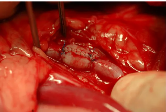
nal incision, both external iliac arteries were dissected from the surrounding tis- sues. After measuring the diameter of the iliac artery, a piece of the internal rectus fascia sheath (3 × 4 cm) was tailored. A standard glass rod (5 mm in diameter) was used to create the ARFS graft as a 20-mm-long tubular graft was sutured with its peritoneal layer inside, using a single-layer simple continuous 6-0 USP polypro- pylene (Prolene, Ethicon) suture. Immediately before clamping the iliac artery, 200 IU/kg heparin sodium injection (Heparin inj., Gedeon Richter) was given in- travenously to prevent thrombosis. The external iliac artery was transected be- tween two ‘de Bakey’ straight vascular clamps, and the ARFS graft was anasto- mosed with the transected arterial ends in an end-to-end manner using a single- layer simple interrupted 6-0 USP polypropylene closure (Fig. 1a). The morpho- logical characteristics of the graft were documented after re-establishment of cir- culation in the artery. The abdomen was closed routinely in four layers. Heparin sodium (200 IU/kg BID) was given subcutaneously as a postoperative anticoagu- lant for two weeks. Meloxicam (0.2 mg/kg sc. SID) was used as a postoperative analgesic for 5 days.
Fig. 1a. Picture of the 20-mm-long ARFS autograft right after end-to-end anastomosis between the bisected ends of the external iliac artery
The morphology and patency of the segments containing the graft were regularly monitored by Doppler ultrasonography using an ESAOTE MEGAS GPX type ultrasonography device with a transducer of 5–7.5 MHz frequency range.
At the end of the 3-month follow-up period, ARFS tubular grafts were surgically removed along with the anastomosed section of the iliac artery, and the arteries proximal and distal to the two anastomosis sites were ligated.
Histopathology and immunohistochemistry
The ARFS graft and a piece of the intact rectus sheath were preserved in 8% formaldehyde solution buffered with phosphate-buffered saline (pH 7.0) and embedded in paraffin. After fixation, 3–4 µm sections were cut from the blocks with a Reichert microtome and were stained with the help of a Shandon Varistain 24-4 continuous automatic slide stainer. Haematoxylin-eosin (HE) and Azan staining was used. The sections were evaluated in a Nikon Optiphot-2 type light microscope and histopathological photographs were taken with a Nikon Coolpix 4500 digital camera. Slides for the immunohistochemical examination were de- paraffinised in two changes of xylene (for 2 × 10 min) and three changes of etha- nol (100%, 70% and 50% for 5 min each), then washed in PBS once and sub- jected to antigen retrieval at high temperature, in a microwave oven (800 W for 30 min), using Target Retrieval Solution (pH 6, DAKO, Glostrup, Denmark).
The activity of endogenous peroxidase was blocked by exposure to a mixture of 0.3% H2O2 and methanol for 5 min. The following antibodies were used for the immunohistochemical reactions: anti-claudin-5 (1:100 dilution, mouse mono- clonal antibody, Zymed Inc.), anti-desmin (1:400 dilution, mouse monoclonal antibody, Novocastra), anti-α-smooth muscle actin (α-SMA) (1:8,000 dilution, mouse monoclonal antibody, Sigma), and anti-pancytokeratin AE1-AE3 (1:100 dilution, mouse monoclonal antibody, DAKO). The positive controls were endo- thelial cells of microvessels in canine mammary gland carcinoma for claudin-5, leiomyoma for α-SMA, arrector pili muscle for desmin, and mammary gland for pancytokeratin. The intensity of the immunohistochemical reactions were evalu- ated on a scale of one to three crosses as follows: (+++) intensive reaction, if the intensity is equivalent with that obtained for the external positive controls; (+) weak reaction, if the membrane positivity is expressly weaker than that in the ex- ternal positive controls; (++) moderate intensity, if the intensity of immunoreac- tivity in the endothelial cell membrane is between the above two.
Electron microscopy (EM)
Tissues of 1 mm3 in size, taken from the grafts, the artery and the rectus sheath, were fixed for 24 h at 4 °C in 4% buffered formaldehyde solution for the EM examination. At the end of the fixation, 1% osmium tetroxide was applied.
The specimens were dehydrated in an ascending series of ethanol and embedded in synthetic resin (Durcupan ACM, Fluka, Switzerland). Ultra-thin sections were cut and contrasted with uranyl acetate and lead citrate, then examined under a transmission electron microscope (JEM 1011, JEOL Ltd., Tokyo). Electron mi-
crographs were obtained and adjusted employing a SIS MegaView III digital im- age acquisition system (Olympus Soft Imaging Solutions, Münster, Germany).
Results
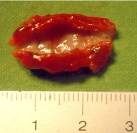
Four out of the eight ARFS grafts were patent and viable with intact lu- minal surface and a thick and rigid wall after three months (Fig. 1b). Stenosis and obstruction of the ARFS graft occurred in two cases during the first week and in two cases during the first month. The dogs did not show any clinical signs related to stenosis or obstruction of the iliac artery. Well-developed collateral vessels were seen around the stenotic graft during the surgical revision. On gross examination, the patent ARFS grafts seemed viable with intact luminal surface and a thick and rigid wall.
Fig. 1b. Picture of the ARFS autograft after harvesting at revision surgery three months later
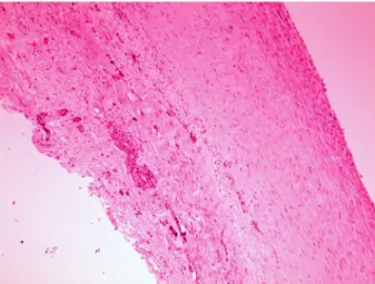
The morphology of ARFS grafts was compared to that of intact arteries and rectus sheath samples. HE staining showed a vessel-like layered wall of the grafts.
The inner surface of the graft was covered with elongated endothel-like cells and the hypocellular wall contained spindle-shaped mesenchymal cells (Fig. 2). Azan staining revealed large amounts of collagen fibres within the graft (Fig. 3). By immunohistochemistry, the endothelial lining of the intact artery samples showed claudin-5 positivity and pancytokeratin negativity, the mesothelial lining of the intact IRFS samples was positive for pancytokeratin and negative for claudin-5, and the luminal surface of the ARFS graft was covered by flat and fusocellular cell lining showing linear claudin-5 positivity and pancytokeratin negativity (Ta- ble 1). Endothelial cells of the vasa vasorum were positive for claudin-5 (internal
positive control) in the ARFS grafts. Smooth muscle cells of the tunica media of intact arteries showed α-SMA and desmin positivity, the wall of the intact IRFS was α-SMA negative and desmin positive, and the spindle-shaped cells of the wall of the ARFS grafts showed moderate α-SMA and desmin positivity (Table 2).
Electron microscopic examination of the ARFS grafts revealed that the wall was rich in thick collagen fibres including large numbers of active fibroblasts. The luminal surface was covered by an endothelial cell lining. In comparison with in- tact arteries, the membrana elastica interna and the lamina elastica externa were absent in the ARFS graft, and smooth muscle cells were not visible either.
Fig. 2. The ARFS graft showed a vessel-like layered structure with a more hypocellular middle layer. Haematoxylin and eosin (HE), × 100
Fig. 3. Azan-positive blue-coloured collagen fibres in the ARFS graft (Azan staining, × 100)
Table 1
Results of immunohistochemistry of the mesothelial/endothelial lining of the three types of tissue samples
External iliac artery Internal rectus sheath ARFS graft
Claudin-5 +++ – ++
Pancytokeratin – +++ –
The intensity of the immunohistochemical reactions is indicated as (+++) in- tensive, (++) moderate, (+) weak or (–) negative
Table 2
Results of immunohistochemistry of the tunica media/wall of the three types of tissue samples External iliac artery Internal rectus sheath ARFS graft
Claudin-5 +++ – ++
Pancytokeratin – +++ –
The intensity of the immunohistochemical reactions is indicated as (+++) in- tensive, (++) moderate, (+) weak or (–) negative
Discussion
The rectus fascia sheath graft is a possible alternative autologous material.
In the first experiment of Kóbori et al. (2000) the ARFS grafts were implanted in seven dogs. The follow-up period was one month, and 5 out of the 7 grafts re- mained patent. The diameter of the grafts was slightly increased. One of the ob- structions was due to technical failure and the other was caused by a septic post- operative complication (Kóbori et al., 2000). In the second experiment of Kóbori et al. (2003), eight grafts were implanted and the follow-up period was six months in 6 cases and 12 months in two cases. The dogs were treated with the immunosuppressant drug cyclosporin A during the whole period in order to mimic the postoperative circumstances of immunosuppressed transplanted pa- tients. All grafts remained patent and only two of them became stenotic. Histo- logical studies (haematoxylin-eosin staining) were done in all cases. The pres- ence of endothelial cells on the luminal surface was detected by the vWf and CD34 expression method. Electron microscopy showed viable smooth muscle filaments and elastin fibres. The degenerative effect of cyclosporin was not de- tected in the ARFS grafts (Kóbori et al., 2003). Based on the promising results of the first two experiments, a third study including a larger group of dogs was per- formed. Forty-one grafts were implanted and immunosuppression was used in separate groups. Thirty-seven grafts remained patent, two cases of thrombosis and two cases of stenosis occurred. There was no evidence of necrosis or aneu-
rysm formation. The histological analysis included conventional light micro- scopic and immunohistochemical examinations for CD34 and factor VIII. The removed ARFS grafts showed signs of arterialisation and the appearance of elastin fibres and smooth muscle cells after six months. Electron microscopy re- vealed intact mitochondrial structures without signs of hypoxia. In conclusion, the autologous graft presented an acceptable long-term patency rate (Kóbori et al., 2008). Based on these previous results we repeated the study in order to fur- ther examine the arterialisation process via a more thorough and detailed mor- phological evaluation protocol. In the present experiment the surgical procedure was the same as in the previous studies, but the results were less satisfactory.
Fifty percent of the ARFS grafts remained patent during the follow-up period, which is a lower percentage than that obtained in the previous experimental stud- ies. The gross examination of the patent grafts showed a rigid, thick wall. The difference between the results may be attributable to technical failures. A part of this may be due to the damage of the sensitive mesothel cell layer on the luminal surface of the grafts. It is presumed that the mesothel cell layer plays an impor- tant role in the prevention of thrombosis in the grafts. This is based on in vitro studies with mesothelial cell seedings on vascular grafts which showed low thrombogenicity (Pronk et al., 1988). Mesothelial cells produce prostacyclin and they have fibrinolytic activity in the same way as endothelial cells (Kent et al., 1988; Sharp et al., 1989; Deutsch et al., 1999; Maeda et al., 2000). Another rea- son for the different patency results can be an improper anticoagulant therapy.
Although heparin sodium (200 IU/kg BID) was given subcutaneously as a post- operative anticoagulant for two weeks, we did not use low-dose acetylsalicylic acid antiplatelet therapy.
Based on our histomorphological examinations, the structure of the rectus sheath used as a vascular graft changed after the implantation. Although the in- ner surface of the intact IRFS is originally covered by claudin-5-negative and pancytokeratin-positive mesothelial cells (Avakian et al., 2008), the internal cells became claudin-5 positive and pancytokeratin negative like in the intact arteries during the follow-up period (Morita et al., 2003; Jakab et al., 2009). Based on this, a complete re-endothelisation process took place during the 3-month follow- up period by the likely migration of endothelial cells from the vessel to the graft (transmigration). Spindle-shaped cells of the wall of ARFS grafts were α-SMA positive like smooth muscle cells of the intact arteries (Meng et al., 2001), but α- SMA immunoreactivity was negative in the intact IRFS. The change in the im- munoreactivity of the ARFS grafts shows that fibroblast cells of the IRFS have changed into myofibroblast cells, which are transdifferentiated fibroblast cells containing smooth muscle actin (Meng et al., 2001). This morphological change may increase the elasticity of the ARFS grafts, enabling them to accommodate to the new circumstances as a vascular substitute. Electron microscopic examina- tion revealed that the inner surface was covered by endothelial cell lining, but the
wall of the grafts did not contain smooth muscle cells and the two definitive lay- ers of elastic fibres were also missing. This may cause less elasticity in the wall of the grafts as compared to an intact artery.
In conclusion, the ARFS autograft may be used as an alternative in arterial replacement, since the graft becomes morphologically and functionally similar to the host vessel via arterialisation, enabling it to behave as a blood vessel. Fur- thermore, rejection is not a possible complication, but the wall of the grafts be- comes thicker and less elastic than that of an intact artery. Nevertheless, in our experiment only half of the grafts remained patent, and the four obstructed grafts suffered from stenosis. Further investigation of the long-term clinical behaviour is recommended before the ARFS graft is routinely used in clinical cases as a tu- bular vascular substitute.
References
Akimaru, K., Onda, M., Tajiri, T., Yoshida, H., Mamada, Y., Taniai, N., Yoshioka, M. and Mineta, S. (2000): Reconstruction of the vena cava with the peritoneum. Am. J. Surg. 179, 289–293.
Avakian, A., Alroy, J., Rozanski, E., Keating, J. and Rosenberg, A. (2008): Lipid-rich pleural mesothelioma in a dog. J. Vet. Diagn. Invest. 20, 665–667.
Bilfinger, T. V., Beere, P. E., Sanderson, C., Glagov, S. and Anagnostopoulos, C. E. (1983): Paral- lel growth of rectus sheath grafts and recipient aorta. Critical role of graft tissue preserva- tion. J. Thorac. Cardiovasc. Surg. 86, 294–299.
Bíró, G., Szabó, A., Szeberin, Z. and Nemes, A. (2001): Arterial reconstructions performed with homograft implantation [in Hungarian]. Magy. Seb. 54, 63–67.
Coltharp, W. H. (1989): Experimental aortic replacement with a vascularised tissue graft. Arch.
Surg. 124, 1331–1334.
Cousar, J. E. and Lam, C. R. (1952): Rectus sheath grafts in vascular repair. Arch. Surg. 65, 471–476.
Csébi, P., Németh, T., Jakab, Cs., Patonai, A., Garamvölgyi, R., Manczur, F., Spitzner, Á., Arany- Tóth, A. and Kóbori, L. (2011): Experimental results of using autologous rectus fascia sheath for venous patch grafts in dogs. Acta Vet. Hung. 59, 373–384.
Deutsch, M., Meinhart, J., Fischlein, T., Presii, P. and Zilla, P. (1999): Clinical autologous in vitro endothelialization of infrainguinal ePTFE grafts in 100 patients: a 9-year experience. Sur- gery 126, 847–885.
Gondolesi, G., Selvaggi, G., Tzakis, A., Rodríguez-Laiz, G., González-Campaña, A., Fauda, M., Angelis, M., Levi, D., Nishida, S., Iyer, K., Sauter, B., Podesta, L. and Kato, T. (2009):
Use of the abdominal rectus fascia as a nonvascularized allograft for abdominal wall clo- sure after liver, intestinal, and multivisceral transplantation. Transplantation 87, 1884–1888.
Hammond, I. G., Obermair, A., Taylor, J. D. and Lawrence-Brown, M. (2004): The control of se- vere intraoperative bleeding using an overlay autogenous tissue (OAT) patch: case reports.
Gynecol. Oncol. 94, 564–566.
Jakab, Cs., Halász, J., Kiss, A., Schaff, Zs., Rusvai, M., Gálfi, P. and Kulka, J. (2009): Claudin-5 protein is a new differential marker for histopathological differential diagnosis of canine hemangiosarcoma. Histol. Histopathol. 24, 801–813.
Kent, K. C., Oshima, A., Ikemoto, T. and Whittemore, A. D. (1988): An in vitro model for human endothelial cell seeding of a small diameter vascular graft. ASAIO-Trans. 34, 578–580.
Kóbori, L., Dallos, G., Gouw, A. S. H., Németh, T., Nemes, B., Fehérvári, I., Tegzess, A. M., Sloff, M. J. H., Perner, F. and de Jong, K. P. (2000): Experimental autologous substitue vascular graft for transplantation surgery. Acta Vet. Hung. 48, 355–360.
Kóbori, L., Doros, A., Németh, T., Fazakas, J., Nemes, B., Slooff, M. J., Járay, J. and de Jong, K.
P. (2005): The use of autologous rectus facia sheath for replacement of inferior caval vein defect in orthotopic liver transplantation. Transpl. Int. 18, 1376–1377.
Kóbori, L., Németh, T., Nagy, P., Dallos, G., Sótonyi, P. Jr., Fehérvári, I., Nemes, B., Görög, D., Patonai, A., Monostory, K., Doros, A., Sárváry, E., Fazekas, J., Gerlei, Zs., Benkő, T., Pi- ros, L., Járay, J. and De Jong, K. P. (2008): Experimental results and clinical impact of us- ing autologous rectus fascia sheath for vascular replacement. Acta Vet. Hung. 56, 411–420.
Kóbori, L., Németh, T., Nemes, B., Dallos, G., Sótonyi, P. Jr., Fehérvári, I., Patonai, A., Sloff, M.
J. H., Járay, J. and De Jong, K. P. (2003): Experimental vascular graft for liver transplanta- tion. Acta Vet. Hung. 51, 529–537.
Maeda, M., Fukui, A., Nakamura, T., Inada, Y., Tamaui, S., Tatsumi-Nagano, K., Yamamoto, H., Ogata, S., Iwata, H. and Ikada, Y. (2000): Progenitor endothelial cells on vascular grafts:
an ultrastructural study. J. Biomed. Mater. Res. 51, 55–60.
Meng, Y., Han, X., Huang, L., Bai, D., Yu, H., He, Y. and Jing, Y. (2001): Orthodontic mechanical tension effects on the myofibroblast expression of alpha-smooth muscle actin. Angle Or- thod. 80, 912–918.
Morita, K., Sasaki, H., Furuse, M., Tsukita, S. and Miyachi, Y. (2003): Expression of claudin-5 in dermal vascular endothelia. Experim. Derm. 12, 289–295.
Németh, T., Kóbori, L., Dallos, G., Nemes, B., Manczur, F., Hetyey, Cs., Sloof, M. J. H. and de Jong, K. P. (2005): Experimental study of a vascular autograft made from the internal rec- tus fascia sheath in dogs [in Hungarian, with English abstract]. Magyar Állatorvosok Lapja 127, 99–105.
Pacholewicz, J. K., Daloisio, C., Shawarby, O. M., Dharan, S. M., Gu, J., McGrath, L. B., Ali Ak- kus, A. M., Çifter, C., Ilhan, Y. S., Cetinkaya, M. E. and Bulbuller, N. (1998): Fascioperi- toneal graft with T-tube drainage for patching bile duct defects. Res. Exp. Med. 197, 263–268.
Pronk, A., Bouter, P. K., Hoynick van Papendrecht, A. A. G. M., Hezius, H. C., Verkooyen, R. P., Verbrugh, H. A. and Leguit, P. (1988): Seeding vascular prostheses with mesothelial cells an alternative to endothelial cell seeding (Abstract). Eur. Surg. Res. 20, 3.
Sharp, W. V., Schmidt, S. P., Meerbaum, S. O. and Pippert, T. R. (1989): Derivation of human mi- crovascular endothelial cells for prosthetic vascular graft seeding. Ann. Vasc. Surg. 3, 104–107.
Takayama, Y., Kanamaru, H., Yokoyama, H., Hashimoto, H., Yoshino, G., Toyoda, H., Osawa, Y., Ito, M., Uenoyama, S. and Koda, Y. (1995): Portal vein reconstruction using an internal jugular vein graft: report of a case. Surg. Today 25, 378–380.