www.nrronline.org
1191 NEURAL REGENERATION RESEARCH
PERSPECTIVE
Histone methylation in Huntington’s disease: are bivalent promoters the critical targets?
Huntington’s disease (HD) is a currently incurable, late onset, progressive, ultimately fatal neurological disorder (Bates et al., 2015). We have recently published the results of comprehensive genetic interaction tests aimed at identification of histone methyltransferases and demethylases involved in HD pathogenesis in a Drosophila model of the disease (Song et al., 2018).
The methylation state of histone proteins regulates the accessibility of chromatin structure by which it may influence transcriptional dysregula- tion observed in HD. We found that several factors affecting the methyl- ation state of histone H3 lysine 4 (H3K4) and H3K27 residues influenced HD symptoms and identified the H3K27 specific demethylase, Utx, as a druggable target whose inhibition ameliorated neurodegeneration.
These results in combination with previous findings suggest that bivalent chromatin regions, which are characterized by simultaneous presence of activating trimethylated-H3K4 (H3K4me3) and repressing H3K27me3 chromatin marks, might play a prominent role in HD pathogenesis.
HD is caused by a dominant gain of function mutation of the hunting- tin (HTT) gene, which encodes for the multifunctional Huntingtin (Htt) protein (Bates et al., 2015). The mutation is elongation of a polymorphic CAG trinucleotide repeat located in the first exon of HTT that encodes an expanded polyglutamine (polyQ) domain in the mutant Htt protein. Mu- tant Htt has widespread neuronal effects, however, medium spiny neurons (MSNs) of the striatum are particularly damaged by the disease. Mutant Htt is prone to aggregation and evokes a multifaceted pathology affecting the proteostasis network, intracellular transport and signaling, mitochon- drial functions and transcriptional regulation (Bates et al., 2015). Due to its dominant monogenic nature HD lends itself to be studied in transgen- ic animal model systems with high genetic validity and became the poster child of those neurodegenerative disorders in which accumulation of aberrant proteins or protein aggregation plays a prominent role.
Transcriptional activity in eukaryotes is regulated by chromatin structure and different states of gene activity are associated with specific modifications of chromatin (Gates et al., 2017). The basic building block of chromatin, the nucleosome, is composed of a hetero-octamer of four histone proteins (H2A, H2B, H3 and H4) that are wrapped around by 146 bp of DNA. Specific amino acid residues of histones can undergo various reversible post-translational modifications, including, among others, acetylation and methylation. These histone marks are deposited by ‘writ- er’ enzymes (e.g., methyltransferases), removed by ‘eraser’ enzymes (e.g., demethylases) and interpreted by ‘reader’ proteins that are able to bind to modified histones (Gates et al., 2017). Specific histone marks are associ- ated with specific transcriptional states (Gates et al., 2017). For example, trimethylation of H3K4 and acetylation of H3K9 are characteristic for active gene promoters, while trimethylation of H3K9, H3K27 or H4K20 residues is characteristic for repressed genes (Gates et al., 2017). However, active and repressive chromatin marks (e.g., H3K4me3 and H3K27me3, respectively) can be present simultaneously at regulatory regions referred to as bivalent chromatin state or bivalency (Harikumar and Meshorer, 2015).
Data accumulated during the last decade indicate that histone methyla- tion is significantly affected in HD. The first study to show altered histone methylation in HD patients was published in 2006 (Ryu et al., 2006). This report showed that levels of H3K9me3 and the H3K9 specific methyl- transferase, ERG-associated protein with SET domain (ESET), were both increased in the striatum and neocortex of HD patients, and also in the striatum of the R6/2 transgenic HD mouse model. Treatment that de- creased ESET expression in R6/2 mice delayed neuronal atrophy, extend- ed lifespan, improved motor performance and increased body weight.
Recently, we published the results of a comprehensive genetic interac- tion study in which we surveyed the effects of protein methyltransferases and demethylases on mutant Huntingtin induced phenotypes in a Dro- sophila model of the disease (Song et al., 2018). Drosophila is a well-suited organism to assess the contribution of genetic factors on HD as several validated transgenic models of the disease exist and the powerful ge- netic tools of Drosophila genetics makes testing for genetic interactions straightforward. We tested protein lysine methyltransferases modifying the H3K4, H3K9, H3K27, H3K36, H3K79 or H4K20 residues, 2 arginine methyltransferases and lysine demethylases acting on H3K4, H3K9,
H3K27 or H3K36 residues in an HD model based on the neuron specific expression of a pathogenic exon1 fragment of human Htt with an elongat- ed polyQ region.
The results of this study imply that enzymes affecting protein methyla- tion might play specific roles in neurodegenerative processes that cannot be simplified to generic effects on gene activation or repression. Analo- gously to our previous result that reduced H3K4me3 specific demethylase activity suppressed mutant Htt induced neurodegeneration (Vashishtha et al., 2013) we found that reduction of two H3K4 specific lysine demeth- ylases, Kdm2 and Su(var)3-3 (orthologs of human KDM2B and LSD1, respectively) also have similar effects. Surprisingly, however, reduction of two of the three H3K4 specific Drosophila methyltransferases, Trithorax and Trithorax-related (orthologs of MLL1/2 and MLL3/4, respectively), also ameliorated neurodegeneration (Song et al., 2018).
Previously we reported that the level of H3K4me3, a characteristic mark of active transcriptional starts sites (TSS), was decreased on pro- moters of downregulated genes both in R6/2 mice and human samples, and the expression level of JARID1C, a H3K4me3 specific demethylase, was increased (Vashishtha et al., 2013). In R6/2 mice genes with reduced H3K4 trimethylation were enriched for gene ontology terms important for neurological functions and interestingly, more than half of down-reg- ulated genes in the cortex of 12 weeks old mice were associated with a specific methylation pattern characterized by a broad distribution of H3K4me3 downstream of TSS. Partial knock-down of the JARID1C ho- molog, little imaginal disks (lid), ameliorated mutant Htt induced pheno- types in Drosophila in vivo, reducing both neurodegeneration and overt toxicity.
By comparing human HD and control prefrontal cortex samples by ChIP-Seq 720 differentially H3K4 trimethylated TSS proximal peaks were identified by Dong et al. (2015), most of which were lower in HD samples.
Surprisingly, however, in spite of the overall positive correlation between H3K4me3 levels and gene expression in these samples, only 58 of those 720 genes that had differential enrichment of H3K4me3 at TSS proximal regions also showed differential gene expression levels, and only one third of these gene expression changes were in the expected regulatory direc- tion. The above results combined suggest that H3K4me3 marks might be either improperly deposited or their regulatory influence on transcription might be debilitated or misinterpreted, and factors participating in H3K4 methylation might affect neurodegeneration by setting the methylation state of specific genes or gene groups rather than by universally influenc- ing transcriptional output of genes.
In our test we also found that factors affecting the H3K27me3 epigene- tic mark characteristic for suppressed regions influence HD pathogenesis (Song et al., 2018). Heterozygous loss of three core components (extra sex combs, Suppressor of zeste 12 and the catalytic subunit Enhancer of zeste (E(z)), orthologs of EED, SUZ12 and EZH1/EZH2, respectively) of the H3K27me3 specific methyltransferase complex, Polycomb repressive complex 2 (PRC2) enhanced neurodegeneration. On the contrary, reduc- tion of the H3K27me3 specific demethylase Utx (ortholog of UTX) by ge- netic means or by treatment with the inhibitor drug GSK-J4 ameliorated neurodegeneration. These findings are in line with previous results show- ing on the one hand the involvement of Htt in PRC2 functions and on the other hand the neurodegenerative consequences of PRC2 impairment.
In the nuclei of mouse embryoid bodies wild-type Htt was found to physically interact with EZH2 and SUZ12 in vivo and in vitro and co-lo- calized with EZH2 (Seong et al., 2010). This interaction bears functional consequences evidenced by reduced H3K27me3 levels on the Hoxb9 gene and reduced global trimethylation of H3K27me3 in Hdh (mouse homolog of HTT) null embryoid bodies, suggesting that Htt facilitates PRC2 func- tions. Accordingly, loss of Htt in Hdh null embryos led to ectopic expres- sion of Hox genes that are under PRC2–dependent repression. The stim- ulatory role that Htt exerts on PRC2 was also demonstrated with human proteins in vitro (Seong et al., 2010). Interestingly, in methylation assays Htt increased the histone methyltransferase activity of human PRC2 in a polyQ repeat length dependent manner, Htt proteins with longer repeats having stronger stimulatory effect.
In a complex study, von Schimmelmann et al. (2016) showed that PRC2-mediated chromatin repression in adult neurons is critical for the maintenance of neuron-type-specific gene expression and neuronal survival, and revealed a key role of PRC2 complex in protecting neurons against degeneration. In mice, elimination of PRC2 histone methyltrans- ferase activity in striatal medium spiny neurons resulted in upregulation of factors promoting cell death, as well as genes encoding transcriptional regulators (including numerous Hox genes) normally suppressed in MSNs. Concordantly, the expression level of genes that are important reg- [Downloaded free from http://www.nrronline.org on Friday, July 20, 2018, IP: 188.6.171.160]
1192
Zsindely N, Bodai L (2018) Histone methylation in Huntington’s disease: are bivalent promoters the critical targets?
Neural Regen Res 13(7):1191-1192. doi:10.4103/1673-5374.235029 ulators of MSN-specific functions, including neurotransmitter receptors, signaling proteins and MSN-specific transcription factors was reduced. In parallel with transcriptional dysregulation PRC2 deficient mice developed a progressive and fatal neurodegenerative phenotype with impaired motor functions and balance, decreased number of striatal MSNs and reduced total brain mass. The observed neurodegeneration and transcriptional changes provoked by loss of PRC2 in MSNs were similar to alterations that are characteristic of HD with 20–30% of upregulated and 50% of downregulated genes in PRC2 deficient MSNs overlapping with genes up- and downregulated in brains of HD patients or mouse models of the disease. The majority of upregulated genes in PRC2 deficient MSNs were associated with a bivalent chromatin state displaying simultaneous pres- ence of H3K4me3 and H3K27me3 epigenetic marks in wild type MSNs, suggesting that bivalent chromatin might have a pivotal role in neurode- generation associated transcriptional dysregulation.
Interestingly, UTX, the H3K27me3 demethylase whose ortholog influ- enced mutant Htt induced neurodegeneration in our analysis (Song et al., 2018) also has a prominent role in the regulation of bivalent promoters (Dhar et al., 2016). Utx was found to be recruited to and required for the activation of several retinoic acid inducible bivalent gene promoters in mouse embryonic stem cells (ESCs). Similarly, UTX was also required for the resolution of the bivalent HOXA-D cluster in human NT2/D1 cells. Furthermore, in mouse ESCs loss of Utx impeded both retinoic acid induced decrease of H3K27me3 and increase of H3K4me3 on bivalent genes (Dhar et al., 2016). UTX and the H3K4 specific methyltransferase, MLL2, are subunits of the ALR/MLL multiprotein complex that mecha- nistically links the removal of repressive and addition of positive chroma- tin marks (Issaeva et al., 2007).
Analysis of chromatin signatures in HD and control human cortical samples support the potential interplay of the H3K4me3 and H3K27me3 marks. In these samples around one third of H3K4me3 enriched regions were found to be located distally of known TSSs. These distal H3K4me3 peak regions are enriched for regulatory chromatin marks and partially overlap with known enhancers. In those distal H3K4me3 peak regions, which had elevated H3K4me3 levels in HD, binding sites of two PRC2 subunits, EZH2 and SUZ12, were found to be enriched (Dong et al., 2015). This suggests that debilitated PRC2 functions might contribute to increased H3K4 trimethylation at these regions in HD.
In conclusion, emerging data indicate that altered histone methylation patterns contribute to transcriptional dysregulation observed in HD.
The involvement of both factors affecting activating H3K4me3 and ones affecting repressive H3K27me3 marks in HD pathology imply that per- turbation of the dynamic interplay of H3K4 and H3K27 (de)methylation that fundamentally influences the activity of bivalent genes might con- tribute to pathogenesis. We hypothesize, that decreased level of available soluble Htt in HD might lead to reduced PRC2 activity and consequent decrease of H3K27me3 levels on bivalent promoters. In turn, improper release of bivalent promoters from repression might result in the dysregu- lation of cell-type-specific transcriptional programs that might contribute to degeneration (Figure 1). If this hypothesis proves to be true that will greatly improve our understanding of the transcriptional effects of HD by connecting epigenetic alterations that are currently considered separately.
To achieve this, analysis of the effects of mutant Htt on bivalent chromatin with direct assays is imperative Furthermore, pharmacological inhibition of UTX, that regulates bivalent promoters by H3K27 demethylation as a subunit of a H3K4 methyltransferase complex, might correct these alter- ations and holds promise as a therapeutic approach.
This work was supported by Hungarian National Research, Development and Innovation Office (NKFIH) grants K-112294, GINOP-2.3.2-15-2016-00032 and GINOP-2.3.2-15-2016-00034 to LB.
Nóra Zsindely, László Bodai*
Department of Biochemistry and Molecular Biology, University of Szeged, Szeged, Hungary
*Correspondence to: László Bodai, Ph.D., bodai@bio.u-szeged.hu.
orcid: 0000-0001-8411-626X (László Bodai) Accepted: 2018-05-08
doi: 10.4103/1673-5374.235029
Copyright transfer agreement: The Copyright License Agreement has been signed by all authors before publication.
Plagiarism check: Checked twice by iThenticate.
Peer review: Externally peer reviewed.
Open access statement: This is an open access journal, and articles are distributed under the terms of the Creative Commons Attribution-NonCommercial-Share- Alike 4.0 License, which allows others to remix, tweak, and build upon the work
non-commercially, as long as appropriate credit is given and the new creations are licensed under the identical terms.
Open peer reviewers: Jing Jin, Johns Hopkins University School of Medicine, USA;
Kyle D. Fink, University of California Davis Health System, USA.
Additional file: Open peer review reports 1 and 2.
References
Bates GP, Dorsey R, Gusella JF, Hayden MR, Kay C, Leavitt BR, Nance M, Ross CA, Scahill RI, Wetzel R, Wild EJ, Tabrizi SJ (2015) Huntington disease. Nat Rev Dis Primers 1:15005.
Dhar SS, Lee S-H, Chen K, Zhu G, Oh W, Allton K, Gafni O, Kim YZ, Tomoiga AS, Barton MC, Hanna JH, Wang Z, Li W, Lee MG (2016) An essential role for UTX in resolution and activation of bivalent promoters. Nucleic Acids Res 44:3659-3674.
Dong X, Tsuji J, Labadorf A, Roussos P, Chen JF, Myers RH, Akbarian S, Weng Z (2015) The role of H3K4me3 in transcriptional regulation is altered in Hun- tington’s disease. PLoS One 10:e0144398.
Gates LA, Foulds CE, O’Malley BW (2017) Histone marks in the ‘Driver’s Seat’:
functional roles in steering the transcription cycle. Trends Biochem Sci 42:977-989.
Harikumar A, Meshorer E (2015) Chromatin remodeling and bivalent histone modifications in embryonic stem cells. EMBO Rep 16:1609-1619.
Issaeva I, Zonis Y, Rozovskaia T, Orlovsky K, Croce CM, Nakamura T, Mazo A, Eisenbach L, Canaani E (2007) Knockdown of ALR (MLL2) Reveals ALR target genes and leads to alterations in cell adhesion and growth. Mol Cell Biol 27:1889-1903.
Ryu H, Lee J, Hagerty SW, Soh BY, McAlpin SE, Cormier KA, Smith KM, Ferran- te RJ (2006) ESET/SETDB1 gene expression and histone H3 (K9) trimethyla- tion in Huntington’s disease. Proc Natl Acad Sci U S A 103:19176-19181.
Seong IS, Woda JM, Song JJ, Lloret A, Abeyrathne PD, Woo CJ, Gregory G, Lee JM, Wheeler VC, Walz T, Kingston RE, Gusella JF, Conlon RA, MacDonald ME (2010) Huntingtin facilitates polycomb repressive complex 2. Hum Mol Genet 19:573-583.
Song W, Zsindely N, Farago A, Marsh JL, Bodai L (2018) Systematic genetic interaction studies identify histone demethylase Utx as potential target for ameliorating Huntington’s disease. Hum Mol Genet 27:649-666.
Vashishtha M, Ng CW, Yildirim F, Gipson TA, Kratter IH, Bodai L, Song W, Lau A, Labadorf A, Vogel-Ciernia A, Troncosco J, Ross CA, Bates GP, Krainc D, Sadri-Vakili G, Finkbeiner S, Marsh JL, Housman DE, Fraenkel E, Thompson LM (2013) Targeting H3K4 trimethylation in Huntington disease. Proc Natl Acad Sci U S A 110:E3027-3036.
von Schimmelmann M, Feinberg PA, Sullivan JM, Ku SM, Badimon A, Duff MK, Wang Z, Lachmann A, Dewell S, Ma’ayan A, Han MH, Tarakhovsky A, Schae- fer A (2016) Polycomb repressive complex 2 (PRC2) silences genes responsible for neurodegeneration. Nat Neurosci 19:1321-1330.
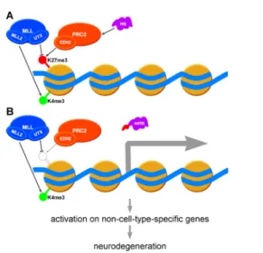
Figure 1 Activation of bivalent promoters might contribute to Huntington’s disease pathogenesis.
(A) Activating trimethylated-histone H3 lysine 4 (H3K4me3) (green semaphore disc) and silencing H3K27me3 (red semaphore disc) chromatin marks are simul- taneously present on bivalent promoters resulting in no or very low transcrip- tion. Activity of the Polycomb repressive complex 2 (PRC2) protein complex, that contains the EZH2 H3K27 specific methyltransferase subunit, is facilitated by Huntingtin. The MLL multiprotein complex antagonizes PRC2 by removing H3K27me3 by the UTX demethylase subunit and depositing H3K4me3 mark by the MLL2 methyltransferase subunit. (B) Data suggest that in Huntington’s dis- ease PRC2 activity might be debilitated resulting in reduced H3K27 trimethylation and consequent activation of bivalent promoters leading to expression of non-cell- type-specific genes and finally neurodegeneration. In a Drosophila model of the disease mutant Huntingtin induced neurodegeneration was enhanced by reduced Enhancer of zeste (E(z)) (homolog of EZH2) and suppressed by reduced Utx (ho- molog of UTX) and Trx (homolog of MLL2) supporting the above model.
[Downloaded free from http://www.nrronline.org on Friday, July 20, 2018, IP: 188.6.171.160]