Morphological reconsideration of the araphid genus Pseudostaurosira (Bacillariophyceae), a revision of Gedaniella, Popovskayella and
Serratifera, and a description of a new Nanofrustulum species
Eduardo A. Morales
1,2,3,*, Carlos E. Wetzel
3, Maria Helena Novais
1,2, Krisztina Buczkó
4, M. Manuela Morais
1,2& Luc Ector
31Water Laboratory, University of Évora, Rua da Barba Rala No. 1, Parque Industrial e Tecnológico, Évora 7005-345, Portugal
2Institute of Earth Sciences - ICT, University of Évora, Rua Romão Ramalho nº59, Évora 7000-671, Portugal
3Environmental Research and Innovation Department (ERIN), Luxembourg Institute of Science and Technology (LIST), 41 rue du Brill, 4422 Belvaux, Luxembourg
4Department of Botany, Hungarian Natural History Museum, Könyves Kálmán krt. 40, 1087, Budapest and MTA Centre for Ecological Research, Danube Research Institute, Karolina St 29, 1113 Budapest, Hungary
*Author for correspondence: edu_mora123@outlook.com
REGULAR PAPER
Background and aims – As a result of the description of many new species, reanalyses of type material, and information becoming available on valve morphogenesis in small araphid diatoms lacking a rimoportulae, the existing classification scheme at the genus level needs revision. Because morphological information has increased manyfold since the system provided by Williams & Round (1987), it may now be possible to find distinguishing characters in order to produce a more stable and useful framework, encompassing a morphogenetic perspective, which could then guide the placement of newly discovered taxa. This new framework could also be used to help assess the molecular information generated for the group, based on which many new genera are being erected, but perhaps without proper pondering of morphological data.
Methods – A thorough review was made of available published information on the ultrastructure of small- celled araphid diatoms lacking rimoportulae. In addition, image databases were searched, and new light and scanning electron microscopical observations made of some hitherto undescribed species.
Key results and conclusions – We provide a table of putative distinguishing features for nine genera (Nanofrustulum, Opephora, Pseudostaurosira, Pseudostaurosiropsis, Punctastriata, Sarcophagodes, Stauroforma, Staurosira and Staurosirella), together with a discussion on their value for discriminating these small diatoms using a morphogenetic perspective. Based on our findings, we amend the genus Pseudostaurosira, establishing wide and short vimines as its most characteristic feature. We use our system in describing a new species from Bolivia, which we place in Nanofrustulum based on its quasifract copulae, the distinguishing trait of the genus. The new species is distinguished from its congeners by its heteropolar valves, apical pore field features, and the multiseriate areolae. We also examine the three genera Popovskayella, Gedaniella, and Serratifera, the latter two recently erected based on molecular information. Since none of these latter genera pass the morphogenetic evaluation we think is essential, we place them in synonymy with other genera and provide the consequent nomenclatural changes. Finally, we make several new combinations in Nanofrustulum, Pseudostaurosira, Sarcophagodes and Staurosirella.
Key words – Araphids, Bolivian Altiplano, Fragilariaceae, molecular phylogenies, Pantocsek, systematics, type material.
© 2019 The Authors. This article is published and distributed in Open Access under the terms of the Creative Commons Attribution License (CC BY 4.0), which permits use, distribution, and reproduction in any medium, provided the original work (author and source) is properly cited.
Plant Ecology and Evolution is published by Meise Botanic Garden and Royal Botanical Society of Belgium ISSN: 2032-3913 (print) – 2032-3921 (online)
INTRODUCTION
While the many features currently used for small araphids lacking rimoportulae might be appropriate for species level delimitation, synapomorphies at the genus level are more complicated to single out (see Williams 2011, 2013 and Kociolek & Williams 2015). This difficulty has generated an alternate view to the taxonomy proposed by Williams &
Round (1987), which is expressed, for example, in Lange- Bertalot (1989, 1993), Krammer & Lange-Bertalot (1991) and Ognjanova-Rumenova et al. (1994). This alternative has gained partial support from molecular analyses (Medlin et al.
2012). Nevertheless, the Williams & Round (1987) proposal, based completely on morphology and later complemented with additional genera (Stauroforma Flower, V.J.Jones &
Round, Pseudostaurosiropsis E.Morales and Sarcophago
des E.Morales), has become widely used. Perhaps, one of the reasons for acceptance comes from the fact that the mor- phological diversity found in samples can be relatively eas- ily ascribed to these genera. This, of course, is helpful when diatoms are used in applied fields such as bioindication.
While molecular analyses strive to seek the best way to ex- press evolutionary relationships among existing genera (e.g.
Medlin et al. 1993, 2008, 2012, Medlin & Desdevises 2016), morphological data continue to be the most trusted basis for identifying taxa and indeed for classifying them, as shown through the plethora of new taxa described in recent decades using a combination of light (LM) and scanning electron mi- croscopy (SEM) (e.g. Witkowski et al. 2010, Morales et al.
2012, Grana et al. 2018).
In the last 30 years, the number of araphid species has increased significantly (Guiry & Guiry 2018). Newly de- scribed features have broadened the range of morphological characters used for genera and species delimitation. There- fore, there is now a need to start modifying the definitions of taxa in order to reflect what is known about their morpholo- gy (Morales & Manoylov 2006b, Morales et al. 2012, García et al. 2017). One of the genera in need of such reconsidera- tion is Pseudostaurosira D.M.Williams & Round in which at least 40 species, some new, others transferred from Fragi
laria Lyngb., Opephora P.Petit and Staurosira D.M.Williams
& Round have been included (e.g. Morales & Edlund 2003, Cejudo-Figueiras et al. 2011, Kociolek et al. 2014, Kulikovskiy et al. 2015, García et al. 2017). Taking as a basis the SEM analysis of type material of the generitype, Pseu
dostaurosira brevistriata (Grunow) D.M.Williams & Round (Morales et al. 2015), and published SEM information for the majority of species currently ascribed to this genus, it is now possible to reassess the original protologue and delimit the genus in a more concise manner. Among the features that have been better revealed by studies published after Williams
& Round (1987) are, for example, the spines, which are now known to be absent in some species (e.g. Pseudostaurosira parasitica (W.Sm.) E.Morales; Morales et al. 2015: figs 95–98), growing on the virgae in others (e.g. Pseudostauro
sira pseudoconstruens (Marciniak) D.M.Williams & Round;
Marciniak 1986: pl. 6, figs 229–232), or even occurring along the striae in some others (e.g. Pseudostaurosira micro
striata (Marciniak) Flower; Marciniak 1986: pl. 6, figs 233, 234). Among the totally new features are the flaps covering
the areolae externally (Morales et al. 2012: figs 45, 52, 53) and the stipulae at the base of spines (Morales et al. 2012:
figs 46, 50).
But even with all this new information on Pseudostauro
sira, it remains difficult to find derived characters that would unify all the species within the genus and at the same time separate them from other araphids without rimoportulae.
Certainly, the characters mentioned before (spines and their associated structures) or even the areolae and their substruc- tures (volae, rotae and flaps), the apical pore fields, and the girdle bands, which are traditionally used to separate small araphid genera, are not helpful in this task. Herein, we at- tempt to establish distinguishing traits through an analysis of features that are less frequently mentioned in araphid taxon- omy and systematics, and also try to incorporate a morpho- genetic perspective following the recommendations of Cox (2004, 2012) and Mann (1984, 2006).
During our search for SEM information to build a com- parative table of distingushing features, we reviewed several new genera that were erected in recent years, such as Popov
skayella Kulikovskiy & Lange-Bertalot, Serratifera Chun- lian Li, Witkowski & Ashworth and Gedaniella Chunlian Li, Witkowski & Ashworth, and revise them together with some other araphid species, based on the discriminating charac- ters we propose, making all the required nomenclatural rear- rangements.
We also describe a new species in Nanofrustulum Round, Hallsteinsen & Paasche from the Andes of Bolivia. This is a small-valved diatom that has unusual features and that we use to test our morphological feature analysis.
MATERIALS AND METHODS
The search for characters for all currently described genera of small fragilarioids lacking rimoportulae was done on the basis of published information (cited throughout the manu- script) and through examination of image databases at the Luxembourg Institute of Science and Technology (LIST) and the Water Laboratory, University of Évora, Portugal.
In addition, two samples were analysed specifically for the present work. The material used for the description of the new Nanofrustulum was an epipsammic sample collected from the Desaguadero River, Bolivia, described in Morales et al. (2012). The second sample was diatomite material from Lutila (Hungarian Natural History Museum, HNHM-ALG- DC-0092), Slovakia, described in Morales et al. (2014b).
For LM analyses, an aliquot of the Bolivian material was boiled for 45 min in 70% nitric acid (1:1 by volume with the sample). The mixture was then rinsed with distilled water until neutrality. Permanent slides were mounted using Naph- rax (Brunel Microscopes, Chippenham, UK) and analysed using a Zeiss Universal microscope (Zeiss, Jena) fitted with DIC (Differential Interference Contrast) and a Spot Insight 2 model 18.2 colour digital camera (Diagnostic Instruments Inc., Michigan). Microphotographs were taken at 2000× us- ing a Zeiss PLAN 100×, 1.25 N.A., immersion objective and SpotSoftware version 4.6 (Diagnostic Instruments, Michi- gan). For the Lutila material, a rehydrated raw sample was dried, mounted in Zrax (product no longer available) and an-
alysed with a LEICA DM LB2 microscope (Leica Microsys- tems, Wetzlar), equipped with a FinePix S2 Pro digital cam- era (Fuji Photo Film, Tokyo), at 1000× using a Leica DMLB 100×, 1.40 N.A., oil immersion objective.
For SEM analysis, a 2 ml subsample of the Bolivian clean material was heated with 100 ml of hydrogen peroxide (35%) for 36 h in a sand bath at 210°C. The supernatant was then removed and 1 ml of hydrochloric acid (37%) added, the material allowed to rest for 2 h, and subsequently rinsed with distilled water until neutrality. The Lutila diatomite was used untreated. All materials were then rinsed with deionized water over 3-µm glass fibre filters. Specimens were coated with platinum using a BAL-TEC MED 020 Modular High Vacuum Coating System (Leica Microsystems, Wetzlar) for 30 s at 100 mA and observed with a Hitachi SU-70 electron microscope (Hitachi, Chiyoda), operated at 5 kV and 10 mm working distance. All micrographs were digitally manipulat- ed and plates containing LM and SEM pictures were mount- ed using CorelDraw X6®.
Morphological terminology follows Barber & Haworth (1981) for valve shape and stria pattern; Cox & Ross (1981) and Cox (2012) for lateral extensions from the sternum (or annulus) (virgae) and cross bars developing later (vimines and viminules) that delimit the areolae; and Williams &
Round (1987) and Round et al. (1990) for areolar substruc- tures, spine features, apical pore fields and girdle bands.
RESULTS AND DISCUSSION Finding distinguishing characters at the genus level The analysis of several hundred SEM images of various de- scribed and potentially new taxa from all continents yielded the list of potential traits displayed in table 1. This table was constructed with difficulty due to the large volume of infor- mation available, but also because current genus descriptions in the literature are ambiguous and do not present clear-cut boundaries. The existence of a range of opinions regarding the features to be used for identification and classification of known species is also confusing (for example, see discussion in García et al. 2017, for Staurosira Ehrenb.). Also, many of the species names that are currently used refer to several entities that are morphologically similar (i.e. they belong to complexes) but that are clearly differentiated when SEM in- formation is used (Morales et al. 2013, 2014a). In addition, some widely used reference works (e.g. Metzeltin & Lange- Bertalot 1998) do not include a thorough argumentation, a necessary step in building a strong taxonomic basis (Silva in Hegewald & Silva 1988).
The analysed information shows that the characters tra- ditionally used to distinguish genera of small araphids lack- ing rimoportula (Williams & Round 1987) are in fact prob- lematic, since they are too variable within each genus and often overlapping among different genera. In the case of Pseudostaurosira; for example, Lange-Bertalot et al. (2017) stated that the “most characteristic feature” is the short stri- ae. While this may be true for the small and rather hetero- geneous (regarding spines, apical pore fields, etc.) group of species included by these authors, consideration of charac- ters in other species reveals that stria length is in fact one of
the most variable characters in this genus and that there are overlaps with species in Nanofrustulum, Pseudostaurosirop
sis and Sarcophagodes. It is therefore not surprising that the same authors consider Pseudostaurosira as “weakly circum- scribed” and that its generitype, P. brevistriata, is “a hetero- geneous group of species”.
More robust and natural classifications can be achieved by using features that are shared only among the members of a genus and that do not appear in any other groups (Kociolek
& Williams 2015). Such characters, synapomorphic for the species of the genus and autapomorphic for the genus, are difficult to recognize when limited morphological informa- tion is available. Fortunately, as many new species have been erected in recent years (e.g. Witkowski et al. 2010, Morales et al. 2012, Kulikovskiy et al. 2015) and type material has been reanalysed with both high resolution LM and SEM (e.g.
Edlund et al. 2006, Cejudo-Figueiras et al. 2011, Morales et al. 2015), the ranges of variation of many features have be- come clearer and the status of particular characters – whether or not they are synapomorphies – can be assessed. Herein, we present only a first step in the recognition of these unique characters that separate genera. Later on we will present tests to evaluate their status as synapomorphies.
It must be added that having a well-delimited generi- type gives an idea of the Bauplan (body plan) of that species and its evolutionary associates within the genus. However, as stated by Cox (2010), analysing characters that are fully formed can lead to a misappreciation of evolutionary rela- tionship, since the morphogenetic basis for character vari- ation remains hidden when only the end products of valve morphogenesis are considered. Therefore, pondering devel- opmental pathways underlying character formation may be useful to establish the nature of these characters, why they vary, and whether they are homologous.
Unfortunately, there are not very many studies of valve morphogenesis in araphid diatoms, certainly not sufficient to represent the great diversity within the group. But some in- formation available for diatoms in general lays a foundation for understanding the group of araphid diatoms treated here.
For example, Cox & Kennaway (2004) and Cox (2010) ana- lysed the origin of the sternum, virgae, vimines, viminules, areolae and associated structures, and the work of Kaluzh- naya & Likhoshway (2007) and Sato et al. (2008, 2009, 2011) showed how these features arise in the specific case of ara- phid diatoms.
Justification of the newly defined distinguishing characters
The characters contained in table 1 help to explain differ- ences among the nine genera described to date, excluding Gedaniella, Popovskayella and Serratifera. These characters are arranged roughly in the order in which they develop dur- ing valve morphogenesis. For example, areolar substructures (volae, rotae and flaps) are the last to form when the valve is being constructed and girdle bands form synchronously with, or towards completion of, valve formation, depending on the species; growth of areolar substructures begins when the areolae are already well defined (e.g. Sato et al. 2011). At least part of the variation in the process of valve construction
FeatureNanofrustulumOpephoraPseudostaurosiraPseudostaurosiropsisPunctastriataSarcophagodesStauroformaStaurosiraStaurosirella
Virgae shape doubly flareddoubly flareddoubly flareddoubly flareddoubly flareddoubly flaredrectangularsingly flared towards mantledoubly flared
Vimen shape
slender and short
without vimines wide and shortslender and shortslender and shortwide and longslender and shortslender and shortslender and long
Viminule prpresent in parts of oductionthe striae without viminules
without viminuleswithout viminulespr
esent in the entire striae without viminules without viminules
present in parts of the striaepresent in parts of the striae Areolae shaperound to transapically ellipticaltransapically ellipticalround to transapically ellipticalround
round to apically elliptical apically reniform round to apically elliptical
round to apically ellipticalapically oriented lineolae Areolae numberalways more than twoonetwo to manytwo to manyalways more than twotwo to manyalways more than twoalways more than twoalways more than two Striae type
uniseriate to multiseriate
uniseriateuniseriateuniseriatemultiseriateuniseriateuniseriate
uniseriate to multiseriate uniseriate to multiseriate
Volae origin inner perimeter of the areolavirgaeinner perimeter of the areolawithout volae inner perimeter of the areola inner perimeter of the areola inner perimeter of the areola inner perimeter of the areola
inner perimeter of the areola
present, originating Rotae prwithout rotaewithout rotaewithout rotaefrom a single point within the aroduction
eolawithout rotaewithout rotaewithout rotaewithout rotaewithout rotae
Girdle element structur
e
valvocopula: open, without fimbriae. copula: quasifract,
open. ligulae in valvocopulae and copulae morphologically
different
valvocopula: open, without fimbriae. copula: entire, open
valvocopula: open, without fimbriae. copula: entire, open
valvocopula: open, without fimbriae. copula: entire, open
valvocopula: open, with fimbriae. copula: entire, open
valvocopula: open, without fimbriae. copula: entire, open
valvocopula: open, without fimbriae. copula: entire, open
valvocopulae: closed, with fimbriae. copula: entire, open
valvocopula: open or closed, with fimbriae. copula: entire, open or closed
SEM refer
ences
Lee et al. 1980, Round et al. 1990, W
itkowski et al. 2010, Li et al. 2018
Round et al. 1990, Sabbe & V
yverman 1995, Li et al. 2018
Williams & Round
1987, Cejudo- Figueiras et al. 2011, Morales et al. 2015
Morales 2001, Morales 2002
Williams &
Round 1987, Hamilton & Siver 2008
Morales 2002
Flower et al. 1996, T
algatti et al. 2014
Williams &
Round 1987, Morales 2006
Williams &
Round 1987, Morales, 2006, Morales & Manoylov 2006a, 2006b
Table 1 – Comparison of genera of small-celled fragilarioid diatoms lacking rimoportulae. Bold indicates important distinguishing features.
could arise from interactions with the environment through selection and adaptation, generating reproducible, inheritable patterns of valve formation (see discussions by Mann 1999, 2010); at least some of these variations could represent fea- tures expressing evolutionary relationship among the differ- ent genera.
We hypothesize that the formation of striae begins soon after the formation of the virgae (in Opephora), vimines (in Pseudostaurosira, Pseudostaurosiropsis, Sarcophagodes and Stauroforma) and viminules (in Punctastriata D.M.Williams
& Round). We are herein using a restricted concept of Ope
phora, following Round et al. (1990) and Morales (2002). In exceptional cases, species of Nanofrustulum, Staurosirella, and Staurosira produce striae that are partially or completely multiseriate (Morales 2005), but in these genera the striae appear with the formation of the vimines, the production of viminules being rather sporadic and insufficient to character- ize these genera as multiseriate as a whole. Only in the case of Punctastriata are all the striae always and entirely multi- seriate invariably in all species.
During stria development, the areolae near the axial area form first and the ones near the valve margin form last (Cox 1999, 2010, Sato et al. 2011). In all the genera considered here, the areolar size increases toward the valve face-man- tle junction and from there to the margin the size decreases in size again. In the case of Opephora with a single areola running uninterrupted from the valve face to the mantle, the ends on both valve face and mantle taper so that a transapical elliptical shape is formed (Round et al. 1990: 382, 383, figs d–j). In genera having a multiseriate areolar pattern, there are fewer areolae towards the extremes of the striae, so more or less elliptical striae shapes are produced (see illustrations for Punctastriata in Round et al. 1990; the case of the new spe- cies placed in Nanofrustulum is discussed below).
Because of this difference in the diameter or number of the areolae within the striae, the virgae in the genera consid- ered should tend to be flared (i.e. they should become wider) at both ends; however, this is not always the case and genera tend to differ in specific patterns. We define the “shape of virgae” to be the geometrical pattern that the virgae assume in relation to neighbouring striae. Some taxa, such as Stau
roforma, always have rectangular virgae and this is because these structures are thickened and raised in internal and ex- ternal views, and remain somewhat independent from stria shape (Van de Vijver et al. 2002: pl. 15, figs 24–26, Genkal et al. 2011: pl. 19, figs 11, 12). Staurosira also has a unique virga shape. Here there is an enlargement of the valve face areolae as the striae proceed towards the valve margin, but the influence of smaller areolae on the shape of the virgae is compensated by the elevation of the latter. On the valve mantle, the virgae are less raised and therefore the smaller areolae found at the end of the striae make these virgae ap- pear subtly flared (Lange-Bertalot 1989: pl. 5, figs 1, 2).
As stated before, there is an underlying mechanism for virga and areola formation, where the timing of areola pro- duction is the result of how the virgae and their associated structures (vimines and viminules) develop. The thickening of the virgae, as the striae and associated features develop, also has an influence on stria formation, especially on their
internal structure (Sato et al. 2011). The majority of genera possess a conspicuous depression along each stria, the end result of virgae and sternum reinforcing in a trans and per- valvar direction. The thickened structures grow up above the striae often giving the impression of a semi-chambered in- ternal structure like an alveolus (see for example Morales et al. 2015: fig. 141, for Pseudostaurosira; Round et al. 1990:
359, fig. E, for Punctastriata; Grana et al. 2018: figs 47, 48, 51 for Staurosira; and Morales et al. 2010c: fig. 47, for Stau
rosirella).
The features of the vimines and viminules determine the characteristics of the areolae they delimit, except in Opep
hora where the single areolar aperture runs from valve face to mantle. Here, therefore, the extension and shape of this single opening is determined solely by the degree of devel- opment of the virgae and sternum (Round et al. 1990: 382, 383, figs d–j). In the case of Staurosirella, the virgae are well separated from each other and the vimines are long (length expressed as the apical distance between virgae) and narrow, delimiting closely spaced, elongated openings or lineolae.
The length and reduced width of the vimines in Staurosire
lla are unique, while in the case of Pseudostaurosira and Sarcophagodes the vimines are also long and wide, but they are thicker (thickness expressed as the transapical distance between areolae) in the former. The remaining genera either lack vimines or have them slender and short.
As stated previously, the production of viminules is con- stant in all species of Punctastriata, therefore this is a defin- ing characteristic for this genus. Although Nanofrustulum, Staurosira and Staurosirella also produce viminules, in this case the biseriate nature of the striae is expressed only on portions along the same striae or on an entire stria within the same valve (Lange-Bertalot 1989, Morales 2005). Again, since there is an underlying ontogenetic mechanism and the end products are different, areolar shape and the varying fea- tures of vimines and viminules can be used as distinguishing features.
The stated differences in the formation of the virgae also determine the sites of production of structures within the are- olae (Sato et al. 2011, Cox 2012). The volae arise from the virgae in Opephora and define and delimit the areolar open- ings (Round et al. 1990: 382, 383, figs e, f), while in the rest of the genera volae are either absent (e.g. Pseudostaurosi
ropsis: Morales 2001: figs i, j) or they are born from the in- ner perimeter of the areola, either on vimines or viminules (e.g. Pseudostaurosira: Morales et al. 2012: fig. 48). Rotae are produced only by Pseudostaurosiropsis (Morales 2002:
pl. 2, figs 5, 6) and originate from a single point, usually near the base of a spine. In areolae that are not in close proxim- ity to a spine, the rotae arise from a point on the side closest to where the spine is located (Morales 2001: figs 7a–d, g).
The points of origin of the rotae are especially clear when the rotae are beginning to be formed or when they are somewhat eroded (Morales 2001: figs 7e, i, j). Rotae can be confused with ‘flaps’, but flaps originate from the base of spines or from other regions around the areolar perimeter, and a single areola can have several flaps (Morales et al. 2012: fig. 45).
Flaps also differ from rotae in that they are produced on top of volae (Morales et al. 2012: figs 51–53), whereas rotae de-
velop in isolation within the areolar opening (Morales 2001:
figs 7i, j).
Volae are very similar in the eight genera included in ta- ble 1 (excluding Pseudostaurosiropsis). The only difference is in the degree of thickening and development. In general, these are highly (dichotomously) branched structures that originate from the inner areolar opening, at different points in Pseudostaurosira (Morales 2003: figs 60–65). Smaller, thinner and less branched volae are produced in Punctastri
ata (Lilitskaya 2016: pl. 4, fig. 2), while larger, thicker, and profusely branched are produced in the large-valved species of Pseudostaurosira (Morales & Edlund 2003: figs 45–50).
Although there is not sufficient information on girdle band formation to permit any generalization, as shown by Tiffany (2015), these structures begin their formation early in valve development, but the end of the process terminates after the margin of the valve has formed. The latter begins its solidification as the last mantle areolae complete their defini- tion, once the distal ends of the virgae fuse (Sato et al. 2011).
Though there are no specific studies for these rimoportula- lacking araphids showing the formation of individual girdle elements, it is known that for diatoms, in general, each of these elements forms within its own silica deposition vesi- cle and that there are differential interactions with previously formed parts of the valve, i.e. the mantle as a whole, com- posed of virgae and striae (Mann 1984, Round et al. 1990).
Therefore, there is a morphogenetic basis for the structural difference between those girdle elements. The valvocopula, for example, has a pars interior that is recessed to accom- modate the valve mantle and to attach internally to it, some- times directly (as in, for example, Nanofrustulum, Opephora, Pseudostaurosira; e.g. Morales et al. 2012: fig. 49), other- wise by means of delicate, narrow extensions called fim- briae, that attach to the internal faces of the virgae (as in, for example, Staurosira and Staurosirella; Morales 2005: fig.
103). The rest of the girdle elements in small araphids lack- ing a rimoportula tend to be plainer, but they can be open or closed. Sometimes, the same girdle is composed of open and closed bands as in the case of Staurosira (Morales 2006), or the girdle may contain all closed or all open elements, as in Staurosirella (Morales 2006). In other cases, the morpho- logical difference in the copulae is even more extreme, as in Nanofrustulum, in which the copulae are much smaller than the valvocopulae, arranged around the girdle and attached to other copulae by well developed ligulae (Round et al. 1999, Morales 2001, Witkowski et al. 2010, Wetzel et al. 2013).
Such differences among elements of the same girdle are not exclusive to small araphids. For example, the large araphid Glyphodesmis Grev. (Round et al. 1990: 240, 241, figs c, i, j) and the raphid Tursiocola R.W.Holmes, S.Nagas. & Takano (Frankovich et al. 2015) also have these differences between valvocopulae and copulae.
This view (that the nature of the girdle bands is variable among closely related taxa) conflicts with the current consid- eration of the open and closed nature of girdle bands as a dis- tinguishing feature in Fragilaria and Ulnaria (Nitzsch) Com- père (Compère 2001) (closed in the latter) (Williams 2011).
Molecular phylogenies using four genes show a weak differ- ence between the two genera (2.5%), insufficient to justify a morphological separation based on known characteristics, as
stated by Medlin et al. (2012). The latter authors also stated that looking to other sources of information such as repro- duction might reveal further differences. Until such studies are done, the open or closed feature of girdle bands remains a practical way of separating species in the two genera even though the synapomorphic nature of this feature is challenged from a morphogenetic standpoint. Besides, a wider analysis of other morphological features, in the light of the numerous new species that are being published for both genera, it could be possible to find synapomorphies; we reserve this task for later.
Are there any other distinguishing characters?
There are perhaps other sources of distinguishing features among the nine genera delimited in table 1. For example, morphogenesis of apical pore fields and spines might hold useful information. We have detected several variations in the structure of apical pore fields such as presence/absence, external and internal depressions, internal and external troughs, rims surrounding the pores, etc. It is known that api- cal pore fields appear earlier in the formation of the valve of araphids possessing a rimoportula, and they complete their formation before the completion of silicification of the valve (Tiffany 2002, Kaluzhnaya & Likhoshway 2007, Sato et al. 2011). However, the process of formation of associated structures such as the troughs seen in Pseudostaurosira pa
rasitica (Morales 2003), the “volcano” shaped domes seen in individual poroids of Stauroforma inermis Flower, V.J.Jones
& Round (Morales 2001), or the whitish rims frequently seen in species of Staurosira and Staurosirella, remain unknown.
Likewise, there are several character states for spines in araphids, which seem to be interesting from the ontogenetic standpoint. For example, certain taxa are reported as having hollow or solid spines (see discussion in Morales & Manoy- lov 2006b), some even have spines with a soft core (Morales et al. 2015: fig. 45). Yet it is difficult to determine whether spine features can be regarded as distinguishing at some level, simply because we do not know the mechanism of their formation and the actual processes determining whether spines are solid or lack a “filling” material altogether. It is possible that at least some of the reports of hollow or solid spines may be more related to stages of development than to actual character states (e.g. Morales et al. 2010b: figs 45, 47). Whether spines are formed in a fashion similar to centric diatoms, in which there is an active deposition of siliceous material guided from within the cell (Pickett-Heaps 1998), merits study. What is known thus far about cell wall forma- tion in araphids, however, suggests that a centric-like mecha- nism might be more likely when spines are produced along striae, since holes would be readily available for cytoplasmic expansion from within the cell. In the case of spines grow- ing on the virgae, it is surprising that one never finds open- ings within virgae in internal views of the valve face–mantle junction under SEM. Since sites of production of features such as areolae and apical pore fields are fixed within the organic apparatus that originates the new valves, it is logi- cal to assume that there are also sites for spine production.
The suppression of such sites would be a relatively simple evolutionary step, perhaps triggered by genetic, environmen-
tally-induced, or even mechanical influences. Explaining the change of location of sites for spine production from striae to virgae or vice versa appears more complex. Yet, there are genera that contain spiny and spineless species (e.g. Stauro
sira and Staurosirella; Morales & Manoylov 2006a, 2006b, Morales et al. 2010a) and species that have spines along the stria or growing from the virgae as in Pseudostaurosira.
Studying the mechanism of spine production-suppression, therefore, could elucidate evolutionary relatedness at differ- ent taxonomic levels.
We disregarded the features of the sternum as a source of distinguishing traits because the formation of the new valves begins with the formation of the sternum and the process seems to be common not only to all araphids, but to all pen- nates (Cox 2012).
Redefinition of the genus Pseudostaurosira Pseudostaurosira D.M.Williams & Round emend.
E.Morales
Generitype – Pseudostaurosira brevistriata (Grunow) D.M.Williams & Round.
Microscopical study of generitype material – Morales et al. (2015: figs 107–127 (LM), 128–143 (SEM)).
Description – Frustules symmetrical in side view, forming chains in taxa that possess spines. Taxa with incipient or no spines, presumably attached by mucilage stalks. Valves cru- ciform, bigibbous, lanceolate, rhombic or elliptic. Sternum of variable width and shape. Transition from valve face to mantle varies from abrupt to more gradual, with formation of a transition zone. Striae composed of one to (rarely) a few rows of wide, round, transapically elliptical, or irregu- larly polygonal areolae running from valve face to mantle.
Internally, areolae open into a single depression running from valve face to mantle, which may become chamber-like.
Volae highly branched, usually dichotomously, arising from the inner perimeter of the areolae and at different depths. Vir- gae rectangular or flared at their proximal and distal ends.
Vimines short and wide, the one at the valve face-mantle transition usually being wider, giving the impression of a single apical rib running along the valve face margin. Vimi- nules seldom produced and, when formed, occupying only a part of a stria. Flaps or flat siliceous growths originate from several points around the external perimeter of the areolae, close to their external surface. Concentric discs sometimes present, partially occluding the depression into which the areolae open internally. Solid spines can grow from the en- larged vimen that connects the virgae along the valve-face edge or from the virgae themselves, or they can be absent.
Spines of varying shape, but usually spatulate, with or with- out terminal branching, with a cylindrical base, and often bearing stipulae. Stipules produced near the base of spines and projecting downward. Apical pore fields absent, reduced or more fully developed, of the ocellulimbus type; in many cases they are sunken into the apical portion of the valve.
They are composed of round poroids, which are sometimes arranged along external troughs parallel to the apical axis of the valve. Mantle plaques present in many species and situ- ated along the abvalvar edge of the valve mantle. Cingulum
composed of a larger valvocopula and few to many ligulate copulae, always open, lacking fimbriae and perforations.
Distinguishing character – Wide and short vimines.
Though these vimines are also found in other genera having round areolae, such as Nanofrustulum (Li et al. 2018: figs 286–289), Pseudostaurosiropsis (Morales 2001: figs 7a–d, g, i, j, l) and Sarcophagodes (Morales 2002: pl. 5, figs 1–3), in Pseudostaurosira they are wider due to transapical expan- sion during valve ontogeny, and shorter due to the areolae being transapically expanded, thus having their shortest axis parallel to the longitudinal axis of the vimines (Morales &
Edlund 2003: figs 39, 45, Cejudo-Figueiras et al. 2011: figs 108–111, Morales et al. 2012: figs 42, 48).
Other salient, but not unique features – Rectangular or doubly flared virgae. The degree of flaring varies depending on the size reduction of terminal vimines (those located to- wards the axial area of the valve and towards the abvalvar edge of the mantle) and the shape of terminal areolae along a stria (Morales et al. 2015: figs 128, 129, 137, 141, 142).
The striae lie within an internal depression. This depres- sion varies among species from shallow (Witkowski et al.
2010: figs 50–55) to deep, the striae appearing semi-cham- bered (Morales 2002: pl. 4, fig. 2).
There is a high variability in spine presence, position and associated structures. Spines can be absent (Morales et al.
2015: figs 95, 96, 99, 100, 103, 104), or produced on virgae (Round et al. 1990: 356, 357, figs d, e) or on vimines (Ceju- do-Figueiras et al. 2011: figs 100, 101, Morales et al. 2015:
figs 130, 132, 135). Spines are always solid, although the presence of a soft core in some species gives the impression of spines being hollow after breakage and erosion (Edlund et al. 2006: figs 18, 20).
Stipules and flaps are produced. The stipules, originating at the base of the spines and projecting at an angle, away from the valve mantle, are a rather common feature among spiny species of Pseudostaurosira (Morales et al. 2012: fig.
50). The shape of these structures varies among species (Mo- rales 2002: pl. 4, fig. 6). Stipules are also present in other genera such as Nanofrustulum (Li et al. 2018: fig. 288, as
“lateral projections”) and Punctastriata (Williams & Round 1987: fig. 43; Flower 2005, fig. 6). Stipules are frequently helpful in the SEM identification of chains in valve view, which can easily be confused with chains of Staurosira, but where stipulae are always lacking.
Flaps originate from different points along the inner pe- rimeter of the areolae and sometimes resemble disks lying atop the volae. Morales et al. (2012: fig. 45) show a valve in which some mantle flaps are broken, while others (that origi- nated very close to the basis of the spines on the valve face) are still attached. Figure 50 of the same publication (Morales et al. 2012) is helpful in the distinction of stipules and flaps;
stipules can be seen projecting downward along the mantle and originate from the body of the spine.
At least two species (Pseudostaurosira decipiens E.Morales, G.Chávez & Ector and Pseudostaurosira laucen
sis var. vulpina Lange-Bert. & Rumrich) have been found to produce internal accumulations of siliceous material on the volae, depositions that appear as two concentric disks (Mo- rales et al. 2012: figs 40, 44; E. Morales, unpublished obs.).
The functions of these are unknown, but they can be used to help identify the two species. However, in P. laucensis var.
vulpina, eroded valves lack depositions (e.g. Rumrich et al.
2000: pl. 10, fig. 9).
Apical pore fields are variable among different Pseudos
taurosira. In this genus they invariably occupy the mantle portion of the apex and never subtend striae, unlike in e.g.
Staurosira (Morales et al. 2012: figs 57, 58, 62). Their devel- opment ranges from absent or present even in the same spe- cies (e.g. Pseudostaurosira sajamaensis E.Morales & Ector:
Morales et al. 2012 and may be slightly developed (e.g. P.
brevistriata: Morales et al. 2015) or fully developed (P. pa
rasitica: Morales 2003). Internally, poroids open into a sin- gle, circular depression, as seen in the type of P. brevistriata (Morales et al. 2015: figs 140–142), but this feature might also be present in species of Staurosirella (e.g. Kulikovskiy et al. 2015: pl. 10, fig. 22). In some cases there are external transapical troughs along which there are lines of pores (Mo- rales 2003: figs 54–58, 64).
Lange-Bertalot et al. (2017) considered that the width of the sternum is a diagnostic feature of Pseudostaurosira (see also the dichotomous key presented in Li et al. 2018). They even placed species such as Pseudostaurosira subsalina (Hust.) E.Morales in Staurosira due to their narrower axial area. There is no support for this, however, and such an area should be regarded as a variable character that can be diag- nostic, but at the species level, e.g. to distinguish Pseudostau
rosira elliptica (Schum.) Edlund, E.Morales & S.A.Spauld.
(Edlund et al. 2006) from Pseudostaurosira americana E.Morales (Cejudo-Figueiras et al. 2011) (see discussion in Grana et al. 2018). Pseudostaurosira and Staurosira each have their distinguishing features and are well delimited gen- era (table 1, and compare our emended description herein with the discussion in García et al. 2017). Moreover, the ster- num width is also variable in genera such as Staurosirella (Morales et al. 2010c) and Staurosira (Grana et al. 2018).
The placement of a newly discovered diatom in the nine genera scheme
Nanofrustulum rarissimum E.Morales, Novais, C.E.Wetzel
& Ector, sp. nov.
Figs 1 & 2
Type material – Bolivia, Desaguadero River, Department of Oruro (17°23′51″S, 68º14′33″W, 3701 m a.s.l.), 5 Jul. 2009, G. Chávez s.n. (holo-: ANSP, slide ANSP GC 26815 partially illustrated here in figs 1A–F (LM), 1G–K, 2A–D (SEM);
iso-: Diatomotheca Boliviensis, Cochabamba, Bolivia, slide DBOL-0246)
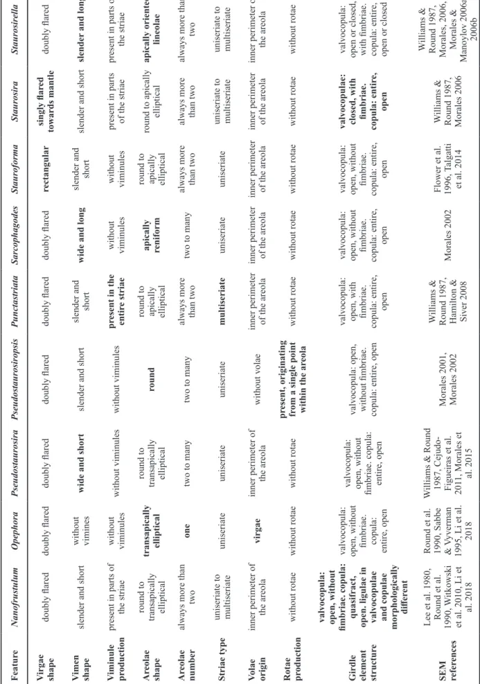
Description – Frustules rectangular in girdle view. Valves ovoid with broadly rounded head poles and narrower, almost cuneate foot poles (figs 1A–H, 2B & 2D). Length 7.3–9.5 µm, width 2.5–3.3 µm, stria density 13–14 in 10 µm. Ster- num narrowly lanceolate (figs 1A, 2B & D). Virgae doubly flared raised externally and internally (figs 1G, 2A, 2D).
Transition between valve face and valve mantle abrupt (figs 1G–K & 2A–C). Valve mantle edge parallel to valve face–
mantle junction (fig. 2A). Siliceous plaques along valve mantle edge present (fig. 2A). One to several rows of are-
olae present on the same stria, progressing uninterruptedly towards valve mantle (figs 1G–K & 2A–C). Areolae square, trapezoid or elliptical, apically or transapically elongated, all opening internally into a single depression (figs 1G–K &
2A–D). Volae robust relative to areolar opening and arising from the inner periphery of each areola (figs 1G, I–K, 2B
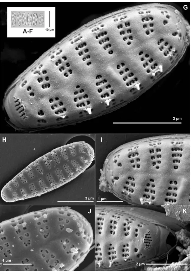
& 2D). Spines located on vimines, arising from at least two points along the width of striae (figs 1G–K, 2A & B). A wide, laminar, skirt-like stipula is present near the base of each spine (fig. 1G–K). Wart-like, whitish depositions sometimes present on the valve face, more numerous at the valve head pole (figs 1H, J & 2B). Apical pore fields composed of vari- able rows of poroids, present at both apices, but more devel- oped at the foot pole (figs 1G–I, 1K & 2B–D). Pore field at head pole located closer to the mantle abvalvar edge (fig. 1G, I & J), while foot pole pore field lies at the transition between valve face and mantle (figs 1G, H, K, 2B & D). Poroids of latter pore field sit within cavernous troughs carved deep into the valve (figs 1G, K & 2C). Girdle composed of an open, ligulated valcocopula lacking perforations, and quasifract, ligulated copulae, also lacking perforations (fig. 2A).
Etymology – “Rarissimum” refers to the fact that the fea- tures of the new taxon are rather infrequent within the genus.
Taxonomic remarks – Using traditional morphological characters, the new taxon shows several features that link it to Pseudostaurosira. The individual areolae bearing well developed volae, the well developed apical pore fields com- posed of round poroids, and even the position of these po- roids within troughs have all been described in other species of the genus. Regarding the areola structure, the new taxon resembles P. brevistriata. Observing figs 128–130 and 132 in Morales et al. (2015) and comparing with figures presented herein, the resemblance in areolar openings and the manner in which the volae are born and developing to the valve inte- rior can be seen. Likewise, figs 140–142 in the same publica- tion show how volae project into the valve interior and even collect extra siliceous material, similarly as what happens in the new Nanofrustulum species. Other features of the new taxon, similar to those in taxa currently ascribed to Pseu
dostaurosira, are the uneven development of the apical pore fields and the development of troughs at the foot pole, which have also been seen in Pseudostaurosira clavatum E.Morales (compare Morales 2002: pl. 4, figs 1, 4, with our figs 1G, K & 2C) and Pseudostaurosira perminuta (Grunow) Sabbe
& Vyverman (Sabbe & Vyverman 1995: figs 54, 57, 58), though not as developed as in the new taxon.
However, as shown in table 1, N. rarissimum has a fea- ture not present in Pseudostaurosira as defined here, namely quasifract valvocopulae (“segmented copulae” in Li et al.
2018), only present in representatives of Nanofrustulum.
Quasifract copulae are present in the type of the genus N.
shiloi (J.J.Lee, Reimer & McEnery) Round, Hallsteinsen &
Paasche (Round et al. 1999: fig. 8). Quasifract copulae are also reported for Pseudostaurosira cataractarum (Hust.) C.E.Wetzel, E.Morales & Ector (Wetzel et al. 2013: figs 2F, G), but this species needs to be transferred to Nanofrustulum (see below). Viminules are lacking in Pseudostaurosira and Nanofrustulum, as currently circumscribed, but the new tax- on produces them regularly. Due to the complexity of girdle band morphogenesis, the nature of this structure can be con-
Figure 1 – Nanofrustulum rarissimum, LM and SEM images taken from type material (ANSP GC 26815): A–F, size diminution series in LM; G–K, SEM images of external details; G & H, view of entire outer surface showing details of sternum, virgae, striae, spines and apical pore fields. Notice heterovalvarity, also expressed in the apical pore fields, and the skirt-like appearance of the stipulae; I & J, detail of head pole. Notice small apical pore fields near the abvalvar edge of the mantle, and the volae; K, detail of foot pole showing the apical pore field with deep throughs, the closed extreme of the valvocopula and ends of some copulae.
Figure 2 – Nanofrustulum rarissimum, SEM images taken from type material (ANSP GC 26815): A, detail of a broken frustule showing girdle structure. Notice entire valvocopula and quasifract copulae, as well as siliceous depositions or plaques on the abvalvar edge of the mantle; B & D, contrast of surfaces. Notice deposition of material on the volae in internal view, and the depressions in which the internal openings of the areolae are contained; C, detail of foot pole showing variability of areolar shape and infrequency of viminule formation.
sidered to have more weight as that of viminules, especially considering that viminule formation may simply require a mechanical reason , i.e. a wider spacing between virgae (Cox 2012). Therefore, N. rarissimum can be ascribed to Nanofru
stulum as the only taxon that produces viminules, thus far.
Another genus that produces viminules is Punctastriata.
However, the species of this genus have entirely multiseriate striae, with no partial production of viminules as in N. raris
simum. Additionally, Punctastriata lacks quasifract bands and its valvocopula have fimbriae.
The following combined features differentiating N. raris
simum from other taxa currently assigned to Nanofrustulum are: heteropolarity of the valves, production of developed apical pore fields and multiseriate areolae, the latter being the most prominent feature characterizing the new taxon. Anoth- er salient feature is the heteropolarity of the apical pore fields and the cavernous structure of the one at the foot pole.
The epipsammic sample from Bolivia in which N. raris
simum was found contains several new araphid taxa (Mo- rales et al. 2012). It is characterized by a high taxon richness (228 species and varieties), with a high proportion of un- knowns. Nanofrustulum rarissimum was rare in the material, and does not appear in a 500 valve count made by Morales et al. (2012: table 1). Analysis of nearby localities, other than the Desaguadero River (Morales, unpublished data) did not yield additional valves of the new taxon, suggesting that it is restricted to the river. One reason could be that the river is dominated by high sediment in the water and the entire river bed at the site of collection is dominated by sand and finer sediments, with no development of aquatic vegetation.
The characteristics of sites nearby are quite different, varying from small streams with high flow of clear water (with beds covered by rock, moss and other High Andean vegetation), to shallow lentic systems that are ephemeral or permanent and fed by ice melting or ground water (with emergent vege- tation and with much lower deposition of fine sediments and sand). These latter waterbodies are strongly affected by met- amorphic rock due to their proximity to the Sajama volcano, while although the Desaguadero has the same rock bottom, it is isolated under several metres of fine sediments on top.
The richness of diatom taxa in the Desaguadero River sample is not uncommon for samples collected in the pristine area of the Titicaca-Sajama Volcano region, far from human- related activities (Morales et al. 2012, 2014c) and contrasting with the European view of the Andean diatom flora (Metzel- tin & Lange-Bertalot 1998, Rumrich et al. 2000). The con- trast could be explained by the areas that have been common- ly collected by foreign scientists, which tend to be located more along roads and close to populated areas, where human activities affect ecosystems and tend to make habitats more uniform and similar in the assemblages they contain (Morales et al. 2012, Goldenberg Vilar et al. 2014).
Assessing the validity of Popovskayella
SEM images of Popovskayella from Lake Baikal are reminis- cent of Pseudostaurosira. In fact, Kulikovskiy et al. (2015) state that the latter is the most closely related genus. The distinguishing features of Popovskayella are the presence of
“one apical row of very small areolae” (which we interpret
as one row on the valve face and one on the valve mantle on each stria, based on the fact that none of its species has a sin- gle areola composing the striae), the lack of branched volae, the presence of three areolae on the transapical striae, and the presence of a single internal groove running from valve face to mantle, through which, the external openings of individual areolae can be observed. The protologue also mentions the presence of sponge-like silica membranes. Let us consider each of these features.
Because striae are formed independently from each other during valve ontogeny, it is difficult to conceive the exist- ence of “apical striae”. That is, in araphid diatoms striae are morphogenetically transapical structures. Therefore, it does not seem useful, at least for small araphids, to consider that areolae are arranged in apical rows and use this as a taxo- nomic criterion. What is more, the single apical alignments of areolae (one on valve face and one on valve mantle) are also present in several representatives of Pseudostaurosira, such as Pseudostaurosira tenuis E.Morales & Edlund (Mo- rales & Edlund 2003), Pseudostaurosira elliptica (Schu- mann) Edlund, E.Morales & S.A.Spauld., Pseudostaurosira microstriata (Marciniak) Flower, and the generitype Pseu
dostaurosira brevistriata (Grunow) D.M.Williams & Round (Morales et al. 2015). The areolar opening in the latter taxon is much wider than in any of the Popovskayella taxa, but areolar diameter is not a distinguishing character since all the genera in table 1 vary widely in the size of the areola opening. Regarding volae, it is difficult to make out why Ku- likovskiy et al. (2015) stated that there are no closing plates since these structures occur in the generitype Popovskayella nanobaculum Kulikovskiy & Lange-Bert. (Kulikovskiy et al.
2015: pl. 11, fig. 15), Popovskayella simplex Kulikovskiy &
Lange-Bert. (Kulikovskiy et al. 2015: pl. 12, fig. 8), Popov
skayella minutula Kulikovskiy & Lange-Bert. (Kulikovskiy et al. 2015: pl. 13, fig. 8) and Popovskayella tenerrima Ku- likovskiy & Lange-Bert. (Kulikovskiy et al. 2015: pl. 14, fig.
8). These volae can obviously not be highly branched since the areolar aperture is narrow in all these taxa, impeding the profuse development of volae, but it is evident that they are in fact dichotomously branched (e.g. see Kulikovskiy et al.
2015: pl. 13, fig. 8).
What Kulikovskyi et al. (2015) regarded as a single are- ola in internal view is in fact the extreme expression of the depressed striae that are present in all nine genera in table 1. What they term an areola in internal view does not cor- respond to such a structure from a morphogenetic perspec- tive. Areolae are formed by production of vimines, which in conjunction with the virgae delimit the areolar openings. A single internal opening, therefore, corresponds to a depres- sion where individual areolae open to the valve interior, in this case much deeper due to heavy silicification of sternum and virgae. Because of the small size of the valves in all the Popovskayella species, it is expected that the stria depression becomes deeply marked as the sternum and the virgae thick- en in a transapical and pervalvar direction. In small-valved taxa of Pseudostaurosira such deep depressions can be seen, as in, for example, Pseudostaurosira microstriata (Marcini- ak 1982: pl. 2, fig. 6) and Pseudostaurosira tenuis (Morales
& Edlund 2003: fig. 41).
The sponge-like silica membranes seem to be a misinter- pretation of the structure shown in Kulikovskiy et al. (2015:
pl. 12, fig. 5). This figure shows small volae projecting in- ward from the inner periphery of the areolae. The apical por- tions of the valve in this figure are covered by material which could correspond to excess amounts of the coating applied for SEM analysis or to an organic layer impregnated with mineral depositions, frequent in material that is only partially clean.
Using the information for Pseudostaurosira in table 1, we conclude that Popovskayella species are in fact members of the former genus and should be transferred to this genus (see section New combinations). They have as distinguishing features wide and short vimines, they lack viminules, their areolae are round to transapically ellipsoid and arranged in a single series along the striae. The volae originate from the inner perimeter of the areolae, and there is no evidence of rotae. Unfortunately, there is no information on girdle band structure for any of the species.
Evaluation of genera based on molecular information The molecular phylogenies presented to date for small ara- phids lacking rimoportulae are based on very limited taxon sampling. Genera such as Pseudostaurosira, Staurosira and Staurosirella now contain dozens of taxa (Guiry & Guiry 2018) and including even ten species per genus in any phy- logeny (many are currently represented by only one in most trees) might still be unrepresentative of the larger groupings.
Thus far, the separation of small araphids from the larger fragilarioids having a rimoportula seems to be convincing- ly resolved; this seems to hold regardless of the genes used (Medlin et al. 2008). A more recent tree, used for many im- portant taxonomic decisions (Li et al. 2018), does not offer better resolution than that of older phylogenies.
Ninety-nine strains were used by Li et al. (2018: table 3), representing 33 taxa (six of which are unknown) in eight known and one unknown genera. Eight of the nine genera included in our table 1 appear in that phylogeny (Li et al.
2018, S1), where they are represented by ten known species (with several strains) and nine undetermined strains. Of the strains used, 119 belong to eightteen genera, but twelve of them are represented by three strains or less. Although the phylogeny presented by Li et al. (2018) is informative, taxon sampling can be a serious shortcoming in any phylogenetic analysis (Theriot 2008, Theriot et al. 2010) and perceptions of relationships may change drastically as more taxa and genes are added (Sato et al. 2008, Theriot 2008). Addition- ally, the approach adopted by Medlin et al. (2012), Medlin &
Desdevises (2016), and Li et al. (2016), which all have tried to fit morphological data into inferred molecular phylogenies a posteriori is risky (Theriot 2008, Williams 2013), because phylogenies and morphology are not treated using a com- mon methodology, or because morphological data are not analysed at all before they are incorporated into phylogenetic reconstructions. This may explain why the morphogenetic approach we follow here, yielding a clear-cut pattern among the nine genera presented in table 1, appears to be in conflict with the conclusions of previous authors (Medlin et al. 2012, Medlin & Desdevises 2016, Li et al. 2018).
Superimposing ecological information a posteriori onto phylogenies also requires further and careful consideration.
For example, Medlin & Desdevises (2016) stated that the members of their new family Staurosiraceae (comprising Nanofrustulum, Opephora, Plagiostriata and Staurosira) are bottom-dwelling or planktonic, never epiphytic. How- ever, Frenguelli (1945) reported that, for example, Opephora schwartzii (Grunow) P.Petit ex Pelletan grows on marine littoral algae, and Sullivan (1978) listed the same species and two additional ones in the same genus from salt marsh spermatophytes. We add that ecological information should also be analysed using a common methodology, before being added to phylogenetic interpretations.
Williams (2013) took the phylogeny-morphology dis- cussion a step further and expressed that the phylogenies for araphid diatoms presented in the literature do not pro- vide evidence for the nodes formed during tree construction.
For us, this deficiency is hard to justify given the amount of available morphological information on fossil and extant araphid diatoms. Indeed, the simultaneous treatment of mor- phological and molecular data is possible as demonstrated by Frankovich et al. (2018), and this approach might produce a more parsimonious consideration of species and their rela- tionships.
Perhaps, one of the most conspicuous shortcomings in the a posteriori attempt to merge morphological and molec- ular data is that the morphological characterization of taxa used in phylogenies is poorly done. In Li et al. (2018), for example, the size diminution series for taxa presented in figs 39–129 contains a mixture of morphological variants that do not seem to fit with each other. Since the authors stated that all microscope analyses were performed using old and new cultures, as well as field collected material, their arrangement of LM photographs and SEM plates seems rather haphazard.
This of course, is counterproductive for users of plates and figures at the bench, with the risk of producing misidentifica- tions and ecological misinterpretations. This is precisely one of the implications of Theriot’s (2008) and Williams’ (2013) reasoning: the degree of attention given to the molecular part of these studies is not the same as that given to the morpho- logical data, to the point that it seems rather impractical (and risky) to join them together.
As stated before, the large amount of work done on anal- yses of type material of many araphid species has greatly clarified the boundaries of taxa widely cited in the literature and used for applied purposes (e.g. Morales et al. 2015).
Therefore, the rather hasty use of names for identification of strains used for phylogenetic reconstruction (e.g. Medlin et al. 2008) is completely unjustified (see also argumentation by Williams 2013). Thus, it seems that investing more effort in the so called “total evidence approach”, combining molec- ular, physiological and ecological information, together with data from nomenclatural types, would add value to current phylogenies.
Clearly then, the elucidation of molecular phylogenies and the search of robust trees that would indicate stable, trustable and statistically plausible relationships among taxa is work in progress. Meanwhile, it is possible to turn our eyes to alternative, equally trustable and testable hypotheses that