CORONAVIRUS INFECTION RETARDS THE DEVELOPMENT OF THE CORTICO-MEDULLARY CAPILLARY NETWORK
IN THE BURSA OF FABRICIUS OF CHICKEN
Attila FARSANG1#*, Ildikó BÓDI2, Orsolya FÖLKER2, Krisztina MINKÓ2, Zsófia BENYEDA3, Ádám BÁLINT4, Anna L.KISS2 and Imre OLÁH2
1National Food Chain Safety Office, Directorate of Veterinary Medicinal Products, Budapest, Hungary; #present address: Ceva-Phylaxia Co. Ltd., Szállás u. 5, H-1107 Budapest, Hungary; 2Department of Anatomy, Histology and Embryology,
Faculty of Medicine, Semmelweis University, Budapest, Hungary;
3Prophyl Ltd., Mohács, Hungary; 4National Food Chain Safety Office, Veterinary Diagnostic Directorate, Budapest, Hungary
(Received 26 September 2017; accepted 12 February 2018)
Coronavirus infection delays the development of the cortico-medullary (CM) capillary network which results in retarded development of bursal follicles.
The smaller size of the medulla in the coronavirus-infected birds may lead to a lower number of B lymphocytes and bursal secretory dendritic cells, which nega- tively affects the reactivity and efficacy of the immune system. Contrary to the wild-type infectious bronchitis virus (IBV) strain, infection induced by H120 vac- cine virus exerts only a moderate influence on caveolin-1 expression of the CM capillary web and on follicular development compared to the untreated controls.
Key words: Coronavirus, cortico-medullary capillary network, caveolin-1, retarded development, bursa of Fabricius, chicken
The rudiment of the bursal follicles (medullary part of the follicle) emerg- es during embryonic days 11–14. The bursal epithelium produces 12–15 plicae, and in the centre of each plica there is a middle plical vessel (MPV). Small inter- follicular arterioles from the MPV give offshoots to the developing follicles (Schoenwolf et al., 1981) which give rise to cortico-medullary (CM) capillaries (Abbate et al., 2007). During the development of the epithelial bud, the CM ca- pillaries form a chalice-like capillary web at the CM border. The development and role of the bursal follicles depend on the state of the CM capillary network, which distributes oxygen, nutritive materials, hormones and signalling molecules to tissues and also collects metabolic waste products. It is a dynamic organ, which is able to react and adapt rapidly to internal and external demands. The physiological importance of capillaries is well known as most of the exchange of water and respiratory gases takes place through the capillary wall via transcytosis
*Corresponding author; E-mail: attila.farsang@ceva.com
in the endothelial cells (van Deurs et al., 2003; Navarro et al., 2004). Transcyto- sis is based on the caveola, which is a flask-like invagination of the cell mem- brane (Pelkmans et al., 2001). Caveolin-1 (Cav-1), an integral membrane protein, is associated with cholesterol in 1 to 1 ratio. Recently it has been published that Cav-1 and cholesterol are expressed in the interfollicular epithelial cells and in the CM capillary web (Bódi et al., 2018).
Coronavirus (infectious bronchitis virus, IBV) infection mainly causes up- per respiratory disease in chickens (King and Cavanagh, 1991; Farsang et al., 2002), but the virus can also replicate in other non-respiratory epithelial cells (Dhinakar Raj and Jones, 1996; Benyeda et al., 2009). Information about the ef- fect of coronavirus infection on the lymphoid organs of chicken is very limited as the research on IBV has been focused on the main clinically affected organs such as the respiratory tract, the kidney and the genital organs. Bursal growth is very fast during the post-hatch period, which requires a continuous re-shaping of the CM capillary network. In this work we investigated the effect of IBV infec- tion using virulent IBV field isolates and H120, a common Massachusetts-type vaccine strain (de Wit et al., 2011) on the development of bursal follicles, which might also have immunological implications.
Materials and methods
Birds
One-day-old specific pathogen free (SPF) White Leghorn chickens (Gal- lus gallus) were obtained from Prophyl Ltd. (Mohács, Hungary) and placed in an isolator (Montair HM 1500, Koenenberg, The Netherlands). Feed and drinking water were provided ad libitum. All studies were conducted according to Di- rective 2010/63/EU and the Hungarian national regulations about animal welfare and ethics [Act 1988/XXVIII modified by Act 2011/CLVIII and Conduct of An- imal Trials (Government Regulation 40/2010)]. The National Food Chain Safety Office is authorised by the Pest County Government Office to perform animal tests for experimental purposes. Three test groups (containing 12 birds each) were formed, together with two untreated control groups for monitoring the de- velopment of the cortico-medullary capillary network. The control groups con- sisted of 20-day-old embryos as well as 1- and 8-day-old birds. Ten one-day-old birds were oronasally infected with 0.05 ml of QX strain isolate and the H120 vaccine strain. The same volume of phosphate-buffered saline (PBS) was applied oronasally to the control birds (n = 10).
Virus isolates
Two field isolates (strains 11518 and 11638) and a vaccine strain (H120) of IBV were used for the treated groups. The isolates proved to be QX strains based on genetic analysis of the whole S1 gene (Kiss et al., 2015). IBV field iso- lates were provided by the Veterinary Diagnostic Directorate of the National Food Chain Safety Office, while the vaccine strain originated from the veterinary vaccine bank of the Directorate of Veterinary Medicinal Products. Virus titres were determined by egg titration of all IBV isolates and H120. Briefly, serial di- lutions of each examined infectious material were prepared. A 0.1-ml volume of diluted virus was inoculated in the allantoic cavity of 9-day-old embryonated eggs. Eight embryonated eggs per dilution were used. The titres were as follows:
106.77 EID50/0.1 ml (strain 11518), 102.7 EID50/0.1 ml (strain 11638), and 106.36 EID50/0.1 ml (strain H120).
Immunohistochemistry
Tissue samples of the bursa of Fabricius were embedded in liver and fro- zen in liquid nitrogen. Briefly, liver embedding began with labelling an approx- imately 1.5 × 1.5 cm2 piece of cardboard with the necessary information. After labelling, the other side of the cardboard was covered with a flat piece of liver cut from the same experimental bird. The tissue samples were placed on the liver and covered by the rest of the liver to protect the samples from sudden cold.
Before starting the experiment, a thermo-flask (about one litre in volume) was filled with liquid nitrogen, and a piece of polystyrene foam (1 cm thick and 8–10 cm in diameter) was placed on the surface of the liquid nitrogen. The float- ing polystyrene foam cooled down in a few minutes. The liver-embedded tissue samples attached to the cardboard were placed onto the floating polystyrene foam. When the blocks became frozen (the colour of the liver changed) and tilted the polystyrene, the block slid into the liquid nitrogen. The blocks were then put in a capped tube, which was stored at –80 °C until cryostat sectioning.
The 10-µm cryostat sections were fixed in cold acetone for 15 min and re- hydrated in PBS. To detect caveolin expression, the sections were incubated with primary antibody (Polyclonal Rabbit Anti-Caveolin, BD Transduction Laborato- ries) at room temperature for 45 min. After washing with PBS, isotype-specific biotinylated secondary antibodies were applied. The endogenous peroxidase re- action was blocked with 3% H2O2 for 10 min. An ABC kit was used to visualise the signals of primary antibodies. The binding sites of primary antibodies were detected with 4-chloro-naphthol. For negative control staining, the primary or secondary antibodies were replaced with PBS.
Fig. 1. Twenty-day-old chicken embryo. The bursal lumen between the plicae is very narrow (arrow). In the axis of the place the middle plical vessel cannot be distinguished from the fibroblasts expressing caveolin-1 (Cav-1;
arrowhead). Higher magnification of follicle shows the budding of cortico-medullary (CM) capillary (inset).
Fig. 2. One-day-old control chick. The bursal lumen is ‘opened’ (arrow). The interfollicular arterioles in the connective tissue are heavily stained with anti-caveolin-1 antibody. Scattered Cav-1-positive cells are found in the interfollicular epithelium (IFE). Fig. 3. Eight-day-old control chick. The bursal lumen between the plicae is wide (arrow), the CM capillary web is well developed and the CM border (C, M) is clearly outlined. Numerous Cav-1-positive cells occur in the interfollicular epithelium (IFE). The magnification is identical in Figs 3, 4, 5 and 6, and thus the actual size of the follicles can be visually scaled and compared. Fig. 4. Eight-day-old chick infected with vaccine virus (H120 strain). The development of the CM capillary web is variable and coincides with the size of the follicle. Fig. 5. Eight-day-old chick infected with virulent field isolate of coronavirus (strain
11518). The bursal lumen is still narrow (arrow). The CM capillary web has started to develop. Fig. 6. Eight- day-old chick infected with coronavirus (strain 11638). The CM capillary network is more developed than in the group infected with strain 11518, but the size of the follicles is comparable to that in group 11518.
Figs 1–6: Entire Cav-1 immunostaining, cryostat section. Bar: 100 µm
1 2
6 4 3
5
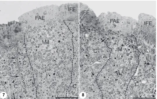
Fig. 7. Eight-day-old control chick. Under the follicle-associated epithelium (FAE) the medulla (M) is wide and the cortex (C) is clearly separated from the interfollicular connective tissue (ar- rows). Few apoptotic cells occur in the medulla. Fig. 8. Eight-day-old chick infected with virulent
virus (strain 11518, the CM border is indicated by a dashed line) The border between the cortex and the interfollicular connective tissue cannot be identified. Under the normal follicle-associated
epithelium (FAE) the medulla is thin. Figs 7–8: Toluidine blue staining, semi-thin section.
Bar: 50 µm
Histology
The tissue samples were cut into small pieces (approximately 1 µm3) fixed in 4% glutaraldehyde overnight. After washing with PBS, the samples were post- fixed in 1% osmium tetroxide, dehydrated in graded ethanol and embedded in Araldite epoxy resin. One µm thick, semi-thin sections were stained with tolui- dine blue and pictures were taken with a Zeiss Axiophot microscope.
Results and discussion
In the 20-day-old embryos the MPV and the interfollicular arterioles had already developed, and in a few follicles the rudiment of the CM capillary web could be observed as a kind of outgrowth of interfollicular arterioles (Fig. 1 and inset). This finding underlines the angiogenesis of CM capillary network for- mation instead of de novo formation (vasculogenesis) (Schoenwolf et al., 1981;
Pardanaud et al., 1989). The angiogenesis is characteristic of blood vessel for- mation in the somatic pleura, which seems to support the ectodermal origin of
7 8
the bursal rudiment (Nagy and Oláh, 2010). The CM capillary web sprouting off from the interfollicular arterioles precedes the development of the cortex (Fig. 2).
The bursal lumen between the plicae is very narrow at this age but Cav-1 is al- ready expressed in a few interfollicular epithelial (IFE) cells (Fig. 1).
In one-day-old birds the Cav-1 expression is highly elevated in the inter- follicular arterioles (Fig. 2), from which the sprouting of the CM capillary web emerged in a few follicles (Fig. 2). The IFE became higher and the bursal lumen
‘opened’ between the plicae. By day 8 the Cav-1 expression in the CM capillary web clearly outlined the CM border and the area between the CM capillary web and the interfollicular connective tissue, indicating the formation of the follicular cortex. This observation suggests that the formation of the cortex and the CM capillary web develop in parallel, but possibly the sprouting of the CM capillary web precedes the establishment of the cortex. The number of Cav-1-positive IFE cells greatly increased during the first week of life (Fig. 3), indicating that the IFE cells participate in cholesterol homeostasis in the early life of chickens (Bódi et al., 2018).
The developmental status of the CM capillary web and the size of the fol- licles are affected by IBV infection. Variation in the development of the CM ca- pillary web and the size of follicles depend on the virulence of IBV strains. In birds treated with the H120 vaccine strain (Fig. 3) the bursal follicles are larger and the CM capillary web is also more developed than in birds infected by wild- type virus (Figs 4, 5 and 6). Interestingly, Cav-1 expression in the budding arte- rioles and the interfollicular epithelial cells seemed to be unaffected by corona- virus infection, but the bursal lumen between the plicae remained narrow in chicks infected by virulent and vaccine viruses (Figs 5–6). The follicle-associated epi- thelium (FAE) did not show histological differences between the control and the infected birds (Figs 7 and 8). However, a decreased number of cells was revealed both in the cortical and the medullary regions of the follicles. In addition to this finding, the medullary region of the follicles in the birds infected with virulent field isolate showed some cellular disintegration (Fig. 8), which may influence the cell migration between the medulla and the cortex.
The negative effects of virulent IBV infection on bursal development and in a broader context on the immune system of the birds might result in elevated susceptibility of young chickens to coronavirus infection after hatching. This is in accordance with the finding that the adaptive immune function develops dur- ing the first three weeks of life, and therefore the young chicks are more suscep- tible than chickens of more advanced ages (de Wit, 2000). In this paper we report that the CM capillary web develops rapidly during the first week of life, preced- ing the formation of the cortex. Early coronavirus infection retarded the budding (angiogenesis) of interfollicular arterioles, which subsequently resulted in the underdevelopment of follicles. Smaller follicles obviously have a limited number of B cells, which demands fewer bursal secretory dendritic cells (BSDC) for
providing a healthy microenvironment. It can be assumed that the lower number of B cells and BSDC also contributes to the higher virus susceptibility of young chickens.
IBV infection still remains a significant animal health issue in spite of in- tensive vaccination programmes (Gelb et al., 1991; Jia et al., 1995; Farsang et al., 2002; Zanella et al., 2003; Ignjatovic et al., 2006; Liu et al., 2009; Mase et al., 2010; Kiss et al., 2015). Although the pathogenesis of IBV infection, espe- cially with regard to the upper respiratory tract, has been thoroughly studied (Dhinakar-Raj and Jones, 1997), much less attention was paid to the effects of IBV on the lymphoid organs. The aim of our study was to fill this gap and com- plement our knowledge on how IBV affects the development of bursal follicles.
The potential immunological implications need further studies, which could lead to more efficient vaccines and more precisely designed vaccination programmes.
Acknowledgements
This work was carried out in the framework of the FA COST Action Towards Control of Avian Coronaviruses (FA1207). Attila Farsang’s contribution was kindly sup- ported by the ‘János Bolyai’ Research Fellowship of the Hungarian Academy of Sciences (BO/125/13).
References
Abbate, F., Pfarrer, C., Jones, C. J., Ciriaco, E., Germanà, G. and Leiser, R. (2007): Age-dependent changes in the pigeon bursa of Fabricius vasculature: a comparative study using light mi- croscopy and scanning electron microscopy of vessel casts. J. Anat. 211, 387–398.
Benyeda, Z., Mató, T., Süveges, T., Szabó, E., Kardi, V., Abonyi-Tóth, Z., Rusvai, M. and Palya, V. (2009): Comparison of the pathogenicity of QX-like, M41 and 793/B infectious bron- chitis strains from different pathological conditions. Avian Pathol. 38, 449–456.
Bódi, I., Minkó, K., Fölker, O., Benyeda, Zs., Felföldi, B., Magyar, A., Kiss, A., Palya, V. and Oláh, I. (2018): Expression of caveolin-1 in the interfollicular but not the follicle- associated epithelial cells in the bursa of Fabricius of chickens. J. Morphol. 279, 17–26.
de Wit, J. J. (2000): Detection of infectious bronchitis virus. Avian Pathol. 29, 71–93.
de Wit, J. J., Cook, J. K. and van der Heijden, H. M. (2011): Infectious bronchitis virus variants: a review of the history, current situation and control measures. Avian Pathol. 40, 223–235.
Dhinakar Raj, G. and Jones, R. C. (1996): Immunopathogenesis of infection in SPF chicks and commercial broilers of a variant infectious bronchitis virus of economic importance. Avian Pathol. 25, 481–501.
Dhinakar Raj, G. and Jones, R. C. (1997): Infectious bronchitis virus: Immunopathogenesis of in- fection in the chicken. Avian Pathol. 26, 677–706.
Farsang, A., Ros, C., Renström, L. H., Baule, C., Soós, T. and Belák, S. (2002): Molecular epizo- otiology of infectious bronchitis virus in Sweden indicating the involvement of a vaccine strain. Avian Pathol. 31, 229–236.
Gelb, J., Lunt, R. L., Metz, A. L. and Fries, P. A. (1991): Attenuation of avian infectious bronchitis virus isolated from commercial layer and broiler chickens. Avian Dis. 35, 82–87.
Ignjatovic, J., Gould, G. and Sapats, S. (2006): Isolation of a variant infectious bronchitis virus in Australia that further illustrates diversity among emerging strains. Arch. Virol. 151, 1567–1585.
Jia, W., Karaca, K., Parrish, C. R. and Naqi, S. A. (1995): A novel variant of avian infectious bron- chitis virus resulting from recombination among three different strains. Arch. Virol. 140, 259–271.
King, D. J. and Cavanagh, D. (1991): Infectious bronchitis. In: Calnek, B. W., Barnes, H. J., Beard, C. W., Reid, W. M. and Yoder, H. W. Jr. (eds) Diseases of Poultry. Iowa State University Press, Ames, Iowa, USA. pp. 471–484.
Kiss, I., Mató, T., Homonnay, Z. G., Kojer, J., Farsang, A., Bálint, Á. and Palya, V. (2015): Survey indicates circulation of 4/91 and QX-type infectious bronchitis viruses in Hungary in 2014 – Short communication. Acta Vet. Hung. 63, 382–388.
Liu, X. L., Su, J. L., Zhao, J. X. and Zhang, G. Z. (2009): Complete genome sequence analysis of a predominant infectious bronchitis virus (IBV) strain in China. Virus Genes 38, 56–65.
Mase, M., Kawanishi, N., Ootani, Y., Murayama, K., Karino, A. and Inoue, T. (2010): A novel genotype of avian infectious bronchitis virus isolated in Japan in 2009. J. Vet. Med. Sci.
72, 1265–1268.
Nagy, N. and Oláh, I. (2010): Experimental evidence for the ectodermal origin of the epithelial an- lage of the chicken bursa of Fabricius. Development 137, 3019–3023.
Navarro, A., Anand-Apte, B. and Parat, M. O. (2004): A role for caveolae in cell migration.
FASEB J. 18, 1801–1811.
Pardanaud, L., Yassine, F. and Dieterlen-Lievre, F. (1989): Relationship between vasculogenesis, angiogenesis and haemopoiesis during avian ontogeny. Development 105, 473–485.
Pelkmans, L., Kartenbeck, J. and Helenius, A. (2001): Caveolar endocytosis of simian virus 40 re- veals a new two-step vesicular-transport pathway to the ER. Nat. Cell Biol. 3, 473–483.
Schoenwolf, G. C., Bell, L. A. and Watterson, R. L. (1981): Vasculogenesis of the bursa cloacalis (bursa of Fabricius) of the chick embryo. J. Morphol. 167, 35–42.
van Deurs, B., Roepstorff, K., Hommelgaard, A. M. and Sandvig, K. (2003): Caveolae: anchored, multifunctional platforms in the lipid ocean. Trends Cell Biol. 13, 92–100.
Zanella, A., Lavazza, A., Marchi, R., Moreno Martin, A. and Paganelli, F. (2003): Avian infectious bronchitis: characterization of new isolates from Italy. Avian Dis. 47, 180–185.