Contents lists available atScienceDirect
Biochemical Pharmacology
journal homepage:www.elsevier.com/locate/biochempharm
Chronic treatment with rofecoxib but not ischemic preconditioning of the myocardium ameliorates early intestinal damage following cardiac
ischemia/reperfusion injury in rats
Szilvia B. László
a, Bernadette Lázár
a, Gábor B. Brenner
a, András Makkos
a, Mihály Balogh
a, Mahmoud Al-Khrasani
a, Barbara Hutka
a, Amir Mohammadzadeh
a, Ágnes Kemény
b,c, Terézia László
d, Bálint Scheich
d, Tamara Szabados
e, Éva Kenyeres
e, Zoltán Giricz
a,
Péter Bencsik
e,f, Zoltán V. Varga
a,g, Julianna Novák
a,g, Zsuzsanna Helyes
c, Péter Ferdinandy
a,f, Klára Gyires
a, Zoltán S. Zádori
a,⁎aDepartment of Pharmacology and Pharmacotherapy, Semmelweis University, 1089 Budapest, Hungary
bDepartment of Medical Biology, University of Pécs, 7624 Pécs, Hungary
cDepartment of Pharmacology and Pharmacotherapy, Medical School & Szentágothai Research Centre, University of Pécs, 7624 Pécs, Hungary
d1st Department of Pathology and Experimental Cancer Research, Semmelweis University, 1085 Budapest, Hungary
eDepartment of Pharmacology and Pharmacotherapy, University of Szeged, 6720 Szeged, Hungary
fPharmahungary Group, 6722 Szeged, Hungary
gHCEMM-SU Cardiometabolic Immunology Research Group, 1089 Budapest, Hungary
A R T I C L E I N F O
Keywords:
Remote ischemia/reperfusion injury Myocardial infarction
Small intestine Cyclooxygenase-2 Ischemic preconditioning Matrix metalloproteinase
A B S T R A C T
There is some recent evidence that cardiac ischemia/reperfusion (I/R) injury induces intestinal damage within days, which contributes to adverse cardiovascular outcomes after myocardial infarction. However, it is not clear whether remote gut injury has any detectable early signs, and whether different interventions aiming to reduce cardiac damage are also effective at protecting the intestine. Previously, we found that chronic treatment with rofecoxib, a selective inhibitor of cyclooxygenase-2 (COX-2), limited myocardial infarct size to a comparable extent as cardiac ischemic preconditioning (IPC) in rats subjected to 30-min coronary artery occlusion and 120- min reperfusion. In the present study, we aimed to analyse the early intestinal alterations caused by cardiac I/R injury, with or without the above-mentioned infart size-limiting interventions. We found that cardiac I/R injury induced histological changes in the small intestine within 2 h, which were accompanied by elevated tissue level of COX-2 and showed positive correlation with the activity of matrix metalloproteinase-2 (MMP-2), but not of MMP-9 in the plasma. All these changes were prevented by rofecoxib treatment. By contrast, cardiac IPC failed to reduce intestinal injury and plasma MMP-2 activity, although it prevented the transient reduction in jejunal bloodflow in response to cardiac I/R. Our results demonstrate for thefirst time that rapid development of intestinal damage follows cardiac I/R, and that two similarly effective infarct size-limiting interventions, rofe- coxib treatment and cardiac IPC, have different impacts on cardiac I/R-induced gut injury. Furthermore, in- testinal damage correlates with plasma MMP-2 activity, which may be a biomarker for its early diagnosis.
1. Introduction
Myocardial infarction (MI) is one of the leading causes of death worldwide, and early reperfusion of the myocardium is the most ef- fective treatment to reduce cardiac injury and infarct size [1]. Re- perfusion of the ischemic myocardium, however, can paradoxically
induce further tissue damage, a phenomenon called ischemia/reperfu- sion (I/R) injury, which has been described and characterized in nu- merous organs. The pathogenesis of I/R injury involves common factors in all tissues, including endothelial dysfunction, activation of leuko- cytes, generation of reactive oxygen and nitrogen species (RONS) and the release of cytokines and other mediators like matrix
https://doi.org/10.1016/j.bcp.2020.114099
Received 6 March 2020; Received in revised form 10 June 2020; Accepted 10 June 2020
Abbreviations:COX, cyclooxygenase; IPC, ischemic preconditioning; I/R, ischemia/reperfusion; LAD, left anterior descending coronary; MDA, malondialdehyde; MI, myocardial infarction; MMP, matrix metalloproteinase; RONS, reactive oxygen and nitrogen species; SOD, superoxide dismutase
⁎Corresponding author at: Department of Pharmacology and Pharmacotherapy, Semmelweis University, Nagyvárad tér 4, 1089 Budapest, Hungary.
E-mail address:zadori.zoltan@med.semmelweis-univ.hu(Z.S. Zádori).
Available online 12 June 2020
0006-2952/ © 2020 The Author(s). Published by Elsevier Inc. This is an open access article under the CC BY license (http://creativecommons.org/licenses/BY/4.0/).
T
metalloproteinases (MMPs)[2]. MMPs are a family of zinc-containing neutral endopeptidases, which, besides degrading components of the extracellular matrix, also regulate the release or activation of cytokines, chemokines, various growth factors and numerous other bioactive molecules in a variety of physiological and pathophysiological condi- tions[3]. Distinct types of MMPs, particularly MMP-2 and MMP-9, are released rapidly upon I/R and contribute to tissue injury[4,5]. In ad- dition, upon the reperfusion of ischemic tissues all these mediators[2]
including MMPs[6,7]are released into the bloodstream and delivered to distant organs, where they may induce damage as well (remote organ injury).
The intestinal mucosa is particularly vulnerable to the deleterious effects of local I/R[8]. Intestinal injury and impaired mucosal barrier integrity enable luminal aggressive factors like bacteria and bile acids to enter the gut tissue and trigger an inflammatory response at both local and remote sites. On the other hand, intestinal damage may be an early consequence of I/R injury of distant organs as well. Histological alterations of the gut with increased permeability have been observed as early as 2 h after reperfusion of the ischemic limbs [9,10]. Inter- estingly, much attention have been paid to remote gut injury following I/R of the limb or kidney[9–12], but little is known about the intestinal effects of cardiac reperfusion after MI. In an animal model of MI in- testinal barrier was impaired 17 days after reperfusion[13]. In a more recent study increased intestinal permeability was reported on days 1–7 of symptom onset in ST-segment elevation MI patients undergoing primary percutaneous coronary intervention. Plasma levels of gut-de- rived bacterial products correlated with the severity of systemic in- flammation and predicted adverse cardiovascular events, suggesting that intestinal damage is a major determinant of cardiovascular out- comes after MI[14]. Hence, early detection of remote intestinal injury following cardiac I/R may help to estimate the risk of future cardio- vascular events and therapeutic interventions targeting intestinal da- mage may help to prevent the development of systemic inflammation.
Over the past decades, numerous treatment strategies have been proposed to protect the heart against the detrimental effects of acute I/
R injury[1], but much less information is available on the impact of these interventions on the remote effects of cardiac I/R, especially in the intestines.
A possible pharmacological approach to reduce cardiac I/R injury- induced inflammation is the inhibition of cyclooxygenase-2 (COX-2), which is upregulated following permanent or transient myocardial ischemia[15–17]. Although under some circumstances the increased generation of COX-2-derived prostanoids may represent an adaptive response that protects the cells from I/R injury[16,18], in the majority of studies COX-2 inhibition decreased cardiac injury following perma- nent or transient ischemia, pointing to a rather detrimental role of COX- 2[17,19–23]. Our recentfinding that chronic rofecoxib treatment re- duced the infarct size in the myocardium after I/R concurs with these studies, although we also demonstrated that its infarct size-limiting effect was counterbalanced by higher mortality rate due to its pro-ar- rhythmic property[24].
Rapid upregulation of COX-2 in response to local I/R has also been demonstrated in the gastrointestinal tract[25,26], and COX-2 inhibi- tion in most[26–28], though not all[29]studies reduced intestinal I/R injury. In addition, there is some evidence that COX-2 inhibitors can mitigate remote organ injury following limb or kidney I/R as well [30–32]. It is, however, still not clear whether pharmacological blockade of COX-2 confers any protection against remote intestinal injury following cardiac I/R.
Another well-documented cardioprotective intervention is ischemic preconditioning (IPC), in which the myocardium is subjected to brief periods of ischemia prior to the induction of a prolonged ischemia in order to initiate intrinsic cell-survival programs [33]. Although the underlying mechanism is complex and still not fully understood, IPC has been shown to target multiple factors involved in I/R injury such as endothelial dysfunction, leukocyte activation and oxidative stress[34].
Of note, in our previous study cardiac IPC limited myocardial infarct size to a comparable extent as rofecoxib treatment[24].
The tissue-protective effect of IPC has also been demonstrated in other organs, including the gut[8,35]. Moreover, IPC of the organs subjected to I/R may also provide protection against remote intestinal damage[36]. However, it is not known whether IPC prior to cardiac I/R can mitigate the remote damage of the gut.
Here we show for thefirst time the rapid development of histolo- gical alterations and mild inflammation in the rat small intestine fol- lowing cardiac I/R, and that two similarly effective infarct size-limiting interventions, pharmacological blockade of COX-2 with rofecoxib and IPC of the heart, have different impacts on them. Our study also iden- tifies plasma MMP-2 activity as a potential biomarker for the detection and estimation of cardiac I/R-induced early remote intestinal injury.
2. Materials and methods 2.1. Animals
Experiments were carried out on male Wistar rats weighing 180–280 g (Study I.) and 320–440 g (Study II.) (Semmelweis University, Budapest, Hungary). Animals were housed in a temperature (22 ± 2 °C)- and humidity-controlled room at a 12 h light/dark cycle.
Food and water were available ad libitum.
2.2. Materials
Rofecoxib [4-(4′-methylsulfonylphenyl)-3-phenyl-2-(5H)-furanone]
was purchased from MedChem Express (Sollentuna, Sweden). All other chemicals, unless otherwise stated, were obtained from Sigma-Aldrich (St. Louis, MO, USA).
2.3. Ethical considerations
All efforts were made to minimize animal suffering and to reduce the number of animals used in the experiments. All procedures con- formed to the Directive 2010/63/EU on European Convention for the protection of animals used for scientific purposes. The experiments were approved by the National Scientific Ethical Committee on Animal Experimentation and permitted by the government (Food Chain Safety and Animal Health Directorate of the Government Office for Pest County (PEI/001/1493-4/2015 and PE/EA/1784-7/2017)).
2.4. In vivo studies
2.4.1. Study I. Evaluating the impact of COX-2 inhibition and IPC on the remote intestinal effects of cardiac I/R injury
In this study the same male Wistar rats were used as in our two parallel studies focusing on the effects of long-term rofecoxib treatment on myocardial ischemia/reperfusion injury[24]and on the composi- tion of intestinal microbiota[37], which complies with the principles of replacement, reduction and refinement of animal experiments[38]. In the present study 7–11 rats were allocated to each treatment group.
Rats were treated intragastrically with either vehicle (1% hydro- xyethylcellulose) or rofecoxib (5 mg/kg) in a volume of 0.33 ml/100 g once daily for 28 days. The applied dose of rofecoxib was chosen partly based on previous animal studies[39], based on the pharmacokinetic similarities of rofecoxib in rats and humans[40]and extrapolation from the maximal recommended daily dose (50 mg) used earlier in the clinical practice, calculating with a 60-kg weight individual, according to Reagan-Shaw et al.[41]. However, we have also confirmed the po- tency of this rofecoxib dose and its selectivity for COX-2, as our results showed that it inhibited COX-2-derived PGE2 synthesis by almost 100%, without having any effect on the gastrointestinal PGE2 levels produced by COX-1[37]. Of note, chronic rofecoxib treatment did not increase the mortality of animals, and did not reduce the cardiac level
of 6-keto prostaglandin F1α (the stable metabolite of prostacyclin), which has been implicated in rofecoxib-induced cardiotoxicity [42]
(vehicle: 40.6 ± 6.3 pg/mg tissue, rofecoxib: 49.4 ± 7.3 pg/mg tissue, n = 7/group, p = 0.38).
On the 29th day, i.e. 24 h after thefinal administration of rofecoxib, all rats were anaesthetized with pentobarbital (60 mg/kg in- traperitoneally) and underwent thoracotomy. Rats were ventilated with rodent ventilator (Ugo-Basile, Gemonio, Italy) with 6.2 ml/kg tidal volume at a rate of 69 ± 3 breath/min according to body weight. Their blood pressure was continuously monitored in the carotid artery (AD Instruments, Bella Vista, Australia), and their body temperature was maintained at 37 °C with a heating pad. The right carotid artery was cannulated for the measurement of mean arterial blood pressure (MAP, AD Instruments, Bella Vista, Australia) and forfluid supplementation with saline containing 10 IU/kg heparin. The time point of completing all these procedures was designated as the starting point of experiment (0 min). 40 min later, two groups of rats treated either with vehicle or with rofecoxib, were subjected to sham operation, in which the left anterior descending coronary artery (LAD) was isolated but not oc- cluded (groups 1 and 3, VEH SHAM and ROF SHAM). In two other groups (groups 2 and 4, VEH I/R and ROF I/R) cardiac I/R injury was induced by occluding LAD for 30 min, followed by 120 min of re- perfusion. In a further 5th group, IPC (3 cycles of 5 min LAD occlusion followed by 5 min of reperfusion) was applied on vehicle-treated rats directly before the 30 min LAD occlusion (VEH IPC + I/R).
Additionally, all animals received intraperitoneal injection of 100 IU/
kg heparin 3 times during the surgical procedure, at 35, 65 and 185 min. The study design and experimental protocol are illustrated in Fig. 1. Although this design allowed us to analyze the effect of two different infarct size-limiting interventions on cardiac I/R injury- evoked intestinal responses parallel, for the sake of clarity the results will be presented and discussed separately.
At the end of reperfusion, the rats were sacrificed, plasma samples were collected and small intestines were excised. The mucosa of small intestine wasflushed with cold saline and photographed for subsequent macroscopic analysis. Full-thickness pieces of the distal jejunum were
snap-frozen in liquid nitrogen and stored at−80 °C for further assays.
Other portions of intestinal tissues were fixed in 10% formalin for histological analysis.
Intestinal tissue and plasma samples were collected only from ani- mals surviving the whole protocol. Whereas all animals survived in both VEH SHAM and ROF SHAM groups (n = 8/group), in the VEH- treated I/R group 1 of the 7 animals died due to ventricularfibrillation (14% mortality). Rofecoxib treatment following cardiac I/R was asso- ciated with increased mortality rate, 4 of 9 animals died due to ar- rhythmias (44%). In the VEH-IPC group (n = 11) 4 rats died during the short I/R stimuli of IPC and were excluded from further evaluations, and 1 of the remaining 7 animals died due to sudden drop in blood pressure in the reperfusion period (14%).
In addition, hearts were excised in order to assess the infarct sizes.
These data were the subject of a separate paper[24], and herein we will only refer to them.
2.4.2. Study II. Evaluating the effect of cardiac I/R injury with or without IPC on the small intestinal microcirculation
This study was performed on a separate group of rats with weights matching those of the vehicle-treated animals of Study I. at the day of surgery (320–440 g). Animals were subjected to the same anaesthesia and surgical procedure as before, but also median laparotomy was performed and the distal jejunum of rats was gently exposed. Small intestinal microcirculation was measured using a PeriScan PIM II laser Doppler perfusion imager (Perimed, Stockholm, Sweden) placed 12 cm above the surface of the jejunum. Each scanning was performed on an area of 31 × 31 sampling points within 59 sec, followed by 1 sec pause.
Hence, imaging was performed in every minute, but we also aimed to minimize thefluid and heat loss of animals and to avoid tissue de- siccation [43]. Therefore, during the reperfusion period the opened abdomen was repeatedly covered with saline-moistened gauze for 5 min intervals from the 95th to 180th min (i.e. from 95 to 100 min, from 105 to 110 min, and so forth), and perfusion values for these periods have not been recorded. Perfusion patterns were analyzed using LDPIwin 2.6 software (Perimed, Stockholm, Sweden) and values were
Fig. 1.Experimental protocol. Male Wistar rats were treated with vehicle (VEH, 1% hydroxyethylcellulose) or rofecoxib (ROF, 5 mg/kg) for 28 days once daily (q.d.).
On the 29th day, rats were subjected to sham operation (groups 1 and 3, VEH SHAM and ROF SHAM) or cardiac ischemia (ISC) followed by reperfusion (groups 2 and 4, VEH I/R and ROF I/R). In an additional vehicle-treated group ischemic preconditioning (IPC) was applied (group 5, VEH IPC + I/R). Number of animals surviving the whole protocol: 5–8/group. White arrow: intraperitoneal injection of 100 IU/kg heparin; black arrow: termination of animals and tissue sampling.
expressed as percentage of the basal (pre-occlusion) values (registered at 10 min).
2.5. Macroscopic evaluation of intestinal damage
High-resolution photographs of the entire small intestinal mucosa were thoroughly analyzed and scored in blinded fashion, as follows: 0, no visible morphologic alteration; 1, small (1–2 mm) hyperemic area at one site; 2, small (1–2 mm) hyperemic areas at two or more sites; 3, extensive (> 2 mm) hyperemic area at one site; 4, extensive (> 2 mm) hyperemic areas at two or more sites[37].
2.6. Histological analysis
Samples taken from the distal part of the small intestine werefixed in 10% formalin, embedded in paraffin, sectioned (5 µm), and stained with haematoxylin and eosin. Digital micrographs were taken by an Olympus BX51 microscope and Olympus DP50 camera. Histological injury was assessed in blinded fashion by two histopathologists ac- cording to the scoring system described by Mantyh et al [44] with minor modifications (Table 1). The total histological score (ranging from 0 to 9) was calculated based on the sum of partial scores.
2.7. Cytokine measurements
The jejunal levels of distinct cytokines were measured by either Luminex xMAP technology, or ELISA. Excised and snap-frozen jejunal tissues were pulverized and homogenized according to the manu- facturers’instructions. ELISA kit was used to quantify the protein levels of tumor necrosis factor-α(TNF-α) (Invitrogen, Camarillo, CA, USA), whereas Milliplex Multiplex assay to determine the levels of inter- leukin-1β(IL-1β) and IL-10 by using customized Milliplex Rat Cytokin/
Chemokine Magnetic Bead Panel (Merck Millipore, Burlington, MA, USA). The total protein concentration of samples was determined by using a bicinchoninic acid assay kit (Thermo Scientific Pierce Protein Research Products, Rockford, IL, USA) with bovine serum albumin (BSA) as a standard, and cytokine amounts are given in pg/mg of total protein.
2.8. Superoxide dismutase, catalase and malondialdehyde assays The activities of superoxide dismutase (SOD) and catalase, as well as the concentration of malondialdehyde (MDA) in the jejunum were measured with assay kits according to the manufacturer's instructions (Cayman Chemical, Ann Arbor, MI, USA).
The SOD kit utilizes a tetrazolium salt for the detection of super- oxide radicals generated by xanthine oxidase and hypoxanthine, and measures the activity of all three types of SOD (Cu/Zn, Mn and Fe- SOD). One unit of SOD was defined as the amount of enzyme needed to exhibit 50% dismutation of the superoxide radical. The total protein concentration of supernatants was determined and SOD activity was expressed in unit/mg protein.
The catalase kit utilizes the peroxidatic function of catalase for determination of enzyme activity. The method is based on the reaction of the enzyme with methanol in the presence of an optimal con- centration of H2O2. The produced formaldehyde was measured spec- trophotometrically with 4-amino-3-hydrazino-5-mercapto-1,2,4-tria- zole as the chromogen. One unit of catalase was defined as the amount of enzyme that causes the formation of 1 nmol of formaldehyde per minute at 25 °C. Catalase activity was expressed in nmol/min/mg tissue.
The measurement of jejunal MDA concentration was based on the reaction of MDA with thiobarbituric acid, and the colorimetric quan- tification of the formed adduct at 530 nm. Results were expressed as nmol/mg tissue.
Table1 Criteriaforquantitativeestimationofthesmallintestinalinjury. 0123 Epithelialdamagenonedestructionofvillustipsdestructionofuptoonehalfofvilluscompletevillusdestruction Congestionandedemanoneminimalincreaseincryptspacing,rareRBC-containing vesselsmoderateincreaseincryptspacing,uptoonehalfofvesselscontain RBCswidelyspacedcrypts,numerousRBC-containingvesselsinlamina propria Cellularinfiltrationnonemildcellularinfiltrationmoderatecellularinfiltrationnumerousleukocytesthroughoutthelaminapropria RBC–redbloodcell.
2.9. Immunohistochemistry
After routine FFPE specimen processing, deparaffinized sections underwent antigen retrieval (pH = 6 citrate buffer, at 95 °C for 15 min). After blocking endogenous peroxidase activity (3% H2O2so- lution in PBS), the sections were blocked in appropriate sera (2.5% goat serum in PBS). Then, sections were incubated with primary anti-ni- trotyrosine antibody (06-284, 1:200, Merck Millipore, Burlington, MA, USA) overnight in diluted blocking solution at 4 °C. After primary an- tibody incubations, the sections were washed three times in PBS and incubated for an hour with SignalStain®Boost IHC Detection Reagent (HRP, Rabbit, 8114, Cell Signaling Technology, Danvers, MA, USA).
The secondary antibody was washed 3 times for 10 min and the specific signal was developed with diaminobenzidine (ImmPACT DAB EqV Peroxidase (HRP) Substrate, Vector Laboratories, Burlingame, CA, USA). The specific staining was visualized and images were acquired using a Leica DM3000 microscope (Leica, Wetzlar, Germany).
2.10. Measuring MMP-2 and MMP-9 activities by gelatin zymography Activity of MMP-2 and MMP-9 was assessed by gelatin zymography from plasma samples collected after 120 min of reperfusion.
Gelatinolytic activities of MMPs were examined as previously described [45]. Briefly, 8% polyacrylamide gels were copolymerized with gelatin (2 mg/ml, type A from porcine skin), and 50 µg of protein per lane was loaded. An internal standard (American Type Culture Collection, Manassas, Virginia) was loaded into each gel to normalize activities between gels. After electrophoresis (90 V, 90 min), gels were washed with zymogram renaturation buffer (Novex, Carlsbad, CA, USA) for 40 min. Samples were incubated for 20 h at 37 °C in zymogram de- velopment buffer (Novex, Carlsbad, CA, USA).
In a separate experiment, one plasma sample from each group was loaded into the gel in 4 replicates. After renaturation, the gel was cut into 4 pieces, which were separately incubated in development buffer containing vehicle or rofecoxib at 0.1, 1, or 10μM concentrations, re- spectively, in order to reveal whether rofecoxib has direct inhibitory effect on MMP activity. The concentration of 1 µM was chosen for ro- fecoxib based on the peak plasma concentration (Cmax) measured after a single, 5 mg/kg oral dose of rofecoxib in rats[46].
Gels were then stained with 0.05% Coomassie brilliant blue in a mixture of methanol-acetic acid-water [2.5:1:6.5 (v/v)] and destained in aqueous 4% methanol-8% acetic acid (v/v) to remove aspecific binding of Coomassie. For positive controls, gelatinase zymography standard containing human MMP-2 and MMP-9 (Chemicon Europe Ltd., Southampton, UK) was used. For negative control, lanes containing tissue samples were cut offafter renaturation of the gel and were se- parately incubated for 20 h at 37 °C in development buffer in the pre- sence of the calcium chelator EGTA (ethylene glycol-bis(2-aminoethy- lether)-N,N,N′,N′-tetraacetic acid; 10 mM). Gelatinolytic activities were detected as transparent bands against the dark-blue background. Gels were scanned in a transilluminator and band intensities were quantified by Quantity One software (BioRad, Hercules, CA, USA), and expressed as the ratio to the internal standard, and presented in arbitrary units.
2.11. Western blot measurements
Distal jejunal tissues were homogenized with a TissueLyser (Qiagen, Venlo, The Netherlands) in lysis buffer containing 200 mM NaCl, 5 mM EDTA, 10 mM Tris, 10% glycerine, and 1 µg/ml leupeptin (pH 7.4), supplemented with a protease inhibitor cocktail (cOmplete ULTRA Tablets, Roche, Basel, Switzerland) and PMSF (Sigma, St. Louis, MO, USA). The homogenized lysates were centrifuged twice at 1,500 × g and 4 °C for 15 min, then the supernatants were collected and their protein concentration was measured by the bicinchoninic acid assay (Thermo Fisher Scientific, Waltham, MA, USA). Equal amount of pro- tein (20 µg) was mixed with Pierce Lane Marker reducing sample buffer
(Thermo Fisher Scientific, Waltham, MA, USA), and loaded and sepa- rated in a 4–20% precast Tris-glycine SDS polyacrilamide gel (BioRad, Hercules, CA, USA). Proteins were transferred electrophoretically onto a polyvinylidene difluoride membrane (BioRad, Hercules, CA, USA) at 200 mA overnight. Membranes were blocked with 5% nonfat dry milk (BioRad, Hercules, CA, USA) in Tris-buffered saline containing 0.05%
Tween-20 (0.05% TBS-T; Sigma, St. Louis, MO, USA) at room tem- perature for 2 h. Membranes were incubated with primary antibodies against COX-2 (12282, 1:500) and COX-1 (4841, 1:500) (Cell Signaling Technology, Danvers, MA, USA) overnight at 4 °C, followed by 2 h incubation at room temperature with anti-rabbit HRP-linked secondary antibody. GAPDH was used to control for sample loading and protein transfer and to normalize the content of target protein. Signals were detected with a chemiluminescence kit (BioRad, Hercules, CA, USA) by Chemidoc XRS+ (BioRad, Hercules, CA, USA).
2.12. Evaluation of 6-keto prostaglandin F1α
Tissue levels of 6-keto prostaglandin F1αwere determined by ELISA, according to the manufacturer's instructions (Cayman Chemical, Ann Arbor, MI, USA). Briefly, tissues were homogenized in precooled acetone containing 10 µM indomethacin, and centrifuged at 10,000 g for 10 min at 4 °C. Acetone was evaporated from the supernatants using a vacuum centrifuge, then the residues were resolved in assay buffer and used for determination of 6-keto prostaglandin F1α.
2.13. Statistics
Data are expressed as mean ± SEM. Statistical analysis of the data was performed with one-way or two-way ANOVA, or in case of non- parametric values with Mann-Whitney (pairwise comparison) or Kruskal-Wallis tests. Two-way repeated measures ANOVA was used to compare the time course of blood pressure and jejunal blood flow changes. Correlations between MMP-values and histological scores were calculated by Spearman test. Outliers detected by Grubb’s test were excluded from the analyses. In thein vitroassays all samples were measured at least in duplicates, and most measurements were repeated two times. In all cases, a probability of p < 0.05 was considered sta- tistically significant.
3. Results
3.1. Cardiac I/R induced early histological alterations in the small intestine of vehicle-but not rofecoxib-treated rats
Occlusion of LAD for 30 min, followed by 2 h reperfusion did not cause any appreciable macroscopic alteration in the small intestine of vehicle- or rofecoxib-treated rats (Fig. 2A). By contrast, histological analysis revealed early significant alterations in the jejunal mucosa of vehicle-treated rats subjected to cardiac I/R compared to sham-oper- ated controls. This was mainly characterized by subepithelial edema with dilated vessels containing numerous red blood cells, and was ob- served in almost all animals (in 5 of 6 rats). In addition, in half of the I/
R animals increased number of leukocytes (lymphocytes, macrophages and granulocytes) could be detected in the lamina propria, including extravasated ones, indicating an increase in vascular permeability. The epithelium and structure of villi were generally preserved, although early signs of epithelial distruption at the tips of some villi were also found in 5 of 6 animals. Altogether, the overall histological score was significantly higher in the I/R group compared to the sham-operated group. Such morphological changes, however, were not observed in the I/R group treated with rofecoxib, indicating that inhibition of COX-2 prevented remote intestinal injury after cardiac I/R (Fig. 2B and 2C).
As we reported before, chronic rofecoxib treatment also reduced the myocardial infarct size after cardiac I/R in these animals, at least partly due to a direct cardio-cytoprotective effect, but it also increased acute
mortality by increasing the incidence of arrhythmias (see section 2.4 and Ref.[24]).
3.2. Rofecoxib treatment inhibited the development of mild intestinal inflammation following cardiac I/R injury
Because histological analysis suggested the presence of mild in- testinal inflammation in the vehicle-treated I/R group, in the next step we aimed to characterize it by measuring different inflammatory factors known to be involved in I/R injury. First, we assessed the intestinal level of COX-2, because its upregulation is an early response to I/R. In contrast to sham-operated animals with barely detectable COX-2 ex- pression, vehicle-treated rats following cardiac I/R exhibited sig- nificantly higher COX-2 protein levels in the jejunum. By contrast, in ROF-treated animals cardiac I/R did not increase the intestinal ex- pression of COX-2, compared to that of the respective sham group (Fig. 3A). The protein levels of the constitutive COX-1 isoform, in contrast to those of COX-2, were similar in all groups irrespective of treatment (Fig. 3B).
The expression of COX-2 can be induced by various pro-in- flammatory cytokines such as TNF-αand IL-1β[47]. These cytokines, especially TNF-α, also play an important role in the orchestration of inflammatory response to I/R in most tissues[2]. We found, however, no differences in terms of intestinal TNF-αlevels between the groups,
and IL-1βeven showed a mild reduction in the rofecoxib-treated I/R group compared to the respective sham group (Fig. 3C, 3D).
We also measured the tissue levels of the anti-inflammatory IL-10, because this cytokine is known to be produced in acute inflammation by numerous cells parallel with pro-inflammatory cytokines in order to regulate immune responses[48]. AsFig. 3E shows, there were only moderate differences in IL-10 levels, although those reflected somewhat the changes in histological scores and COX-2 levels. Namely, the highest concentration of IL-10 was measured in the vehicle-treated I/R group, whereas significantly lower levels were detected in rofecoxib- treated animals subjected to I/R.
Similarly, the highest level of 6-keto prostaglandin F1α(the stable metabolite of prostacyclin, which is mainly produced by COX-2 in in- flammation [49]) was measured in the vehicle-treated I/R group (Fig. 3F), but the differences between the groups did not reach statis- tical significance.
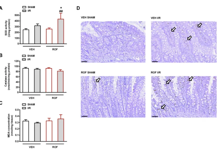
Finally, because oxidative stress and RONS are well-established factors in the pathogenesis of I/R injury, we aimed to determine the tissue activities of SOD and catalase, twofirst line antioxidant enzymes involved in removal of toxic oxygen metabolites and preventing cellular damage[50]. Whereas catalase activities were similar in all groups, total SOD activity tended to rise in the vehicle-treated I/R group, and increased significantly in rofecoxib-treated I/R animals (Fig. 4A and 4B). Superoxide anion, besides nitric oxide, is one of the initial RONS Fig. 2.The effects of 4-week treatment with vehicle (VEH) and rofecoxib (ROF, 5 mg/kg) on macroscopic (A) and histological scores (B) of jejunum. Data are expressed as mean ± SEM. For pairwise comparison of respective treatment groups Mann-Whitney test was used, n = 5–8/group (*P< 0.05 vs. respective VEH,
#P < 0.05 vs. respective SHAM). PanelCdemonstrates representative histological micrographs (scale bar: 100 µM, haematoxylin and eosin staining). Cardiac ischemia/reperfusion (I/R) injury induced histological alterations in vehicle-treated (VEH) rats. White arrows demonstrate dilated capillaries with numerous red blood cells in the lumen, whereas increased cellularity of the lamina propria with increased number of granulocytes, including extravasated ones is marked by asterisk. (For interpretation of the references to colour in thisfigure legend, the reader is referred to the web version of this article.)
Fig. 3.Jejunal levels of cyclooxygenase-2 (COX-2,A), COX-1 (B), tumor necrosis factor-α(TNF-α,C), interleukin-1β(IL-1β,D), IL-10 (E) and 6-keto prostaglandin F1α(6-keto PGF1α,F) in rats treated with vehicle (VEH) or rofecoxib (ROF, 5 mg/kg) for 4 weeks and subjected to sham operation or cardiac I/R injury. Results are expressed as mean ± SEM. For statistical analysis two-way ANOVA was used, followed by Fisher’s LSD post hoc test, n = 5–8/group (*P< 0.05 vs. respective VEH,
#P < 0.05 vs. respective SHAM).
Fig. 4.Jejunal activities of the antioxidant superoxide dismutase (SOD,A) and catalase (B), and the tissue levels of malondialdehyde (MDA,C) in rats treated with vehicle (VEH) or rofecoxib (ROF, 5 mg/kg) for 4 weeks and subjected to sham operation or cardiac I/R injury. Results are expressed as mean ± SEM. For statistical analysis two-way ANOVA was used, followed by Fisher’s LSD post hoc test, n = 5–8/group (*P< 0.05 vs. respective VEH, ##P< 0.01 vs. respective SHAM). Panel D: immunohistochemistry for nitrotyrosine, showing weak and sparse staining (arrows). Scale bar: 50 µM.
generated during I/R[51]and SOD catalyzes its dismutation into hy- drogen peroxide and molecular oxygen. Hence, increased SOD activity in rofecoxib-treated animals may reflect an enhanced defensive re- sponse to elevated intracellular superoxide level[52]. Nevertheless, the level of intestinal MDA (a marker of lipid peroxidation) showed no major difference among the treatment groups (Fig. 4C). We also as- sessed the extent of protein tyrosine nitration (a marker of nitrosative stress), but asFig. 4D demonstrates, nitrotyrosine staining showed only a weak and sparse signal even in the I/R groups (Fig. 4D).
Collectively, these data indicate that 2 h of reperfusion following cardiac ischemia induced only mild responses in the small intestine, but those were prevented by rofecoxib treatment.
3.3. The activity of MMP-2 but not MMP-9 in the plasma correlated with the intestinal histological score
The pivotal role of certain types of MMPs, especially MMP-2 and MMP-9, in I/R injury has been demonstrated in numerous organs in- cluding the heart[4,5]and gastrointestinal tract[53]. Moreover, there is evidence that both types are involved in remote I/R injury as well[6].
Therefore, in the next step we aimed to determine the activities of MMP-2 and MMP-9, the most abundant MMP types in the myocardium, from plasma samples with gelatin zymography. By gelatin zymography, two distinct bands were detected for both MMP-2 (72 and 75 kDa) and MMP-9 (86 and 92 kDa) (Fig. 5I), representing different zymogen and active forms[45]. Analysis of band intensities revealed that cardiac I/R increased the plasma activity of both MMP-2 isoforms in the vehicle- treated group, which was attenuated by rofecoxib treatment (Fig. 5A and 5B). In addition, plasma MMP-2 activities showed a relatively weak, but statistically significant positive correlation with the
histological scores of intestines (Fig. 5E and 5F).
By contrast, although plasma MMP-9 activities showed similar trends, they neither differed between the groups significantly (Fig. 5C and 5D), nor correlated with the histological scores (Fig. 5G and 5H).
These results suggest that increased activity of MMP-2, but not MMP-9 in the circulation is associated with early intestinal injury fol- lowing cardiac I/R, and the protective effect of rofecoxib is associated with lower MMP-2 activity.
3.4. Rofecoxib did not inhibit the activity of MMP-2 and MMP-9 in vitro In order to assess whether lower MMP-2 activity in the rofecoxib- treated I/R group is due to direct inhibition of MMP-2 activity by ro- fecoxib or due to reduced protein expression, gelatinolytic activities of plasma samples from each group were examined in gels incubated with different concentrations of rofecoxib.
AsFig. 6 shows, none of the tested rofecoxib concentrations in- hibited the activity of either MMP isoformsin vitro, suggesting that lower plasma MMP-2 activities of rofecoxib-treated I/R rats are the result of decreased enzyme synthesis.
3.5. Ischemic preconditioning of the myocardium failed to ameliorate the remote intestinal damage following cardiac I/R
We aimed to examine the influence of IPC, another infarct size- limiting intervention, on cardiac I/R injury-evoked intestinal responses as well. For this purpose, all above-mentioned measurements were also performed on intestinal and plasma samples of vehicle-treated rats subjected to 3 cycles of 5 min LAD occlusion followed by 5 min of reperfusion, prior to the prolonged ischemia and reperfusion. This IPC Fig. 5.Panels A-D: Gelatinolytic activities of matrix metalloproteinase-2 (MMP-2, 72 kDa,A; 75 kDa,B) and MMP-9 (86 kDa,C; 92 kDa,D) in plasma samples of animals treated with vehicle (VEH) or rofecoxib (ROF, 5 mg/kg) for 4 weeks and subjected to sham operation or cardiac I/R injury. Results are expressed as mean ± SEM. For statistical analysis two-way ANOVA was used, followed by Fisher’s LSD post hoc test, n = 4–5/group (*P < 0.05 and ***P < 0.001 vs.
respective VEH, ##P< 0.01 vs. respective SHAM). PanelsE-H: Correlations between MMP-2 and MMP-9 activities and histological scores of rats in the VEH SHAM (empty black circles), VEH I/R (greyfilled black circles), ROF SHAM (empty red circles) and ROF I/R (greyfilled red circles) groups, calculated by Spearman test.
PanelI: Representative zymograms of plasma matrix metalloproteinase-2 (MMP-2) (72 and 75 kDa) and MMP-9 activities (86 and 92 kDa) in vehicle- (VEH, 1%
hydroxyethylcellulose) and rofecoxib-treated (ROF, 5 mg/kg) rats subjected to sham operation or cardiac ischemia/reperfusion injury (I/R). (For interpretation of the references to colour in thisfigure legend, the reader is referred to the web version of this article.)
protocol has afforded reliable cardioprotection in our previous works, and also reduced the myocardial infarct size significantly in the present animals[24].
However, asFig. 7shows, IPC of the myocardium failed to inhibit the development of mild intestinal damage and inflammation after cardiac I/R. Namely, histological score and COX-2 protein expression in the IPC + I/R group were significantly elevated and were comparable to those of the I/R group (Fig. 7A and 7B). All other measured para- meters that were unaffected by cardiac I/R (7C-7G) (including macro- scopic scores, IL-1βconcentrations and catalase activities, which are not shown), remained unaltered in the IPC + I/R group as well.
Cardiac IPC had no effect on the cardiac I/R-evoked elevation of plasma MMP-2 activities either. AsFig. 8demonstrates, plasma activ- ities of both MMP-2 isoforms were significantly higher in the IPC + I/R group than in sham-operated animals (Fig. 8A and 8B). When the MMP- 2 activities of all vehicle-treated animals irrespective of surgical pro- cedure were correlated to the respective histological scores, a strong correlation between the two parameters was detected (Fig. 8E and 8F).
By contrast, in terms of MMP-9 activities we found no significant dif- ferences between the groups (Fig. 8C and 8D), and MMP-9 activities showed no correlation with the histological scores (Fig. 8G and 8H).
3.6. IPC prevented the transient jejunal hypoperfusion caused by cardiac I/
R
We addressed the question whether the inability of the applied cardioprotective IPC to prevent cardiac I/R-evoked intestinal damage is due to impaired intestinal microcirculation during the multiple is- chemic periods. Therefore, in a separate study jejunal microcirculation was assessed by laser Doppler perfusion imaging in weight-matched rats and systemic blood pressure was measured in parallel.
In sham-operated animals both the mean arterial blood pressure and jejunal bloodflow remained stable during the entire observation period (Fig. 9). 30 min LAD occlusion without IPC induced prompt reduction in the blood pressure, partly due to transient arrhythmias, which was accompanied by impaired jejunal microcirculation. The latter, how- ever, proved to be moderate (15.5% reduction on average, peaking at 31.6%) and transient, and microcirculation was normalized within 15 min, whereas the systemic blood pressure of animals failed to re- cover completely and remained lower until the end of experiment. On the other hand, brief LAD occlusions during IPC resulted in mild, mostly non-significant reductions in the systemic blood pressure, and atte- nuated the blood pressure changes in response to prolonged I/R. More Fig. 6.The effects of different concentrations of rofecoxib (ROF, 0.1, 1 and 10 µM)in vitroon the gelatinolytic activities of matrix metalloproteinase-2 (MMP-2 72 kDa,A; 75 kDa,B) and MMP-9 (86 kDa,C; 92 kDa,D). Data are expressed as mean ± SEM. For statistical analysis one-way ANOVA was used, followed by Fisher’s LSD post hoc test, n = 5/group.
importantly, IPC completely prevented the transient drop in jejunal microcirculation during the 30 min LAD occlusion, and bloodflow re- mained stable during the whole experiment.
Ourfindings that cardiac I/R caused only mild and temporary in- testinal microcirculatory impairment which was completely prevented by IPC suggest that the histological damage of intestine in the I/R and
IPC + I/R groups was not likely due to intestinal ischemia.
4. Discussion
Here we demonstrate for thefirst time the presence of overt histo- pathological alterations in the rat small intestine as early as 2 h Fig. 7.Intestinal histological scores (A), jejunal tissue levels of cyclooxygenase-2 (COX-2,B), COX-1 (C), tumor necrosis factor-α(TNF-α,D) and 6-keto prostaglandin F1α(6-keto PGF1α,E), tissue activity superoxide dismutase (SOD,F), and concentration of malondialdehyde (MDA,G) in vehicle-treated rats subjected to sham operation and cardiac ischemia/reperfusion injury (I/R) with or without cardiac ischemic preconditioning (IPC). Data are expressed as mean ± SEM. Statistical analysis was performed with Kruskal-Wallis test followed by Dunn’s post hoc test (A), and one-way ANOVA followed by Fisher’s LSD post hoc test (B-G), n = 6–8/
group (#P < 0.05 vs. SHAM).
Fig. 8.Panels A-D: Gelatinolytic activities of matrix metalloproteinase-2 (MMP-2, 72 kDa,A; 75 kDa,B) and MMP-9 (86 kDa,C; 92 kDa,D) in plasma samples of vehicle-treated rats subjected to sham operation or cardiac ischemia/reperfusion injury (I/R) with or without cardiac ischemic preconditioning (IPC). Results are expressed as mean ± SEM. For statistical analysis one-way ANOVA was used, followed by Fisher’s LSD post hoc test, n = 4–5/group (#P< 0.05 and ##P< 0.01 vs. SHAM). PanelsE-H: Correlations between MMP-2 and MMP-9 activities and histological scores of rats in the VEH SHAM (empty black circles), VEH I/R (grey filled black circles) and VEH IPC (blackfilled circles) groups, calculated by Spearman test. PanelI: Representative zymograms of plasma matrix metalloproteinase-2 (MMP-2) (72 and 75 kDa) and MMP-9 activities (86 and 92 kDa) in vehicle-treated (VEH, 1% hydroxyethylcellulose) rats subjected to sham operation, cardiac ischemia/reperfusion injury (I/R), or cardiac ischemic preconditioning (IPC) followed by I/R (IPC + I/R).
following reperfusion of the ischemic myocardium, which show posi- tive correlation with circulatory MMP-2 activity. Our results also sug- gest that different pharmacological or surgical strategies aiming to re- duce the extent of cardiac I/R injury do not necessarily provide protection against remote I/R injury arising in distant organs. Namely, two infart size-limiting interventions, pharmacological blockade of COX-2 with rofecoxib and cardiac IPC, had different effects on remote gut injury as only the former intervention was able to prevent it.
The intestine is particularly sensitive to I/R injury[8]. A few recent studies have demonstrated that remote intestinal injury can occur also in response to permanent or transient coronary artery ligation in rats and mice[13,14,54]and in MI patients[14]. Moreover, gut injury may predispose patients to adverse cardiovascular events post-MI [14], suggesting that early detection and prevention of remote intestinal damage following cardiac I/R is of outmost clinical importance. In these studies, based on the time points of analysis and detection of mor- phological and functional alterations, the appearance of intestinal in- jury was expected in the range of days to weeks. Our results indicate the presence of intestinal damage already 2 h after reperfusion of the is- chemic myocardium, suggesting that gut injury develops much more rapidly, similarly to that after I/R of the limbs[9,10]or the kidney [11].
The remote injury of the intestine in this early phase was char- acterized mainly by histological alterations including mild subepithelial edema and increased vascular permeability, which are early signs of ischemic injury[55]. These changes were accompanied by significant elevation of COX-2 protein expression in the jejunum, which again is an
early response to I/R in numerous organs [16,25,56] including the small intestine[26,27], and occurs in tissues remote to the initial site of injury as well[32,57]. COX-2 and its products thereafter may induce either pro-inflammatory[26,32]or cytoprotective effects[16,25]de- pending on the involved cell types and on the intensity of COX-2 in- duction. Whether upregulation of intestinal COX-2 in the present study confers protection or is rather detrimental and aggravates tissue injury remains to be established. Although long-term treatment with rofecoxib mitigated the intestinal damage after cardiac I/R, our results also in- dicate that COX-2 expression in these intestines remained low. Hence, as discussed below, the protective effect of rofecoxib was not likely due to inhibition of COX-2 at the level of intestine, but was rather initiated at a remote site.
Despite the histological signs of tissue injury the jejunal con- centrations of cytokines and oxidative stress markers showed no or only minor differences in response to cardiac I/R. These results suggest that our cardiac I/R injury model induced only mild intestinal damage being insufficient to trigger significant cytokine elevation or oxidative da- mage, in contrast to other remote intestinal I/R injury models with si- milar durations of reperfusion (1–5 h)[11,58]. Of note, in a recent study 6-min of cardiac arrest followed by cardio-pulmonary resuscita- tion in rats induced histological damage of the jejunum within 6 h, whereas the tissue levels of cytokines increased only after 24 h[59].
Thus, the severity and onset of intestinal inflammation may depend largely on the applied I/R model and histological analysis may reveal mild alterations prior to significant changes of cytokines.
To date, numerous circulating factors have been implicated in the Fig. 9.The effects of SHAM operation and oc- clusion of left anterior descending coronary ar- tery (LAD) for 30 min followed by 120 min of reperfusion, with or without cardiac ischemic preconditioning (IPC), on the mean arterial pressure (MAP,A) and jejunal blood flow (B).
Results are expressed as percentages of the pre- occlusion values registered at 10 min. Circles and error bars represent mean + SEM. n = 6–9/
group, for statistical analysis two-way repeated measures ANOVA was used, followed by Holm- Sidak post hoc test. Filled circles represent P < 0.05 vs. respective SHAM values, whereas + denotes P < 0.05 between re- spective I/R and IPC + I/R values.
pathogenesis of remote organ injury after I/R injury[60,61]. Our pre- sent data indicate that MMP-2 might be one of them, as its plasma activity showed positive correlation with remote intestinal injury after cardiac I/R. Thisfinding is in accordance with previous studies showing that MMP-2 and MMP-9 are involved in remote lung injury after I/R of the limbs[6,7]. However, we found no significant correlation between the gelatinolytic activities of plasma MMP-9 and intestinal histological scores, which implies that only specific types of MMPs play roles in the development of intestinal injury after cardiac I/R, at least under the present experimental circumstances.
We extended our work to analyze the jejunal bloodflow by laser Doppler imaging. Our data revealed that in our model the impairment of local microcirculation is not likely to be involved in the development of remote intestinal damage either. Namely, cardiac I/R induced only moderate (30%) and temporary reduction in jejunal blood flow, and there is evidence that small intestine can sustain even prolonged per- iods of ischemia (2 h) without injury, as long as bloodflow is reduced to levels (> 50% of control) that do not substantially decrease oxygen consumption[55]. Furthermore, cardiac IPC attenuated the changes in blood pressure and prevented completely the reduction in jejunal blood flow after cardiac I/R, but failed to ameliorate intestinal injury, as discussed below.
Another mainfinding of the present study is that two different in- farct size-limiting interventions had diverging effects on the intestine, suggesting that infarct size reduction per se does not automatically translate to remote organ protection, and different therapeutic inter- ventions aiming to limit cardiac injury in MI patients may have sub- stantially different outcomes in distant organs.
Ourfinding that rofecoxib reduces cardiac I/R injury is in agree- ment with previous studies showing that different COX-2 inhibitors exert cardioprotection following permanent or transient myocardial ischemia[17,20–23]. Needless to say, the infarct size-reducing property of rofecoxib has only limited clinical relevance, as this compound was withdrawn from the market due to serious adverse cardiovascular ef- fects observed in the VIGOR and APPROVe trials[62,63]. In fact, our cardiac I/R model also revealed the hidden cardiotoxic, pro-arrhythmic properties of rofecoxib[24]. Nevertheless, it proved to be a valuable test compound to analyze the remote intestinal effects of myocardial protection after cardiac I/R.
The infarct size-limiting effect of rofecoxib was accompanied by significant reduction in intestinal injury. Rofecoxib treatment also prevented the rise of jejunal COX-2 expression in response to cardiac I/
R, therefore it is unlikely that the observed protective effect of rofecoxib was primarily due to inhibition of COX-2 activity at the level of intes- tine. Instead, it may be rather due to modulating the release and/or activity of different circulatory factors that induce remote tissue injury.
This assumption is supported by thefinding that rofecoxib prevented the elevation of plasma MMP-2 activity in response to cardiac I/R, and the decrease in MMP-2 activity correlated with the reduction in in- testinal histological score. The low circulatory MMP-2 activity in rofe- coxib-treated I/R animals is likely due to an inhibition of MMP-2 re- lease or synthesis, and not due to direct inhibition of MMP-2 activity, since rofecoxib at concentrations comparable to those measured in the rat plasma after 5 mg/kg oral dose[46]did not inhibit the gelatinolytic activity of MMP-2in vitro. Ourfinding is corroborated by previous re- ports showing that COX-2 and prostaglandin E2 upregulate MMP-2 expression in tumor cells and atherosclerotic lesions, and pharmacolo- gical inhibition of COX-2 with different compounds including rofecoxib suppresses MMP-2 activity via the inhibition of MMP-2 gene tran- scription[64–66].
The inability of cardiac IPC to mitigate remote intestinal injury despite its robust cardioprotective effect is an issue that remains to be resolved. The lack of protection in the intestine is likely due to the high activity of plasma MMP-2 in the circulation, which, in contrast to ro- fecoxib, was not influenced by IPC and showed strong positive corre- lation with the severity of histological damage. It is well-established
that reperfusion of the ischemic myocardium increases MMP-2 activity in the coronary effluentin vitro[4]and in plasmain vivowithin minutes [67]. In addition, our previous studies have consistently demonstrated that the same IPC protocol used in the present experiment prevented the early release of MMP-2 from the isolated heart, preserved cardiac mechanical function and limited infarct size[68], indicating that in- hibition of early MMP-2 activation is an effector mechanism in IPC- evoked cardioprotection. However, to our best knowledge, there is no available data on the effect of cardiac IPCin vivoon the time-course of plasma MMP-2 activity. Our present study indicates that IPC had no effect on plasma MMP-2 activity at a single time point, after 2 h of reperfusion, but this does not allow us to draw any conclusions on the effects of IPC in thefirst minutes of reperfusion. Moreover, it is also plausible that plasma MMP-2 activity at this later time point originates from the release of MMP-2 from multiple organs as a result of remote organ injury[6], which conceals the inhibitory effect IPC on the cardiac release of MMP-2. Nevertheless, our experiment clearly demonstrates that plasma MMP-2 activities measured at certain time points after cardioprotective interventions may not correlate with cardiac injury, and that IPC-evoked cardioprotection may involve mechanisms in- dependent of MMP-2 inhibition.
Finally, it should be noted that our study also has some limitations.
First, the use of rofecoxib instead of other clinically available selective COX-2 inhibitors has certainly limited translational relevance.
Nevertheless, as this compound has a high pKavalue (in contrast to etoricoxib, which therefore can trigger topical damage[69]) and lacks any direct antibacterial effects (in contrast to celecoxib)[37], it proved to be a useful tool for analysing the impact of COX-2 inhibition on cardiac I/R-induced bowel damage, without having any above-men- tioned, non-COX-2-dependent intestinal effects. Second, only male rats were used in the present study, despite that numerous studies reported gender differences in I/R injury[70,71]. Hence, although the infarct size-limiting and intestinoprotective effects of COX-2 inhibition and IPC have been reported in both genders[21,27,72,73], it remains to be established whether our results can be translated to females. Third, the present study focused only on the analysis of intestinal effects evoked by cardiac I/R injury, and the cardiac effects of rofecoxib and IPC were not investigated. Therefore, at present we cannot explain why the ef- fects of two infarct size-limiting interventions on the plasma MMP ac- tivities and intestinal integrity are different, and careful parallel ana- lysis of both the cardiac and intestinal tissues will be required to decipher it. In addition, further studies will be required to assess the effects of COX-2 inhibition and IPC at another, later time points fol- lowing cardiac I/R, as the short reperfusion period applied in the pre- sent study induced only very mild intestinal responses. Finally, at present the potential role of MMP-2 in the pathogenesis cardiac I/R injury-induced intestinal damage is based only on correlation analysis, and further experiments are required to determine whether MMP-2 contributes to the observed damage.
In conclusion, to our best knowledge this is thefirst demonstration of early histological alterations and mild inflammation in the rat small intestine 2 h after cardiac I/R injury. These changes were associated with increased activity of MMP-2, but not MMP-9 in the circulation, suggesting that plasma MMP-2 activity is a potential biomarker for the detection and estimation of cardiac I/R-induced early gut damage. Two infarct size-limiting interventions, pharmacological blockade of COX-2 by rofecoxib and cardiac IPC, had different effects on the development of remote intestinal injury, as only rofecoxib treatment was able to prevent it. The protective effect of rofecoxib may at least partly rely upon its ability to prevent the rise of MMP-2 activity in the circulation after cardiac I/R. Our study highlights the importance of evaluating the local and systemic effects of cardiac I/R injury in parallel, because different infarct size-limiting interventions may have substantially dif- ferent impacts on remote organs.
CRediT authorship contribution statement
Szilvia B. László: Investigation, Formal analysis, Visualization, Writing - original draft.Bernadette Lázár:Investigation, Formal ana- lysis. Gábor B. Brenner: Investigation, Formal analysis. András Makkos:Investigation, Formal analysis.Mihály Balogh:Investigation.
Mahmoud Al-Khrasani:Conceptualization, Writing - review & editing.
Barbara Hutka:Investigation.Amir Mohammadzadeh:Investigation.
Ágnes Kemény: Investigation. Terézia László: Investigation. Bálint Scheich: Investigation. Tamara Szabados: Investigation. Éva Kenyeres:Investigation.Zoltán Giricz:Investigation, Writing - review
& editing. Péter Bencsik: Investigation, Writing - review & editing.
Zoltán V. Varga:Investigation, Funding acquisition.Julianna Novák:
Investigation.Zsuzsanna Helyes:Writing - review & editing, Funding acquisition.Péter Ferdinandy:Conceptualization, Writing - review &
editing, Funding acquisition.Klára Gyires:Conceptualization, Writing - review & editing.Zoltán S. Zádori:Conceptualization, Writing - re- view & editing, Supervision, Funding acquisition.
Acknowledgements
The authors wish to express their thanks to Veronika Pol-Maruzs, Judit Simon, Dávid Szili, Viktor Sajtos, Bálint Heródek and Anikó Perkecz for their technical assistance. The graphical abstract contains artworks produced by Servier Medical Art (http://smart.servier.com).
The research was supported by the National Research, Development and Innovation Office of Hungary [Grants NKFI FK 124878, NVKP-16- 1-2016-0017 National Heart Program, TÉT_15_IN-1-2016-0068, GINOP-2.3.2.-15-2016-00048 ,Stay Alive”, EFOP 3.6.2. ,Live longer”];
by the European Union’s Horizon 2020 research and innovation pro- gramme under grant agreement No 739593; and by the Higher Education Institutional Excellence Programme of the Ministry of Human Capacities, within the framework of the Therapeutic Development thematic programme, and of the Neurology thematic programme of the Semmelweis University (FIKP 2018). Z. Giricz, Á.
Kemény and Z.V. Varga were supported by the János Bolyai Research Scholarship of the Hungarian Academy of Sciences. Z. Giricz, Á.
Kemény, A. Makkos and Z.V. Varga were supported by the New National Excellence Program of the Ministry of Human Capacities (ÚNKP-18-4, ÚNKP-19-4, ÚNKP-19-3-I-SE-60 and ÚNKP-19-4-I-SE-18).
G.B. Brenner was supported by EFOP-3.6.3-VEKOP-16-2017-00009, Az orvos-, egészségtudományi- és gyógyszerészképzés tudományos műhelyeinek fejlesztése”and Richter Gedeon Nyrt. scholarship.
Conflicts of Interest
Peter Ferdinandy is the founder and CEO of Pharmahungary, a group of R&D companies.
References
[1] D.J. Hausenloy, D. Garcia-Dorado, H.E. Botker, S.M. Davidson, J. Downey, F.B. Engel, et al., Novel targets and future strategies for acute cardioprotection:
Position Paper of the European Society of Cardiology Working Group on Cellular Biology of the Heart, Cardiovasc. Res. 113 (6) (2017) 564–585,https://doi.org/10.
1093/cvr/cvx049.
[2] T. Kalogeris, C.P. Baines, M. Krenz, R.J. Korthuis, Ischemia/reperfusion, Compr Physiol 7 (1) (2016) 113–170,https://doi.org/10.1002/cphy.c160006.
[3] S. Löffek, O. Schilling, C.W. Franzke, Series“matrix metalloproteinases in lung health and disease”: biological role of matrix metalloproteinases: a critical balance, Eur. Respir. J. 38 (1) (2011) 191–208,https://doi.org/10.1183/09031936.
00146510.
[4] P.Y. Cheung, G. Sawicki, M. Wozniak, W. Wang, M.W. Radomski, R. Schulz, Matrix metalloproteinase-2 contributes to ischemia-reperfusion injury in the heart, Circulation 101 (15) (2000) 1833–1839,https://doi.org/10.1161/01.cir.101.15.
1833.
[5] M. Lindsey, K. Wedin, M.D. Brown, C. Keller, A.J. Evans, J. Smolen, et al., Matrix- dependent mechanism of neutrophil-mediated release and activation of matrix metalloproteinase 9 in myocardial ischemia/reperfusion, Circulation 103 (17) (2001) 2181–2187,https://doi.org/10.1161/01.cir.103.17.2181.
[6] D.M. Roach, R.A. Fitridge, P.E. Laws, S.H. Millard, A. Varelias, P.A. Cowled, Up- regulation of MMP-2 and MMP-9 leads to degradation of type IV collagen during skeletal muscle reperfusion injury; protection by the MMP inhibitor, doxycycline, Eur. J. Vasc. Endovasc. Surg. 23 (3) (2002) 260–269,https://doi.org/10.1053/ejvs.
2002.1598.
[7] P.A. Cowled, A. Khanna, P.E. Laws, J.B. Field, R.A. Fitridge, Simvastatin plus nitric oxide synthase inhibition modulates remote organ damage following skeletal muscle ischemia-reperfusion injury, J. Invest. Surg. 21 (3) (2008) 119–126,https://
doi.org/10.1080/08941930802046501.
[8] I.H. Mallick, W. Yang, M.C. Winslet, A.M. Seifalian, Ischemia-reperfusion injury of the intestine and protective strategies against injury, Dig. Dis. Sci. 49 (9) (2004) 1359–1377,https://doi.org/10.1023/b:ddas.0000042232.98927.91.
[9] R.J. Corson, I.S. Paterson, S.T. O'Dwyer, P. Rowland, E. Kirkman, R.A. Little, et al., Lower limb ischaemia and reperfusion alters gut permeability, Eur. J. Vasc. Surg. 6 (2) (1992) 158–163,https://doi.org/10.1016/s0950-821x(05)80234-8.
[10] M.M. Yassin, A.A. Barros D'Sa, T.G. Parks, M.D. McCaigue, P. Leggett, M.I. Halliday, et al., Lower limb ischaemia-reperfusion injury alters gastrointestinal structure and function, Br J Surg 84 (10) (1997) 1425–1429,https://doi.org/10.1111/j.1365- 2168.1997.02772.x.
[11] S.W. Park, S.W. Chen, M. Kim, K.M. Brown, J.K. Kolls, V.D. D'Agati, et al., Cytokines induce small intestine and liver injury after renal ischemia or nephrectomy, Lab.
Invest. 91 (1) (2011) 63–84,https://doi.org/10.1038/labinvest.2010.151.
[12] Z. Turoczi, A. Fulop, Z. Czigany, G. Varga, O. Rosero, T. Tokes, et al., Improvement of small intestinal microcirculation by postconditioning after lower limb ischemia, Microvasc. Res. 98 (2015) 119–125,https://doi.org/10.1016/j.mvr.2015.02.001.
[13] J. Arseneault-Breard, I. Rondeau, K. Gilbert, S.A. Girard, T.A. Tompkins, R. Godbout, et al., Combination of Lactobacillus helveticus R0052 and Bifidobacterium longum R0175 reduces post-myocardial infarction depression symptoms and restores intestinal permeability in a rat model, Br. J. Nutr. 107 (12) (2012) 1793–1799,https://doi.org/10.1017/S0007114511005137.
[14] X. Zhou, J. Li, J. Guo, B. Geng, W. Ji, Q. Zhao, et al., Gut-dependent microbial translocation induces inflammation and cardiovascular events after ST-elevation myocardial infarction, Microbiome 6 (1) (2018) 66,https://doi.org/10.1186/
s40168-018-0441-4.
[15] S.C. Wong, M. Fukuchi, P. Melnyk, I. Rodger, A. Giaid, Induction of cyclooxygenase- 2 and activation of nuclear factor-kappaB in myocardium of patients with con- gestive heart failure, Circulation 98 (2) (1998) 100–103,https://doi.org/10.1161/
01.cir.98.2.100.
[16] K. Shinmura, X.L. Tang, Y. Wang, Y.T. Xuan, S.Q. Liu, H. Takano, et al., Cyclooxygenase-2 mediates the cardioprotective effects of the late phase of is- chemic preconditioning in conscious rabbits, Proc. Natl. Acad. Sci. U.S.A. 97 (18) (2000) 10197–10202,https://doi.org/10.1073/pnas.97.18.10197.
[17] T. Saito, I.W. Rodger, H. Shennib, F. Hu, L. Tayara, A. Giaid, Cyclooxygenase-2 (COX-2) in acute myocardial infarction: cellular expression and use of selective COX-2 inhibitor, Can. J. Physiol. Pharmacol. 81 (2) (2003) 114–119,https://doi.
org/10.1139/y03-023.
[18] M.G. Camitta, S.A. Gabel, P. Chulada, J.A. Bradbury, R. Langenbach, D.C. Zeldin, et al., Cyclooxygenase-1 and -2 knockout mice demonstrate increased cardiac ischemia/reperfusion injury but are protected by acute preconditioning, Circulation 104 (20) (2001) 2453–2458,https://doi.org/10.1161/hc4401.098429.
[19] T. Saito, I.W. Rodger, F. Hu, H. Shennib, A. Giaid, Inhibition of cyclooxygenase-2 improves cardiac function in myocardial infarction, Biochem. Biophys. Res.
Commun. 273 (2) (2000) 772–775,https://doi.org/10.1006/bbrc.2000.3010.
[20] A. Carnieto Jr., P.M. Dourado, P.L. Luz, A.C. Chagas, Selective cyclooxygenase-2 inhibition protects against myocardial damage in experimental acute ischemia, Clinics (Sao Paulo) 64 (3) (2009) 245–252,https://doi.org/10.1590/s1807- 59322009000300016.
[21] L. Lada-Moldovan, S. Kaloustian, T.M. Bah, S.A. Girard, M.A. Dery, G. Rousseau, Chronic pretreatment with celecoxib reduces infarct size, J. Cardiovasc. Pharmacol.
54 (1) (2009) 31–37,https://doi.org/10.1097/FJC.0b013e3181aa3905.
[22] F.N. Salloum, N.N. Hoke, I.M. Seropian, A. Varma, E.D. Ownby, J.E. Houser, et al., Parecoxib inhibits apoptosis in acute myocardial infarction due to permanent cor- onary ligation but not due to ischemia-reperfusion, J. Cardiovasc. Pharmacol. 53 (6) (2009) 495–498,https://doi.org/10.1097/FJC.0b013e3181a7b5b6.
[23] M. Zhao, X. He, M. Zhao, X.Y. Bi, H.L. Zhang, X.J. Yu, et al., Low-dose celecoxib improves coronary function after acute myocardial ischaemia in rabbits, Clin. Exp.
Pharmacol. Physiol. 39 (3) (2012) 233–240,https://doi.org/10.1111/j.1440-1681.
2011.05664.x.
[24] G.B. Brenner, A. Makkos, Cs.T. Nagy, Zs. Onódi, N.V. Sayour, T.G. Gergely, et al., Hidden cardiotoxicity of rofecoxib can be revealed in experimental models of ischemia/reperfusion, Cells 9 (3) (2020),https://doi.org/10.3390/cells9030551.
[25] T. Brzozowski, P.C. Konturek, S.J. Konturek, Z. Sliwowski, D. Drozdowicz, J. Stachura, et al., Role of prostaglandins generated by cyclooxygenase-1 and cy- clooxygenase-2 in healing of ischemia-reperfusion-induced gastric lesions, Eur. J.
Pharmacol. 385 (1) (1999) 47–61,https://doi.org/10.1016/s0014-2999(99) 00681-0.
[26] T. Moses, L. Wagner, S.D. Fleming, TLR4-mediated Cox-2 expression increases in- testinal ischemia/reperfusion-induced damage, J. Leukoc. Biol. 86 (4) (2009) 971–980,https://doi.org/10.1189/jlb.0708396.
[27] T.V. Arumugam, N. Arnold, L.M. Proctor, M. Newman, R.C. Reid, K.A. Hansford, et al., Comparative protection against rat intestinal reperfusion injury by a new inhibitor of sPLA2, COX-1 and COX-2 selective inhibitors, and an LTC4 receptor antagonist, Br. J. Pharmacol. 140 (1) (2003) 71–80,https://doi.org/10.1038/sj.bjp.
0705402.
[28] K. Kawata, I. Takeyoshi, K. Iwanami, Y. Sunose, H. Tsutsumi, S. Ohwada, et al., The effects of a selective cyclooxygenase-2 inhibitor on small bowel ischemia-