UNIVERSITY OF PANNONIA Georgikon Faculty, Keszthely
Department of Animal Sciences and Animal Husbandry
DOCTORAL (PhD) THESIS
Festetics Doctoral School Head of School: Prof. Dr. Angéla Anda
Integrative taxonomic revision of Niphargus spp. and other rare and endemic troglobiont macroinvertebrates from the caves of the Western Mecsek Mts. (South
Hungary)
Supervisor: Dr. Előd Kondorosy Written by: Dorottya Angyal
Keszthely 2015
DOI: 10.18136/PE.2016.602
2
Integrative taxonomic revision of Niphargus spp. and other rare and endemic troglobiont macroinvertebrates from the caves of the Western Mecsek Mts. (South
Hungary)
Értekezés doktori (PhD) fokozat elnyerése érdekében Írta:
ANGYAL DOROTTYA
Készült a Pannon Egyetem Festetics Doktori Iskola keretében
Témavezető: Dr. Kondorosy Előd egyetemi docens, CSc
Elfogadásra javaslom (igen/nem) (aláírás)
A jelölt a doktori szigorlaton ……….%-ot ért el.
Keszthely, 2013.
Szigorlati Bizottság elnöke Az értekezést bírálóként elfogadásra javaslom:
Bíráló neve: igen/nem
Bíráló neve: igen/nem
A jelölt az értekezés nyilvános vitáján ………%-ot érte el.
Keszthely, 2015
Bíráló Bizottság elnöke A doktori (PhD) oklevél minősítése
Az EDT elnöke
3 CONTENTS
ABSTRACT………6
KIVONAT……….……….7
RESUMEN……….……….8
1. INTRODUCTION AND LITERARY OVERVIEW………...9
2. MATERIAL AND METHODS………13
2.1 Introduction of the karstic area and the studied caves……….………14
2.2 Sampling methods………...17
2.3 Morphological studies………..18
2.3.1 Morphological studies on Niphargus molnari and N. gebhardti...18
2.3.2 Morphological studies on Protelsonia hungarica hungarica and P. hungarica robusta………..……….20
2.3.3 Morphometric studies on Bythiospeum hungaricum and B. cf. gebhardti.……..20
2.3.4 Morphological studies on Brachydesmus troglobius………..20
2.3.5 Scanning electron microscopy and multilayer photos……….20
2.4 Molecular studies ………...………20
2.4.1 Molecular methods applied for studies on N. molnari and N. gebhardti……...20
2.4.2 Phylogenetic analysis applied for studies on N. molnari and N. gebhardti...23
2.4.3 Molecular methods applied for studies on B. hungaricum and B. cf. gebhardti..24
2.4.4 Phylogenetic analysis applied for studies on B. hungaricum and B. cf. gebhardti………...26
2.4.5 Molecular methods applied for studies on B. troglobius……….28
2.4.6 Phylogenetic analysis applied for studies on B. troglobius………..…29
2.5 Statistical methods………...31
3. RESULTS……….32
4
3.1 Revision of Niphargus molnari Méhely, 1927 and Niphargus gebhardti
Schellenberg, 1934 (Amphipoda, Niphargidae)………...………...32
3.1.1 Preliminary knowledge related to N. molnari and N. gebhardti……..……...32
3.1.2 Redescription of N. molnari and N. gebhardti……….32
3.1.3 Phylogenetic studies on N. molnari and N. gebhardti ………....52
3.1.4 New distributional data for N. molnari and N. gebhardti and remarks on their ecology………...…...55
3.2 Revision of Protelsonia hungarica hungarica Méhely, 1924 and Protelsonia hungarica robusta Méhely, 1927 (Isopoda, Stenasellidae)………….………..60
3.2.1 Preliminary knowledge related to P. hungarica hungarica and P. hungarica robusta and to the genera Protelsonia Méhely, 1924 and Stenasellus Dollfus, 1897..60
3.2.2 Revision of the Stenasellidae material preserved in the Hungarian Natural History Museum………..………...61
3.2.3 Morphological studies on the newly collected P. hungarica hungarica and P. hungarica robusta material………..………63
3.2.4 New distributional data for P. hungarica hungarica and P. hungarica robusta and remarks on their ecology……….70
3.3 Revison of Bythiospeum hungaricum (Soós, 1927) and Bythiospeum cf. gebhardti (H. Wagner, 1931) (Littorinimorpha, Hydrobiidae)……….………...73
3.3.1 Preliminary knowledge related to B. hungaricum and B. cf. gebhardti………...73
3.3.2 Molecular taxonomic revision of B. hungaricum and B. cf. gebhardti…………73
3.3.3 Shell morphometric studies on B. hungaricum and B. cf. gebhardti……….77
3.3.4 New distributional data for B. hungaricum and B. cf. gebhardti, and remarks on their ecology………..……….….82
3.4 Revison of Brachydesmus troglobius Daday, 1889 (Polydesmida, Polydesmidae)..…..83
3.4.1 Preliminary knowledge related to B. troglobius………...………83 3.4.2 Morphological studies on old museum samples and newly collected material
5
of B. troglobius………..………84
3.4.3 Molecular studies on B. troglobius and other polydesmids……….………88
3.4.4 New distributional data for B. troglobius and remarks on its ecology…...……..89
3.5 Macroinvertebrate diversity of the sampled caves - further faunistic results…….…….91
4. DISCUSSION………...96
4.1 Niphargus studies……….96
4.2 Protelsonia studies………...97
4.3 Bythiospeum studies……….98
4.4 Brachydesmus studies………...……….100
4.5 Further faunistic data……….101
4.6 Conclusion……….102
5. SUMMARY………103
6. NEW SCIENTIFIC RESULTS/THESIS POINTS………..………...106
7. BIBLIOGRAPHY……….…..107
8. ACKNOWLEDGEMENT………...118
9. SUPPLEMENTS………...119
9.1 Data of Niphargus specimens involved in phylogenetic studies and references of studies………...119
9.2 Faunistic lists of the 14 examined caves………..……..126
6 ABSTRACT
Integrative taxonomic revision of Niphargus spp. and other rare and endemic troglobiont macroinvertebrates from the caves of the Western Mecsek Mts. (South
Hungary)
Present thesis focuses on the careful revision of seven rare and endemic obligate cave- dwelling macroinvertebrate taxa, which inhabit some caves of the Western Mecsek Mts. The author provides detailed and richly illustrated redescriptions of Niphargus molnari Méhely, 1927 and Niphargus gebhardti Schellenberg, 1934 with the addition of cytochrome c oxidase subunit I (COI) barcode sequences, and presents phylogenetic relationships of both species within the genus Niphargus using three independent molecular markers. She completes the traditional morphological studies with comparative scanning electron microscopy, applied for the first time on niphargids, Protelsonia hungarica hungarica Méhely, 1924, Protelsonia hungarica robusta Méhely 1927 and Brachydesmus troglobius Daday, 1889. Using further integrative taxonomic methods, like the analysis of COI and 16S rRNA gene sequences as well as shell morphometric studies, the author contributes to the clarification of the taxonomic positions of Bythiospeum hungaricum (Soós, 1927) and Bythiospeum cf. gebhardti (H.
Wagner, 1931). Due to the found distinguishing characters of the Protelsonia morphotypes, the author verifies the validity of the two separate subspecies. Performing phylogenetic studies, she contributes to the knowledge on the relationships of B. hungaricum, B. cf.
gebhardti and B. troglobius to the rest of the rissooid and polydesmid genera, and also participates to the delimitation of their interspecific and intergeneric boundaries. Based on the new distributional data of the studied taxa and the evaluation of the present condition of their habitats, suggestions on their conservation planning are also added. A faunalist of 105 macroinvertebrate species and subspecies (apart from the revised taxa) from the 14 studied caves, including 25 and 7 taxa new for the fauna of the Abaligeti Cave and the Mánfai-kőlyuk Cave is also given.
7 KIVONAT
A Nyugat-Mecsek barlangjaiban élő ritka és endemikus valódi barlanglakó makrogerinctelenek integratív taxonómiai revíziója, különös tekintettel a Niphargus
fajokra
Jelen disszertáció a Nyugat-Mecsek néhány barlangjában élő hét, ritka és endemikus, kizárólagosan barlangokhoz kötődő makrogerinctelen taxonjának körültekintő revíziójára összpontosít. A szerző a Niphargus molnari Méhely, 1927 és Niphargus gebhardti Schellenberg, 1934 fajok részletes és gazdagon illusztrált újraleírásait közli, melyeket a citokróm c-oxidáz I (COI) barcode szekvenciák megadásával és a három független marker felhasználásával készült, Niphargus genuszon belüli filogenetikai elemzéssel egészít ki. A hagyományos morfológiai módszereket a szerző összehasonlító pásztázó elektronmikroszkópia használatával ötvözi, melyre korábban nem volt példa vakbolharákok, valamint a Protelsonia hungarica hungarica Méhely, 1924, a Protelsonia hungarica robusta Méhely 1927 és a Brachydesmus troglobius Daday, 1889 fajok esetében. További integratív taxonómiai módszerek alkalmazásával - mint a COI és a 16S rRNA génszakaszok analízise, valamint a héjmorfometriai elemzések - a szerző hozzájárul a Bythiospeum hungaricum (Soós, 1927) és a Bythiospeum cf. gebhardti (H. Wagner, 1931) taxonómiai helyzetének tisztázásához. A Protelsonia morfotípusok esetében felderített elkülönítő bélyegek segítségével a szerző megerősíti a P. hungarica robusta alfaji elkülönítésének érvényességét.
Az elvégzett filogenetikai vizsgálatok eredményeként a szerző hozzájárul a B. hungaricum, a B. cf. gebhardti és a B. troglobius rissooidea és polydesmida csoportokon belüli rokonsági fokának felderítéséhez és a fajok és génuszok közti határok kijelöléséhez. A szerző javaslatokat tesz a revideált fajok és alfajok védelmét illetően. A 14 vizsgált barlangból egy 105 makrogerinctelen taxonból álló faunalistát is közread, mely többek között 25 illetve 7 fajt és alfajt tartalmaz, melyek az Abaligeti-barlang és a Mánfai-kőlyuk faunájára újak.
8 RESUMEN
Revisión taxonómica integral de Niphargus spp. y otros macroinvertebrados troglobiontes raros y endémicos provenientes de las cuevas de montaña Mecsek
occidental (Hungría del sur)
Esta tesis se enfoca en el revisión de siete táxones de macroinvertebrados troglobiontes obligadas raras y endémicas que habitan en algunas cuevas del Mecsek occidental. Se proveen re-descripciones e ilustraciones detalladas de Niphargus molnari Méhely, 1927 y Niphargus gebhardti Schellenberg, 1934, en conjunto con la secuencia ‘código de barras’ del Citocromo c oxidasa subunidad I (COI). Además presenta las relaciones filogenéticas de ambas especies dentro del género Niphargus utilizando tres marcadores moleculares independientes. Se aplicó el método tradicional de estudio morfológico comparando imágenes de microscopía electrónica de barrido tomadas por primera vez a los niphárgidos, Protelsonia hungarica hungarica Méhely, 1924, Protelsonia hungarica robusta Méhely 1927 y Brachydesmus troglobius Daday, 1889. La autora contribuye a esclarecer las posiciones sistemáticas de Bythiospeum hungaricum (Soós, 1927) y Bythiospeum cf. gebhardti (H. Wagner, 1931) utilizando métodos taxonómicos integrales adicionales como los análisis de COI y la secuenciación genética de 16S rRNA en conjunto con estudios morfométricos del caparazón.
Debido a los caracteres distintivos encontrados en los morfotipos de Protelsonia, este estudio verifica la validez de la separación de ambas subespecies. Al realizar estudios filogenéticos la autora contribuye al conocimiento de las relaciones de B. hungaricum, B. cf. gebhardti y B.
troglobius con el resto de los grupos rissooides y polydesmidos, lo que conduce a su vez a la delimitación de las fronteras interespecíficas y intergenéricas. Se agregan además sugerencias para la planeación de la conservación de los taxa estudiados basadas en nuevos datos de distribución y las condiciones actuales de sus hábitats. Se proporciona asimismo una lista de 105 especies y subespecies de macroinverterbrados (además de los táxones revisados) de las 14 cuevas estudiadas, incluidos 25 y 7 táxones nuevos para la fauna de la Cueva de Abaliget y Cueva Mánfai-kőlyuk.
9
1. INTRODUCTION AND LITERARY OVERVIEW
Over the past decades, the amount of zoospeleological research in Hungary has declined significantly. Due to the special challenges of collecting in caves and the low number of researchers involved in it, only a small proportion of the cavities have been sampled at all.
Bajomi (1977) referred only 49 caves that had been studied in zoological aspect until the 1970s. Only 10 of them had been sampled more or less systematically, while from the remaining ones only sporadic data are available. Aside from a few studies (e.g. Korsós 2000, Seres 2000), between the 1980s and 2010, extensive zoospeleological research had not been performed either, which has resulted in a significant fallback compared to neighboring countries’ such as Slovenia’s or Romania’s results and knowledge in this field.
Although, the Hungarian zoospeleology possessed a rather flourishing epoch too. In the 1920s, a productive period had begun, when illustrious zoologists of the era, like Endre Bokor, Lajos Soós, Lajos Méhely, Antal Gebhardt and Endre Dudich had started their intensive faunistic, taxonomic and ecological survey in some of the caves of the Aggtelek Karst and the Mecsek Mts., with special regard to the Baradla Cave, the Abaligeti Cave and the Mánfai-kőlyuk Cave (e.g. Bokor 1921, Méhely 1924, Soós 1927, Dudich 1932, Gebhardt 1934). Endre Dudich is dubbed as the ‘father of the Hungarian biospeleology’, as the establishment of the biospeleological laboratory in the Baradla Cave and the start of the
‘Biologica Hungarica’ publication series were also thanked to him (Salamon et al. 2014).
From the end of the 1950s, still related to the survey of the Baradla Cave, further - mainly taxonomic - studies had been published dealing with various invertebrate taxa, like Nematoda, Oligochaeta, Palpigradi, Bathynellidae or Copepoda (e.g. Ponyi 1957, Andrássy 1959, Dózsa- Farkas 1970, Dózsa-Farkas & Loksa 1970, Zicsi 1974). Bajomi (1969) had performed the extensive zoological survey of the Meteor Cave and had proved the presence of 90 animal species.
Caves provide unique conditions which affect the development of a highly specific invertebrate fauna. The ecological classification of cave-dwelling animals was rather heterogeneous until the general acceptance of Sket’s category system (Sket 2008). According to that, ‘trogloxene’ is a species that only occurs sporadically in a hypogean habitat, and unable to establish subterranean population. ‘Subtroglophiles’ are species inclined to perpetually or temporarily inhabit subterranean habitats, but are intimately associated with epigean habitats for some biological functions. ‘Eutroglophiles’ are essentially epigean species that are able to maintain a permanent subterranean population, while ‘troglobiont’ is a species which binds solely to hypogean habitats (Sket 2008). The latter group has the greatest importance in biospeleology. Aside from some cases (e.g. the pigmented Duvalius species), common features of the aquatic or terrestrial troglobiont animals are reduction of eyes, depigmentation, elongation of appendices and development of sensory and chemical organs (e.g. Culver et al. 1995, Trontelj et al. 2012). The troglobionts often possess a rather restricted distribution area; sometimes they are bound to a single cave or karstic area (Romero, 2009).
10
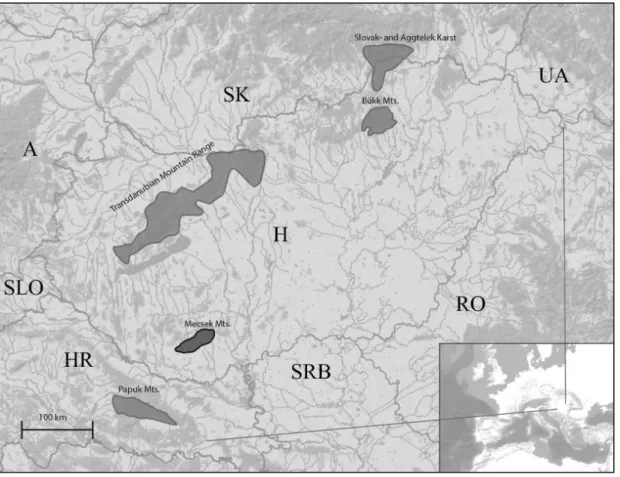
Fragmented mountain areas in East-Central Europe have been suggested to be centers of endemism that evolved through a complex geological history including Eocene marine regression-transgression cycles and Pleistocene glacial cycles (Mamos et al. 2014, Meleg et al. 2013, Hou et al. 2013). Mecsek Mountains is one of these isolated mountain ranges that is situated in South Hungary and surrounded by the Pannonian plains (Figure 1). In biological sense, the area is populated by a relatively high number of endemic species the origin of which may date back to Tertiary and which therefore apparently have survived mass extinctions in glacial periods (Gebhardt 1967). The subterranean environment of the Western Mecsek harbours numerous terrestrial and aquatic highly endemic invertebrates, known only from one or a few caves. Discovery and study of these species dates back to the end of the 19th century, when the first description of a troglobiont, endemic invertebrate species was born (Daday 1889a). Although, the real zoospeleological assessment of both the Abaligeti Cave and the Mánfai-kőlyuk Cave is due to the intensive research of Elemér Bokor, Antal Gebhardt, Lajos Méhely and Endre Dudich, carried out between the 1920s and 1930s (e.g.
Bokor 1924, Dudich 1929, Gebhardt 1931; 1933; 1934, Méhely 1925). The vast majority of the taxa found in the caves had been collected by Antal Gebhardt, who successfully cooperated with the most relevant specialists of the era such as Karl Verhoeff, which had led to the description of some highly endemic species, like the chordeumatid millipede species Hungarosoma bokori Verhoeff, 1928 (Verhoeff 1928). Due to the collectors’ and the taxonomists’ results, until 1931, the presence of 159 and 190 animal species had been revealed from the Mánfai-kőlyuk Cave and the Abaligeti Cave (e.g. Gebhardt 1933, Kolosváry 1928, Mödlinger 1930, Stach 1929). Later on, further records of additional invertebrate species (e.g. Bathynella chappuisi Delachaux, 1920, Farkas 1957) had increased the number of the taxa found in the Abaligeti Cave. According to the last publication of Gebhardt written about the zoological survey of the Mánfai-kőlyuk Cave and the Abaligeti Cave, 159 and 286 animal species, respectively were known from the two caves (Gebhardt 1967), however he included the 92 Protozoa species identified from the Abaligeti Cave by Lajos Varga too (Gebhardt 1964). That time, eight taxa had been treated as endemic of the two caves. The planarians ‘Dendrocoelides pannonicus’ (Dendrocoelum pannonicum Méhely, 1927) and ‘Polycelis tóthi’ (Polycelis tothi Méhely, 1927), the aquatic isopod ‘Stenasellus hungaricus v. robustus’ (Protelsonia hungarica robusta Méhely, 1927) and the hydrobiid
‘Paladilhiopsis gebhardti’ (Bythiospeum cf. gebhardti (H. Wagner, 1931)) were known only from the Mánfai-kőlyuk Cave, while ‘Stenasellus hungaricus’ (Protelsonia hungarica hungarica Méhely, 1924), the amphipod ‘Niphargus foreli gebhardti’ (Niphargus gebhardti Schellenberg, 1934), and the hydrobiid ‘Paladilhiopsis hungarica’ (Bythiospeum hungaricum (Soós, 1927)) were mentioned as the endemisms of the Abaligeti Cave. ‘Niphargus leopoliensis molnári’ (Niphargus molnari Méhely, 1927) was known from both caves. Three millipede species, Hungarosoma bokori Verhoeff, 1928, ‘Orobainosoma hungaricum’, (Haasea hungarica (Verhoeff, 1928)) and Brachydesmus troglobius Daday, 1889 were treated as rare fauna elements known only from a few habitats (Gebhardt 1967). Seven of these rare end endemic taxa had been chosen for further studies as objects of present thesis, namely the amphipods Niphargus molnari and Niphargus gebhardti, the aquatic isopods Protelsonia hungarica hungarica and Protelsonia hungarica robusta, the hydrobiid snails Bythiospeum
11
hungaricum and Bythiospeum cf. gebhardti and the polydesmid millipede Brachydesmus troglobius. All of them can be classified as troglobionts. Previous knowledge related to these taxa is detailed at the beginning of each chapters of the result part of present thesis.
In case of taxa with uncertain taxonomic positions - like the focal rare and endemic species -, revising all possible sources of data might increase the robustness of taxonomic conclusions (Padial et al. 2010). ‘Integrative taxonomy’ is defined as the science that aims to delimit the units of life’s diversity from multiple and complementary perspectives, applying comparative morphology, phylogeny, population genetics, ecology, development, behaviour, etc. (Dayrat 2005). Among some hardly identifiable invertebrate groups, like Niphargus or Hydrobiidae, the use of molecular studies for supplementing or confirming morphological data, spreads rapidly (e.g. Liu et al. 2001, Fehér et al. 2013, Ntakis et al. 2015). Another great advantage of molecular analysis of closely related taxa is the possibility of detecting cryptic species, which might influence future conservation decisions (Trontelj & Fišer 2009). For phylogenetic reconstruction in the order Polydesmida, the use of cladistic analysis based on morphological characters is the generally recognized method (Bueno-Villegas et al. 2008, Djursvoll et al. 2000), however recently, application of molecular taxonomic data for the same purpose also started to be unfolded (Spelda et al. 2011). Adaptation of shell morphometric studies is extremely helpful in cases of snail taxa with remote visible morphological characters (e.g. Harl et al. 2014a).
Applying the methods of modern integrative taxonomy, my aims were:
i) to revise the seven above mentioned rare and endemic troglobiont macroinvertebrate taxa from the caves of the Western Mecsek and to clarify their taxonomic positions;
ii) to contribute to the knowledge on morphology and molecular genetics of the focal taxa with the help of newly applied methods, like scanning electron microscopy, DNA barcoding or phylogenetic analysis;
iii) to record the new distributional data of the focal species and subspecies and to make suggestions on their conservation planning, based on evaluation of their rarity and the present condition of their habitats;
iv) and to create a faunistic list based on the newly collected aquatic and terrestrial macroinvertebrate material other than the revised species and subspecies.
12
Figure 1: Location of the Mecsek Mts. and other karst areas within the Pannonian biogeographical region (Made by G. Balázs).
13
2. MATERIAL AND METHODS
2.1 Introduction of the karstic area and the studied caves
The Mecsek Mountains is relatively small with its approximately 545 km² extension.
The base of the fragmented, creased-structured block mountain is a structural unit of granitoids and metamorphic rocks, formed in Variscan tectonic phase in Precambrium and early Perm. Until the end of the Paleozoic, due to the repeated and altering oriented crust movements, certain parts of the ancient mountain range descended, while others had eroded.
The area belonged to the Pangea ancient continent and had been flooded by the Tethys Sea, which had caused intensive sediment formation. During the Triassic period of the Mesozoic (250-200 million years ago), sedimentary rocks, like sandstone, slaty clay, limestone and dolomite had been stratified on the ‘Jakabhegyi Red Limestone’ formation (Lehmann 2003).
At present, 230 caves are known from the Mecsek Mts., however only 28 of them exceed 50 meters in length (Nyerges & Takácsné Bolner 2011). Mecsek Mts. can be divided into three parts: Western-, Middle- and Eastern Mecsek. Among these, Western Mecsek is the richest in karstic objects. Ten catchment areas are known from there, the most considerable ones are the Abaligeti Cave and the Kispaplika Spring in Abaliget, the Vízfő Spring and the Mészégető Spring in Orfű and the Mánfai-kőlyuk Cave, the Kánya Spring and the Melegmány Spring near Mánfa. The area is rich in surface karst phenomena too, like dolines and springs. Five caves of the area are highly protected by law.
During my investigations conducted between September 2010 and December 2013, 14 caves were studied in biospeleological aspect in the karstic area of the Western Mecsek with the permission of the Duna-Dráva National Park Directorate and the South Transdanubial Environmental Protection and Nature Conservation Inspectorate. Rare and endemic troglobiont macroinvertebrate taxa discussed in present thesis have been found in 8 of them, these caves are introduced below in the text. Basic data of the 14 studied caves are listed in Table 1. The studied caves belong to three different catchment areas. Some of the caves are hydrologically connected with each other according the following model. From the Szajha- felső Cave and the Vadetetős Cave the karstic water flows into the main passage of the Abaligeti Cave somewhere behind the siphon in a yet unknown part of the cave. As a part of the Vízfő catchment area, the karstic water of the Trió Cave, the Gilisztás Cave and the Spirál Cave associates, constituting the same hydrological system. The Mánfai-kőlyuk Cave originally was hydrologically distinct from the other six caves, however from the 1960s to the 1990s it was artificially connected with the Vízfő system for increasing the water supplement of Komló coal-mining city (Rónaki 1972, Tegzes 2012 pers. comm.).
14
Table 1: Basic data of the studied caves. Caves marked with ‘*’ refer habitats of the focal revised rare and endemic macroinvertebrates.
The highly protected Abaligeti Cave is the largest known cave in the Mecsek Mts.
With its three collaterals (Eastern, Western 1 and Western 2) and the main passage, the total length of the cave is 2000 m. Its lowest point below the entrance is 10 m, while its highest point is 38 m (Havasi et al. 2003). The Western 2 collateral is in connection with the Akácos Cave, which serves as a second entrance of the Abaligeti Cave. Hydrological connection of the Western 2 collateral with the periodically active Törökpince Cave had mentioned in the literature too (e.g. Gebhardt 1963), however the latter cave has been dried out for a long time.
The Abaligeti Cave is characterized by both streaming and stagnant water. Streaming water
Name of cave both in English and Hungarian
Type of cave Cadastre number
Entrance’s altitude above sea level (m)
Entrance’s coordinates EOV-Y
Entrance’s coordinates EOV-X
Length of cave (m)
Vertical extension of cave (m) Abaligeti Cave *
(Abaligeti-barlang)
outflow cave 4120-1 218 578 056.429 88 434.520 2000 48
Mánfai-kőlyuk Cave * (Mánfai-kőlyuk)
outflow cave 4120-2 240 585 324.364 89 720.420 360 11
Vadetetős Cave * (Vadetetős- víznyelőbarlang)
inflow cave 4120-27 320 577 872.842 86 795.058 180 35,7
Trió Cave * (Trió-barlang)
inflow cave 4120-71 301 580 722.262 86 347.182 250 58
Gilisztás Cave * (Gilisztás- víznyelőbarlang)
inflow cave 4120-70 307 580 693.262 86 268.727 134 51.1
Spirál Cave * (Spirál-víznyelő)
inflow cave 4120-130 350 582 719.925 87 242.072 1400 86.4
Szajha-felső Cave * (Szajha-felső- víznyelőbarlang)
inflow cave 4120-16 283 578 056.137 88 041.665 148 42
Törökpince Cave * (Törökpince- víznyelőbarlang)
inflow cave 4120-13 275 577 544.640 88 007.391 87 7
Római Cave (Római-zsomboly)
pothole 4120-222 248 578 465.730 88 298.610 26.6 24
Kispaplika Cave (Kispaplika- forrásbarlang)
outflow cave 4120-22 220 578 537.570 88 409.775 40 9.5
Nyárás-völgyi Cave (Nyárás-völgyi- víznyelő)
inflow cave 4120-31 291 578 760.081 86 896.453 34 19
Orfűi Vízfő Cave (Orfűi Vízfő-barlang)
outflow cave 4120-3 211 581 611.158 88 670.206 330 27
Achilles Cave (Achilles- víznyelőbarlang)
inflow cave 4120-90 288 580957.614 87 510.384 140 28
Akácos Cave (Akácos- víznyelőbarlang)
inflow cave - 269 577 686.412 88 185.955 ? ?
15
can be found in the main passage (‘Styx Stream’) and in the two Western collaterals. Shallow pools of water in the cave are of two types: some are formed by dripping water of the speleothems, whereas other are filled during floods and contain residual water. The most significant nutriment source of the cave is the epigean originated vegetal material aggregated in the stream’s alluvium and the decaying wooden remains introduced by human activity. The cave has been opened for the public since 1957. Some of the most attractive speleothem formations are illuminated by lamps, which has caused the evolvement of the ‘lamp flora’, serving alternative type of energy source for the cave-dwelling invertebrates. Bat guano aggregations are not substantial elements of the ecosystem, as the one-time vast bat colonies have been recently reduced (Szatyor 2004). With its 12.6 ºC, the average temperature of the cave is relatively high. Considerable fluctuation can be detected only until 40-50 m distance from the entrance. The relative humidity is 97%.
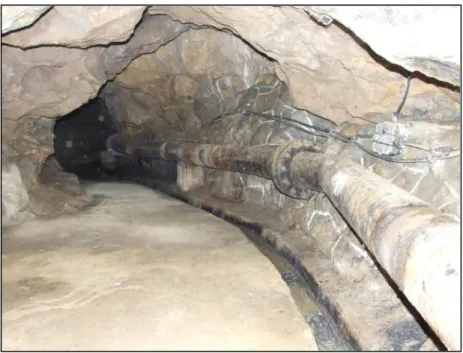
The outflow cave, Mánfai-kőlyuk Cave is situated in the Eastern edge of the Western Mecsek, 3 km from Mánfa village and by today, it is in highly protected status. It opens with a remarkable, spacious entrance. The cave possesses an upper and a lower passage, the former had been discovered until its third siphon, and then the research had to be finished because of the artificial utilization of the cave. In 1957 the local waterworks started to use the cave’s spring for supporting the water supplement of Komló mining city. In 1969, a 58 m long artificial tunnel was scooped in the upper passage, to carry the water to the dam in the Mély- völgy (‘Deep valley’) where the outflow water associated with the water of the nearby springs. 163 m from the entrance of the tunnel, a 10.8 m thick concrete dam had been emerged in order to swell the water behind that (Havasi 2003, Kordos 1984). These interventions had caused the change of the cave’s hydrological system and the destruction of the natural passages with their original formations. Utilization of the cave’s water had stopped in the 1990s, but the artificial pieces, like rusty water pipes and other instruments had been left in the cave (Figure 2). However, the Duna-Dráva National Park took steps towards the rehabilitation of the cave in 2013, it was impossible to set back the original, natural conditions. At present the possibilities for occupying suitable microhabitats in the cave by invertebrates are rather restricted. The water carrier canal in the upper passage and the streaming water in the lower passage serve as aquatic habitats, while the entrance region and the remained small lateral chambers with some organic material (e.g. decaying woods) provide shelter for the terrestrial hypogean fauna. The average temperature of the cave is 10.6 ºC, though, due to the deliberately large entrance, the fluctuation is rather high.
Vadetetős Cave is an inflow cave near Kővágótöttös village. Its present horizontal extension is 180 m, however it is under excavation by the local speleological group, Pro Natura Karst and Cave Research Society. The vast majority of the cave can be characterized by narrow passages, which widen in some certain parts, forming small halls. The cave is relatively rich in calcite and sinter formations. Shallow pools and slowly streaming waters can also be found in the cave. In the entrance region, appreciable amount of plant material (mainly decaying leaves) is deposited.
16
As parts of the same system, the two inflow caves, Trió Cave and Gilisztás Cave are situated in the Szuadó Valley near Orfű village. Their entrances are quite close to each other.
With its 55 m depth, the Trió Cave is the third deepest cave of the area. It is rather rich in speleothem formations and it possesses various combinations of spacious halls, narrow passages and high chimney-stacks. Reminding to a pothole, Gilisztás Cave consists of mainly vertical, descending parts with a siphon at its end. Both caves are built up by steel ladders to ease the movement in the cave. Regarding the water bodies, the two caves can be characterized mainly by small pools formed by dripping water and residual water. Some wooden remains can be found in the caves due to human impact, which could serve as nutrition resource for the cave-dwelling fauna.
The Spirál Cave is situated near Pécs city. With its 86 m depth, it is the deepest cave of the Mecsek Mts. Its horizontal extension is also remarkable, the known length is at present 1400 m and it is still under excavation by the Karst Research Group of Mecsek. The majority of the vertical passages are supplied by ladders, though; some parts of the caves can be visited only by applying rope technique using special equipment. Chimney-stacks, spacious halls, narrow rives and smaller chambers vary in the caves. A streamy branch can also be found at the lowest level, which ends in a siphon. Beautifully developed speleothems and sinter pews can also be observed, the ‘Spirálszíve-terem’ (Spirál’s heart hall) is the richest of them.
The Szajha-felső Cave is situated in the area of a platform right above the Abaligeti Cave and the caves are supposedly connected with each other. The cave entrances are approximately 1 km from each other (Dezső 2011). The present deepest point of the cave is 42 m, though it is still being excavated. The first part of the cave is a vertical shaft, which continues in a narrow horizontal session. The cave is rather poor in formations. A few small, permanent shallow pools can be found; epigean nutrition source is uncharacteristic.
Contrary to the above discussed caves, the Törökpince Cave is formed in conglomerate. The cave opens with an extremely tight entrance aperture, which continues in an 87 m long, narrow horizontal passage. Recently, the cave has proved to be dry in all seasons, has not contained any types of water bodies. As the cave directly opens in deciduous woodland, its first few meters contain massive amount of organic matter in all seasons, which causes the appearance of trogloxene invertebrate species in the entrance zone. The cave is periodically occupied by a badger (Meles meles).
17
Figure 2: Detail of the artificial tunnel of the Mánfai-kőlyuk Cave.
2.2 Sampling methods
The focal 8 caves were regularly visited between September 2010 and December 2013. The most frequently visited cave was the Abaligeti Cave, collection trips had been conducted in all seasons, in total 20 occasions. 8 trips in the Mánfai-kőlyuk Cave had been performed, while the other 6 caves had been visited in total 22 times. Kispaplika Cave, Orfűi Vízfő Cave, Achilles Cave, Római Cave, Akácos Cave and Nyárás-völgyi Cave had been visited less frequently; two collecting trips were conducted in each. Except in case of the show-cave Abaligeti Cave, the collecting-, observation- and documentary trips had happened by the assistance of speleologist colleagues from the Pro Natura Karst and Cave Research Society.
The applied collecting methods were heterogeneous, though, the most frequently used method was the ‘singling’, which means catching the single individual noticed on the spot.
This method has an obvious advantage: it reduces the opportunity of ‘over collecting’ in the sensitive cave ecosystem. Singling can be happened both from aquatic and terrestrial habitats, using entomological (soft) forceps, aspirator or a hand net. Occasionally, pitfall traps and aquatic traps were also used. The former meant 2 dl volume plastic cups filled with ethanol or ethylene glycol. It can be baited or unbaited. As baits, various nutriments can be used, like meat, cheese or beer which should be placed in a vial. Soil traps had been emptied after 5-10 days. Two types of traps for collecting the aquatic fauna were applied. The baited ‘bottle trap’
is suitable for collecting omnivore invertebrates, while ‘leaf litter trap’ - developed by the author - attracts mainly detritivores. The former meant a plastic bottle with a reversed open neck placed meat bait inside, while the latter meant perforated nylon pockets filled with
18
sterilized leaves. Bottle traps had been left in the water for one day, while leaf litter traps had been emptied after two weeks. Four water collectors (0.5 l volume bottles with a funnel and a drain) were placed under stalactites in the Abaligeti Cave in order to collect epikarstic water.
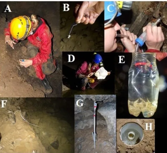
Containers were emptied monthly in total three times; water was filtered by plankton net (Figure 3). Collected specimens were fixed and stored in 70 or 96% ethanol.
Figure 3: Sampling methods applied for collecting the aquatic and terrestrial cave-dwelling macroinvertebrate fauna. A: checking of organic material (bat guano), B: singling by forceps, C: singling by aspirator, D: singling by hand net, E: bottle trap (note that the photo was taken in the Baradla Rövid-alsó Cave), F: leaf litter trap, G: collecting of epikarstic water, H: pitfall trap (Photos: D. Angyal, G. Balázs & A. Illés).
2.3 Morphological studies
2.3.1 Morphological studies on Niphargus molnari and N. gebhardti
Following the instructions of Cene Fišer taxonomist and zoospeleologist, modern Niphargus taxonomic processes had been acquired during my study trips in the Department of
19
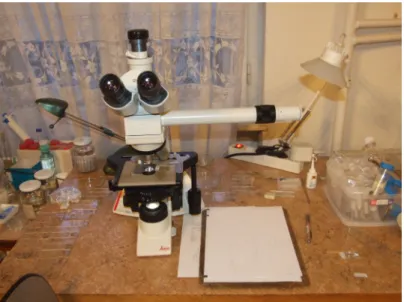
Biology, Biotechnical Faculty, University of Ljubljana. Preparation techniques were used after Fišer et al. (2009). Specimens were cooked in 10% KOH solution, rinsed with HCl and washed in distilled water. Cleared exoskeletons were stained with chlorazol black in glycerol, and then dissected in glycerol gelatin under a Leica MZ75 and a Leica M125 stereomicroscope. Two slides were made of each specimens, one contained the left side appendages and the mouth parts, while the other contained the whole body with the right side appendages. The slides were examined using a Leica DM 1000 light microscope (Figure 4).
Drawings were made using a drawing tube mounted on the light microscope and were computer graphically edited afterwards. Measurements were made using the AnalySIS Program Package, the computer was connected with a Zeiss Axioscope II light microscope.
230 morphological characters on each specimen were examined according to the characters of the DELTA program package (Fišer et al. 2009) which were recorded in an Excel data matrix.
Slides and material preserved in 96% ethanol have been deposited in the Crustacea Collection of the Hungarian Natural History Museum (HNHM).
Figure 4: Leica DM 1000 light microscope with drawing tube used for morphological analysis and for making of drawings.
2.3.2 Morphological studies on Protelsonia hungarica hungarica and P. hungarica robusta
Same procedure was applied for the dissection and for making of drawings as in case of the Niphargus specimens. In some occasions only the pleopods, gnathopods or the uropods were dissected. Slides and material preserved in 96% ethanol have been deposited in the Crustacea Collection of the HNHM.
20
2.3.3 Morphometric studies on Bythiospeum hungaricum and B. cf. gebhardti
Images of the used snail individuals were made by a camera attached to a Zeiss Axioscope II light microscope. Three independent photos were made of each individual in order to perform a repeatability test. ImageJ scientific image analysing program was used for making the measurements, from where the data were converted into an Excel file. Material preserved in 96% ethanol has been deposited in the Mollusca Collection of the HNHM.
2.3.4 Morphological studies on Brachydesmus troglobius
Brachydesmus specimens were examined under a Leica M125 stereomicroscope. In some cases the male’s gonopods were dissected and studied under higher magnification.
Studied specimens and dissected gonopods have been deposited in the Myriapoda Collection of the HNHM.
2.3.5 Scanning electron microscopy and multilayer photos
Scanning electron micrographs of the main characters of a male and a female specimen of N. molnari, N. gebhardti, B. troglobius and P. hungarica hungarica were made with a HITACHI S-2600 N scanning electron microscope in the Department of Botany of the HNHM. Specimens were placed in absolute alcohol for one day, then cleaned in an EMAG Emmi-16 Ultrasonic Cleaner and dried out on air. Dry samples were stuck onto holders and were sputter-coated by gold-palladium. Micrographs were digitally edited.
Multilayer photos of P. hungarica hungarica and B. troglobius were shot by Tamás Németh in the Department of Zoology, HNHM with Nikon D5200 camera using Mitutoyo M Plan Apo 5X microscope lens. Single flash were diffused with a paper cylinder. Exposures were stacked in Zerene Stacker.
2.4 Molecular studies
2.4.1 Molecular methods applied for studies on N. molnari and N. gebhardti
DNA extraction of two specimens of N. molnari from the Abaligeti Cave and six specimens of N. gebhardti from the Abaligeti Cave, Trió Cave, Gilisztás Cave, Vadetetős Cave, Spirál Cave and the Szajha-felső Cave (one specimen of each cave) was performed using QIAamp DNA Microkit® (Qiagen) or Sigma Aldrich GenElute Mammalian Genomic
21
DNA Miniprep Kit® following the manufactrer’s instructions (Figure 5). Only a few pereopods were used for DNA isolation of each animal. The following mitochondrial and nuclear markers were used: cytochrome c oxidase subunit I (COI), two fragmnets of 28S rDNA and histone H3. The primer pairs used for PCR amplifications are as follows: for COI:
LCO 1490 - HCO 2198 (Folmer et al. 1994), for 28S rDNA: 28S lev2 - 28S des2 or 28S rtest2 (Verovnik et al. 2005, Zakšek et al., 2007) and H3aF2- H3aR2 (Colgan et al. 2000) for histone (H3). Data of primers applied during the molecular studies on the Niphargus spp. are listed in Table 2. Protocols and thermo profiles used in PCR were as follows:
1) cytochrome c oxidase subunit I (COI) - N. molnari
Primers: F: LCO 1490, R: HCO 2198
PCR reactions (15 µl) were obtained by mixing 11 µl mQ water, 1.5 µl 10X PCR buffer (with MgCl2) , 1.5 µl dNTP, 0.2 µl of each primers (5µM), 0.07 µl BIOTOOLS DNA Polymerase®
(5U/ µl) and 1 µl DNA extract. PCR temperature conditions were as follows: initial denaturation for 4 min at 95°C, denaturation for 1 min at 95°C, hybridization for 1 min at 45°C, and polymerization for 2 min 30 sec at 72°C. After fourty cycles a final extension for 7 min at 72°C was added.
2) cytochrome c oxidase subunit I (COI) - N. gebhardti
Primers: F: LCO 1490, R: HCO 2198
PCR reactions (25 µl) were obtained by mixing 13.85 µl mQ water, 2.5 µl 10X PCR buffer, 2.5 µl dNTP mix (2mM), 1.5 µl of each primers (5µM), 0.15 µl Fermentas Dream Taq® (5U/
µl) and 3 µl DNA extract. PCR temperature conditions were as follows: initial denaturation for 3 min at 94°C, denaturation for 45 sec at 94°C, hybridization for 45 sec at 48°C, and polymerization for 1 min at 72°C. After thirty cycles a final extension for 3 min at 72°C was added.
3) 28S rDNA - N. molnari, N. gebhardti Primers: F: 28S lev2, R: 28S des2, 28S rtest2
PCR reactions (15 µl) were obtained by mixing 11 µl mQ water, 1.5 µl 10X PCR buffer (with MgCl2), 1.5 µl dNTP, 0.2 µl of each primers (5µM), 0.07 µl BIOTOOLS DNA Polymerase®
(5U/ µl) and 1 µl DNA extract. PCR temperature conditions were as follows: initial denaturation for 3 min at 94°C, denaturation for 30 sec at 94°C, hybridization for 1 min at 45°C, and polymerization for 1 min at 72°C. After fourty cycles a final extension for 5 min at 72°C was added.
4) histone H3- N. molnari, N. gebhardti
22 Primers: F: H3aF2, R: H3aR2
PCR reactions (15 µl) were obtained by mixing 11 µl mQ water, 1.5 µl 10X PCR buffer (with MgCl2) , 1.5 µl dNTP, 0,2 µl of each primers (5µM), 0,07 µl BIOTOOLS DNA Polymerase®
(5U/ µl) and 1 µl DNA extract. PCR temperature conditions were as follows: initial denaturation for 3 min at 94°C, denaturation for 45 sec at 94°C, hybridization for 1 min at 46°C, and polymerization for 1 min at 72°C. After fourty cycles a final extension for 3 min at 72°C was added.
PCR products were cleaned using Roche High Pure Purification Kit® or Exonuclease I and Alkaline Phosphatase (Fermentas, Germany) according to the manufacturer’s instructions.
The fragments were sequenced in both directions using PCR amplification primers using ABI 3130 sequencer in the Laboratory of Molecular Taxonomy of the HNHM or in Macrogen Europe (Amsterdam, The Netherlands). Contigs were assembled and sequences were edited using Geneious Pro 5.5.6. (Biomatters, New Zeland).
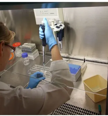
Figure 5: DNA isolation in the Laboratory of Molecular Taxonomy of the HNHM.
23
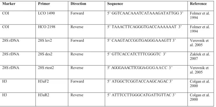
Table 2: Data of primers applied during the molecular studies on the Niphargus spp.
Marker Primer Direction Sequence Reference
COI LCO 1490 Forward 5’ GGTCAACAAATCATAAAGATATTGG 3’ Folmer et al.
1994 COI HCO 2198 Reverse 5’ TAAACTTCAGGGTGACCAAAAAAT 3’ Folmer et al.
1994 28S rDNA 28S lev2 Forward 5’ CAAGTACCGGTGAGGGAAAGTT 3’ Verovnik et
al. 2005 28S rDNA 28S des2 Reverse 5’ GTTCACCATCTTTCGGGTC 3’ Zakšek et al.
2007 28S rDNA 28S rtest2 Reverse 5’ AGGGAAACTTCGGA-GGG AACC 3’ Verovnik et
al. 2005
H3 H3aF2 Forward 5’ ATGGCTCGGTACCAAGCAGAC 3’ Colgan et al.
2000
H3 H3aR2 Reverse 5’ ATTTCCTTGGGCATGATTGTTAC 3’ Colgan et al.
2000
2.4.2 Phylogenetic analysis applied in studies on N. molnari and N. gebhardti
638 base pair long COI sequences of N. gebhardti from 6 caves of the Western Mecsek were compared to study the intraspecific variation. Sequences were edited by BioEdit Sequence Alignment Editor program and were fitted by ClustalW Multiple Sequence Alignments program. In order to recover phylogenetic relationships of N. molnari and N.
gebhardti within the genus Niphargus, a dataset of three molecular markers were complied, using available Niphargus sequences from previous studies (see references among the supplements) and Synurella ambulans as outgroup taxon (Švara et al. 2015, Meleg et al.
2013). Altogether 104 taxa were included in the final dataset. List of taxa and sequences with GenBank accession numbers used in the analyses are listed as supplements at the end of present thesis (chapter 9.1). The sequences were aligned using MAFFT 7 (Katoh & Standley 2013). Each sequence alignment was concatenated to the joint dataset and analysed as a single dataset in phylogenetic analysis. The length of combined dataset, including sequences of COI, 28S rDNA and H3 was 2068bp. A general time-reversible model with a proportion of invariant sites and a gamma distribution of rate heterogeneity (GTR+I+Γ) assuming six discrete gamma categories was chosen as the most appropriate model according to AIC and BIC criteria, using ModelGenerator (Keane et al. 2006). Phylogenetic relationships were reconstructed with Bayesian inference (BA) using MrBayes v3.2 (Ronquist & Huelsenbeck 2003). Two parallel searches with four chains each were run for 20 million generations, sampled every 1000th generation. After discarding the first 25% of the sampled trees, the final tree was constructed according to the 50% majority rule. MrBayes phylogenetic analysis was run on the CIPRES Science Gateway, www.phylo.org (Miller et al. 2012). COI sequences of
24
one individual of N. molnari from the Abaligeti Cave and two specimens of N. gebhardti from the Abaligeti Cave and the Szajha-felső Cave (one of each) have been uploaded to the GenBank (http://www.ncbi.nlm.nih.gov/) with the accession numbers KP967552 (N. molnari) and KP967553 (N. gebhardti, Abaligeti Cave) and KP967554 (N. gebhardti, Szajha-felső Cave).
2.4.3 Molecular methods applied for studies on B. hungaricum and B. cf. gebhardti
First stage:
First stage of the molecular studies on Bythiospeum taxa performed in the Laboratory of Molecular Taxonomy of the HNHM. One specimen from the Abaligeti Cave (collected in 11. 10. 2010 in the main passage’s stream from stones, 230 m from the entrance) and four individuals from the Mánfai-kőlyuk Cave (collected in 21. 10. 2011 in the upper passage from the water carrier canal) were used for the analysis. Samples were dried by vacuum centrifuge, then shells were removed under stereomicroscope and the dry body was smashed using Polycar AT reducer. DNA extraction was performed using QIAamp DNA Microcit®
(Qiagen) following the manufactrer’s instructions. The following primer pairs were used for PCR amplifications of cytochrome c oxidase subunit I (COI): LCO 1490 (Folmer et al. 1994) and COI-H (Machodrom et al. 2003). PCR reactions (25 µl) were obtained by using the following concentrations: 0.25 mM dNTP, 0.4 uM primer, 2 mM MgCl2, 2.128 ug/ul BSA, 50 ng DNS and 1 U Fermentas Dream Taq DNA Polymerase®. PCR temperature conditions were as follows: initial denaturation for 1 min at 94°C, hybridization for 1 min 30 sec at 40°C, and polymerization for 1 min 30 sec at 72°C. After fourtyone cycles a final extension for 6 min at 72°C was added. PCR products were cleaned using Roche High Pure Purification Kit®
according to the manufacturer’s instruction. The fragments were sequenced in both directions using PCR amplification primers, by Big-Dye fluorescent sequencing kit on ABI 3130 sequencer.
Second stage:
Second stage of the molecular studies was done in the Laboratory of Molecular Taxonomy of the HNHM and in the Laboratory of Molecular Systematics, Museum of Natural History, Vienna. Data of the involved specimens (10 from the Abaligeti Cave and 11 from the Mánfai-kőlyuk Cave) are listed in Table 3. Shells of the snails were removed under stereomicroscope. Only the soft bodies were used for DNA extraction, except sample BG_Man 12, which individual was smashed together with the shell because of experimental purpose. DNA extraction was performed using QIAamp DNA Microcit® (Qiagen) following the manufactrer’s instructions with a single change: instead of 20 µl Protainase K, 25 µl was added. After the overnight lysis, QIAshredder (50) was used in case of the imperfectly lysed samples. For PCR amplifications of cytochrome c oxidase subunit I (COI) LCO 1490 and HCO 2198 (Folmer et al. 1994) primers were used. PCR reactions (25 µl) were obtained by
25
mixing 13.8 µl mQ water, 2.5 µl 10X PCR buffer (with MgCl2), 2.5 µl dNTP, 1 µl 10 µg/ µl BSA, 1.5 µl of each primers (5µM), 0.2 µl Fermentas Dream Taq DNA Polymerase® (5U/
µl) and 2 µl DNA extract. PCR temperature conditions were as follows: initial denaturation for 1 min at 96°C, denaturation for 1 min at 94°C, hybridization for 1 min at 40°C, and polymerization for 1 min 30 sec at 72°C. After thirtyfive cycles a final extension for 10 min at 72°C was added.
For PCR amplifications of the 16S ribosomal RNA (16S) 16 sar - 16 sbr (Palumbi et al. 1991) and 16SLOrc2_fwd - 16SLOrc_rev (Harl et al. 2014b) primers were used. Data of the primers used during the molecular studies on Bythiospeum are listed in Table 4. PCR reactions with the ‘Palumbi primers’ were obtained for 25 µl reaction volume by mixing 11.9 µl mQ water, 2.5 µl 10X PCR buffer, 2 µl 25 mM MgCl2, 2.5 µl 2 mM dNTP, 2 µl 10 µg/ µl BSA, 1 µl of each primers (5µM), 0.1 µl Fermentas Taq DNA Polymerase® (5U/ µl) and 2 µl DNA extract. PCR temperature conditions were as follows: initial denaturation for 4 min at 92°C, denaturation for 1 min at 92°C, hybridization for 1 min at 52°C, and polymerization for 1 min at 72°C. After thirty-five cycles a final extension for 5 min at 72°C was added. In case of the ‘Harl primers’, the PCR reactions were obtained for 25 µl reaction volume by mixing 17.875 µl mQ water, 5 µl 10X PCR buffer (with MgCl2), 0.5 µl 2 mM dNTP, 0.25 µl of each primers (5µM), 0.125 µl Fermentas Taq DNA Polymerase® (5U/ µl) and 1 µl DNA extract.
PCR temperature conditions were as follows: initial denaturation for 3 min at 94°C, denaturation for 30 sec at 94°C, hybridization for 30 sec at 52°C, and polymerization for 30 sec at 60°C. After thirty-five cycles a final extension for 7 min at 72°C was added. Uncleaned PCR products had been sent to LGC Genomics (Berlin, Germany), where the Microtitre plate sequencing includes the clean-up treatment of all PCR products.
Table 3: Data of ’Hungarian blind snail’ samples used for the second stage of the molecular studies.
Sample code Cave Locality within cave Date of
collection BH_Aba_21, BH_Aba_22, BH_Aba_23, BH_Aba_24,
BH_Aba_25, BH_Aba_26, BH_Aba_27, BH_Aba_28, BH_Aba_29, BH_Aba_30
Abaligeti Cave main passage, stream, on stones, 200-300 m from the entrance
14. 04. 2014
BG_Man_01, BG_Man_02, BG_Man_03, BG_Man_04, BG_Man_05, BG_Man_06, BG_Man_07, BG_Man_08, BG_Man_09, BG_Man_10, BG_Man_12
Mánfai-kőlyuk Cave
upper (artificial) tunnel, from the water carrier canal
14. 04. 2014
26
Table 4: Data of primers used in molecular studies on Bythiospeum species.
Marker Primer Direction Sequence Reference
COI LCO 1490 Forward 5’ GGTCAACAAATCATAAAGATATTGG 3’ Folmer et al. 1994 COI HCO 2198 Reverse 5’ TAAACTTCAGGGTGACCAAAAAAT 3’ Folmer et al. 1994 COI COI-H Reverse 5′ TCAGGGTGACCAAAAAATCA 3′ Machodrom et al.
2003
16S 16 sar Forward 5’ CGCCTGTTTATCAAAACAT 3’ Palumbi et al. 1991
16S 16 sbr Reverse 5’ CCGGTCTGAACTCAGATCACG’ Palumbi et al. 1991 16S 16SLOrc2_fwd Forward 5’ TTACCTTTTGCATAATGGTTAAATTA 3’ Harl et al. 2014b 16S 16SLOrc_rev Reverse 5’ CGGTCTGAACTCAGATCATG 3’ Harl et al. 2014b
2.4.4 Phylogenetic analysis applied for studies on B. hungaricum and B. cf. gebhardti
First stage:
COI sequences were edited using Bio Edit Sequence Alignment Editor. Alignments were fitted by ClustalW Multiple Sequence Alignments program. Further sequence analysis was performed by MEGA 6 software (Tamura et al. 2013). 638 bp COI sequences of one individual of B. hungaricum and B. cf. gebhardti have been uploaded to the GenBank with the accession numbers KP296923 (B. hungaricum) and KP296922 (B. cf. gebhardti).
Second stage:
Sequences were edited using Bio Edit Sequence Alignment Editor. Alignments were fitted by ClustalW Multiple Sequence Alignments program. In order to study the phylogenetic relationships within the genus Bythiospeum and within the superfamily Rissooidea, a dataset was compiled using the own sequences and sequences downloaded from the GenBank database (http://www.ncbi.nlm.nih.gov/). Data of the downloaded COI sequences are listed in Table 5. In total 43 taxa have been involved in the analysis, including 31 Bythiospeum taxa and 12 species from 10 other rissooid genera. The major part of the genera were chosen based on the phylogenetic study of Wilke et al. (2001). Moitessieria cf. puteana was used as outgroup taxon. Distance estimation of the 43 taxa was performed by variance estimation method, using p-distance model in MEGA6 (Tamura et al. 2013). Bayesian phylogeny analysis was performed too, including the Bythiospeum COI sequences and the M. cf. puteana sequence. Saturation of phylogenetic information was examined using Xia’s test (Xia et al.
2003 and Xia & Lemey 2009) employed in DAMBE v5.3.8 (Xia & Xie 2001). The alignments showed only little substitution saturation, with ISS.C values (P= 0.000): Iss.c 0.7377
> Iss 0.1537. The most appropriate model (HKY+I) was selected with jModeltest v.0.1.1
27
(Posada 2008), under the Bayesian Information Criterion. Baysian inference was calculated with MrBayes 3.2.2 (Huelsenbeck & Ronquist 2001, Ronquist & Huelsenbeck 2003) for 5 x 10 6 generations (samplefreq = 100, nchains = 4, burnin = 10%).
Table 5: Data of rissooid sequences downloaded from National Center for Biotechnology Information homepage used in phylogenetic analysis.
Genbank accession number
Species Valid name Length of
COI fragment
Author (uploaded by)
Publication Distribution of species HM.107133.1 Bythiospeum
francomontanum
Bythiospeum francomontanum Bernasconi, 1973
603 bp Hirsch et al. 2010
unpublished France, Switzerland HM.107121.1 Bythiospeum acutum Bythiospeum
suevicum (Geyer, 1905)
603 bp Hirsch et al. 2010
unpublished Germany
HM107134.1 Bythiospeum husmanni Bythiospeum husmanni (C.
Boettger, 1963)
603 bp Hirsch et al. 2010
unpublished Germany, The Netherlands (doubtful) HM107127.1 Bythiospeum sp.
(Wasserflaare)
603 bp Hirsch et al. 2010
unpublished HM107126.1 Bythiospeum sp.
(Blautopf)
603 bp Hirsch et al. 2010
unpublished HM107125.1 Bythiospeum saxigenum
saxigenum
Bythiospeum saxigenum (Geyer, 1905)
603 bp Hirsch et al. 2010
unpublished Germany
HM107123.1 Bythiospeum pellucidum
Bythiospeum pellucidum (Seckendorf, 1846)
603 bp Hirsch et al. 2010
unpublished Germany
HM107118.1 B. suevicum Bythiospeum suevicum (Geyer, 1905)
603 bp Hirsch et al. 2010
unpublished Germany
HM107115.1 B. quenstedti quenstedti Bythiospeum quenstedti quenstedti (Wiedersheim, 1873)
603 bp Hirsch et al. 2010
unpublished Germany
AF367634.1 B. sp. (France) 638 bp Wilke
2001
Wilke et al.
2001 AF367635.1 Moitessieria cf. puteana Spiralix puteana
(Coutagne, 1883)
638 bp Wilke 2001
Wilke et al.
2001
France FJ029100.1 Bythinella carinulata Bythinella
carinulata (Drouet, 1867)
638 bp Benke et al. 2009
Benke et al.
2009
France
AY222649.1 Bythinella schmidtii Bythinella schmidtii (Kuster, 1852)
638 bp Szarowska
& Wilke 2004
Szarowska &
Wilke 2004
Slovenia
AF213340.1 Erhaia jianouensis Erhaia jianouensis Liu & Zang, 1979
638 bp Wilke et al. 2000
Wilke et al.
2000
China, India AF213348.1 Amnicola limosa Amnicola limosus
(Say, 1817)
638 bp Wilke et al. 2000
Wilke et al.
2000
Atlantic Ocean, Gulf of Maine