171
myo-Inositol
Arthur Weissbach Principle
A crude inositol dehydrogenase preparation obtained from Aerobacter aerogenes which has been grown on a medium containing inositol, catalyses the oxidation of m y o i n o s i t o l by diphospho
pyridine nucleotide ( D P N ) .
With this system microgram amounts of m y o i n o s i t o l can be estimated rapidly and simply by follow
ing the reduction of D P N spectrophotometrically
] )
. Other enzymic methods for the microdeter- mination of m y o i n o s i t o l have been described by Kean and Charalampous
2
) and Charalampous and Abrahams
3
^.
Inositol dehydrogenase catalyses the reaction:
(1) myo-Inositol -f D P N
+
^ ^ s c y / / o m y o i n o s o s e + D P N H + H
+
Under the conditions of the method described here the reaction does not go to completion, but the maximum formation of D P N H is proportional to the m y o i n o s i t o l concentration. Presumably the initial oxidation product is s c y / / o m y o i n o s o s e , although in the presence of crude inositol dehydro
genase further reactions may occur
4
). Under suitable conditions scy/Zomyoinosose is quantitatively reduced by D P N H and inositol dehydrogenase
5
).
Reagents
1 . Sodium carbonate, Na2CC>3
2 . Sodium hydrogen carbonate, NaHCC>3 3. Diphosphopyridine nucleotide, DPN
free acid; commercial preparation, see p. 1010.
4 . rajw-Inositol
commercial preparation, see p. 1023.
5. Inositol dehydrogenase
a crude preparation obtained from Aerobacter aerogenes
1
). See Appendix, p. 174.
Preparation of Solutions
I. Sodium carbonate buffer ( 0 . 5 M; pH 9 . 5 ) :
Add 0 . 5 M sodium hydrogen carbonate solution ( 4 2 g. NaHCC >3/1000 ml.) to 0 . 5 M sodium carbonate solution ( 5 3 g. Na2CO3/1000 ml.) until the pH reaches 9 . 5 (glass electrode).
II. Diphosphopyridine nucleotide ( 2 X 1 0 ~ 2
M (3-DPN):
Dissolve 7 3 . 5 mg. DPN in 5 ml. distilled water.
III. myoinositol ( 2 x 1 0 - 3 M ) :
Dissolve 1 8 . 0 mg. myoinositol in distilled water and make up to 5 0 ml.
IV. Inositol dehydrogenase (about 3 0 mg. protein/ml.)
D A. Weissbach, Biochim. biophysica Acta 77, 608 [1958].
2)
E. L. Kean and F. C. Charalampous, Biochim. biophysica Acta 36, 1 [1959].
3) F. C. Charalampous and P. Abrahams, J. biol. Chemistry 225, 575 [1956].
4
> /. M. Goldstone and B. Magasanik, Feder. Proc. J , 218 [1954].
5) A. Weissbach, unpublished.
172 Section B : Estimation of Substrates
Stability of the s o l u t i o n s
Crude inositol dehydrogenase is stable for at least four months at — 20° C. If during this period a precipitate forms it can be centrifuged down and discarded without affecting the enzyme activity.
D P N and m y o i n o s i t o l solutions are stable for many months at — 20° C. The carbonate buffer is stable indefinitely at r o o m temperature if kept in a tightly stoppered bottle.
Procedure
Experimental material
In a few cases crude extracts from heat killed bacteria inhibit the determination about 10—20%. It is recommended that when the samples for assay are very crude extracts, the bulk of the impurities should be removed by the Barium-Zinc method of
Agranoffet al.
6 ) : Mix a solution of the sample, containing 0.01 to 1.0 (jimole inositol, with
1 ml. 0.15 M Ba(OH) 2 solution, heat 15 min. at 100° C, add
1 ml. 5% ZnS0 4 -7 H 2 0 solution and centrifuge. Treat supernatant with
2.0 ml. of an aqueous slurry containing 1ml. Amberlite 1RA-400 (OH~-form) (as packed wet resin),
centrifuge again, concentrate the supernatant
in vacuoto 1 ml. and use a portion of this solution for estimation of myoinositol.
Standard curve
The myoinositol content of the sample may be read directly from a standard curve deter
mined for each enzyme preparation. However, it is usual to set up control cuvettes contain
ing 0.02, 0.06 and 0.1 pimoles myoinositol ( = 0.01, 0.03 and 0.05 ml. solution III) when unknown samples are to be assayed. Optical densities of approximately 0.080, 0.240 and 0.400 should be obtained.
Spectrophotometric m e a s u r e m e n t s
Wavelength: 340 mpi; light path: 1 cm.; final volume: 1.0 ml.
Read against a control cuvette containing enzyme solution since this absorbs slightly at 340irtfji. The sample should not contain more than 0.1 pimole inositol. The standard curve is only linear up to a concentration of 10~
4
M inositol.
Pipette the solutions in the given order into the cuvettes:
Experimental cuvette Control cuvette
0.20 ml. buffer (solution I) 0.20 ml. buffer (solution I) 0.10 ml. DPN solution (II) 0.10 ml. DPN solution (II) 0.05 ml. enzyme solution (IV) 0.05 ml. enzyme solution (IV) Sample + dist. water to give 1.00 ml. total volume 0.65 ml. dist. water
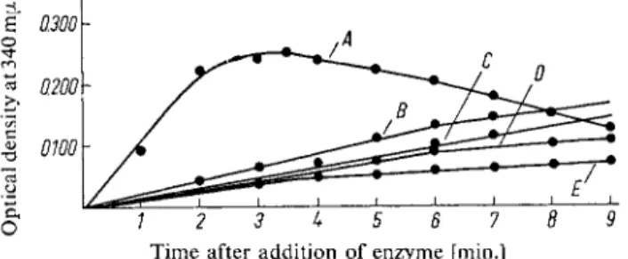
The absorption due to formation of DPNH reaches a maximum value 3 to 4 min. after the start of the reaction. This value is used. The fall in optical density which occurs with longer reaction times is at least partly due to the presence of DPNH oxidase activity in the enzyme preparation (Fig. 1).
6) B. W. Agranoff, R. M. Bradley and R. O. Brady, J. biol. Chemistry 233, 1077 [1958].
1.2.0 Myo-Inositol
173 Calculations
The value for m y o i n o s i t o l in (jimoles/ml. assay mixture is read from the standard curve by use of the optical density obtained approximately 3 to 4 min. after the start of the reaction. T o calculate the m y o i n o s i t o l concentration of the sample it is necessary to divide by the volume of the sample.
A n y dilution of the sample occurring during the preliminary treatment has to be taken into account.
0.1 pimole Inositol = 18 u.g.
Time after addition of enzyme [min.]
Fig. 1. Time curve for the oxidation of A : 0.07 [xmoles m y o i n o s i t o l — B : 0.07 xmoles « e o i n o s i t o l — C : 1.0 (jimole glucose — D : 0.07 pimoles D-inositol — E : 0.10 (xmoles scyllitol.
Enzyme: Inositol dehydrogenase.
Sources of Error and Specificity
The substrate specificity of crude inositol dehydrogenase (Table 1) in general reflects the specificity of whole cells
7
). Sequoyitol ( = m y o i n o s i t o l monomethyl ether) reacts like m y o i n o s i t o l itself.
Glucose in low concentrations does not react; above 10~
3
M, glucose, dihydroxyacetone and glycer
aldehyde react slowly (Fig. 1). It is surprising that scyllitol which contains no axial O H group reacts Table 1. Substrate specificity of a crude inositol dehydrogenase preparation from A.aerogenes
Increase in Substrate u.mole optical density
at 340 mpi*)
m y o i n o s i t o l 0.07 0.255
Pinitol 0.07 0.031
c/s-lnositol 0.07 0
e/?/-Inositol 0.07 0.010
tf//olnositol 0.07 0
Dambonitol 0.07 0
Quebrachitol 0.07 0
L-Inositol 0.07 0
/?eolnositol 0.07 0.075
D-Inositol 0.07 0.050
Scyllitol 0.10 0.050
DL- 1 -O-methyl-myoinositol 0.07 0.010
Sequoyitol 0.08 0.300
Ribose 0.05 0
Glucose 0.05 0
Galactinol**) 0.10 0
Inositol m o n o p h o s p h a t e * * * ) 0.08 0 Inositol monophosphate after treatment with
wheat phytase) 0.08 0.286
*) 3
J
/z minutes after the addition o f the enzyme
**) a-D-galactosyl-myoinositol
***) From the California Foundation for Biochemical Research.
7
) B. Magasanik, J. biol. Chemistry 205, 1007 [1953].
174 Section B: Estimation o f Substrates
slowly. Possibly the preparation was contaminated with a small amount o f m y o i n o s i t o l , which could also explain the slow reaction of D-inositol and Myoinositol. N o n e of the substrates listed in Table 1 inhibit the oxidation of m y o i n o s i t o l . However 1 0 ~
4
colchicine gave a 5 0 % inhibition of the assay, which is to be expected from the studies of Franzl and Char gaff®.
Other Methods
Charalampous et al.
3
) determined m y o i n o s i t o l by anaerobic oxidation with 2,6-dichlorophenolindo- phenol catalysed by inositol dehydrogenase and pig heart diaphorase. Inositol oxidase converts m y o i n o s i t o l to glucuronic acid, which m a y be estimated by the orcinol reaction or by T P N H and a T P N H - l i n k e d dehydrogenase
2
).
Appendix
Inositol dehydrogenase*) Aerobacter aerogenes, strain 4 1 1 2 4 * ) ; culture m e d i u m
7
) : 0.27% N a
2
H P 04
; 1.58% K H2
P 04
; 0.02%M g S 0
4
- 7 H2
0 ; 0.001 % C a C l2
; 0 . 2 % ( N H4
)2
S 04
; 5 x 10~4
M myo-inositol; p H 6.0. Shake culture for 18 hours at 37° C with vigorous aeration. Wash the cells at the centrifuge with cold distilled water.
Shake 3 g. cell paste with 10 ml. 0.1 M tris buffer (pH 7.5) and 7 g. glass powder in a Nossal shaker for 1 min. at 0 ° C (frequency: 15/sec). Alternatively grind 3 g. cell paste with 9 g. alumina powder A-301 for 15 min. at.0°C, then add 15 ml. 0.1 M tris buffer (pH 7.5). Centrifuge the suspension for 30 min. at 0 ° C and 2 0 0 0 0 g. Store the supernatant at - 2 0 ° C .
*) From the N e w Y o r k State Department of Health.
8) R. Franzl and E. Chargaff, Nature [London] 168, 955 [1951].