Research Article
Study of the Potential Endocrine-Disrupting Effects of Phenylurea Compounds on Neurohypophysis Cells In Vitro
Krisztián Sepp ,1Zsolt Molnár,2Anna M. László,3Tünde Alapi,4László Tóth,2 Andrea Serester,2Zsuzsanna Valkusz,1Márta Gálfi,2and Marianna Radács2
1First Department of Internal Medicine, Faculty of Medicine, University of Szeged, Szeged, Hungary
2Institute of Applied Science, Department of Environmental Biology and Education, Gyula Juhász Faculty of Education, University of Szeged, Szeged, Hungary
3Department of Biometrics and Agricultural Informatics, Faculty of Horticultural Science, Szent István University, Budapest, Hungary
4Department of Inorganic and Analytical Chemistry, Faculty of Science and Informatics, University of Szeged, Szeged, Hungary
Correspondence should be addressed to Krisztián Sepp; sepp.krisztian@med.u-szeged.hu
Received 20 September 2018; Revised 1 November 2018; Accepted 15 November 2018; Published 10 February 2019
Academic Editor: Rosaria Meccariello
Copyright © 2019 Krisztián Sepp et al. This is an open access article distributed under the Creative Commons Attribution License, which permits unrestricted use, distribution, and reproduction in any medium, provided the original work is properly cited.
Homeostatic disruptor agents, and endocrine disruptor compounds (EDC) specifically, can originate from agricultural and industrial chemicals. If they modify the adaptation of living organisms as direct (e.g., by altering hormone regulation, membrane functions) and/or indirect (e.g., cell transformation mechanisms) factors, they are classified as EDC. We aimed to examine the potential endocrine-disrupting effects of phenylurea herbicides (phenuron, monuron, and diuron) on the oxytocin (OT) and arginine-vasopressin (AVP) release of neurohypophysis cell cultures (NH). In our experiments, monoamine-activated receptor functions of neurohypophyseal cells were used as a model.In vitroNH were prepared by enzymatic (trypsin, collagenase) and mechanical dissociation. In the experimental protocol, the basal levels of OT and AVP were determined as controls. Later, monoamine (epinephrine, norepinephrine, serotonin, histamine, and dopamine) activation (10−6M, 30 min) and the effects of phenylurea (10−6M, 60 min) alone and in combination (monoamines 10−6M, 30 min + phenylureas 10−6M, 60 min) with monoamine were studied. OT and AVP hormone contents in the supernatant media were measured by radioimmunoassay. The monoamine-activated receptor functions of neurohypophyseal cells were modified by the applied doses of phenuron, monuron, and diuron. It is concluded that the applied phenylurea herbicides are endocrine disruptor agents, at least in vitro for neurohypophysis function.
1. Introduction
Nowadays, society is the most complex evolutionary scene, in which transactions are realized between human beings and their environment (including the atmosphere, hydro- sphere, and lithosphere). In this relation, people modify their environment so as to maintain their existence (e.g., chemisation) [1]. Chemisation came to be as a result of the biological and ecological problems caused by the vast and ever increasing amounts of chemicals used in agriculture (pesticides, herbicides, and fertilizers) [2]. These chemicals are xenobiotics [3]. These days, persistent organic pollutants (POPs) are placed in the focus. Endocrine disruptor
compounds (EDC) are a group of heterogeneous POPs, which can interfere with endocrine communication [4, 5].
Phenylurea herbicides as POPs inhibit photosynthesis and can be found as contaminants in surface water and ground- water [6, 7]. Phenylureas have been used in agriculture extensively because of their herbicidal properties [8]. Sev- eral of the chlorinated benzenes are known to be por- phyrogenic, carcinogenic, and mutagenic in animals and humans [9]; EDC lead to anxiety and aggression in rats [10] and to predator avoidance and reproductive and social behaviors in fish [7].
Biological objects, in order to maintain their inner homeostatic balance, take inputs from their environment,
Volume 2019, Article ID 1546131, 9 pages https://doi.org/10.1155/2019/1546131
of the pituitary gland store these peptide hormones and release them into blood vessels to provoke their biological functions [12, 13].
OT secretion induced by stimuli is inversely propor- tional to the inhibition of food intake and gastric motility and stimulates the release of somatostatin and gastrin [14].
On the other hand, the actions of OT modulate neuroendo- crine reflexes to establish complex social behaviors [15].
Beyond its peripheral effects on reproductive organs, OT might have an effect on neurons responsible for the cogni- tive feelings of organisms [16]. It is known that AVP and OT affect social cognition, particularly the acquisition and retention of social recognition in rats and mice [17]. Both hypotension and extracellular fluid volume expansion appear to stimulate hypothalamic oxytocinergic neurons and OT secretion; in rats, OT increases blood pressure and pulse rate [18]. There are several published studies investi- gating the prosocial effects of OT in humans: behaviors that facilitate interpersonal relations, including trusting behav- ior, generosity, and cooperation [19, 20]. In the female reproductive system, the pregnant uterus is one of the tra- ditional targets of OT [21]. Systematic OT could also act peripherally, stimulating smooth muscle cells of the male reproductive tract [20].
The peptidergic hormone AVP can also be released by external stimuli and plays roles as both neurotransmitter and neuromodulator in the brain [22]. AVP participates in the regulation of the cardiovascular system and water-electrolyte balance [23]. As an antidiuretic hormone, AVP regulates body osmolality, blood volume, and blood pressure [24]. From these studies, it is postulated that OT and AVP are crucial in the adaptation processes of living organisms [25].
In homeostatic balance, biogenic amines play an essen- tial role in the regulation of AVP and OT. Biogenic amines (with the ability to activate or inhibit OT and AVP) are important in the response mechanisms to external inputs.
It is well documented that neuronal systems contain bio- genic amines, such as dopamine (DA), epinephrine (E), nor- epinephrine (NE), histamine (HA), and serotonin (5-HT).
These mediators may contribute reinforcement information to influence memory performance [26, 27].
The monoamine-regulated OT and AVP release serves as a model for the study of neuroendocrine cell function [28–32].
The integrity of neuroendocrine regulation can be con- trolled by following endocrine function attractors inin vitro models [33, 34]. Such regulation attractor in the OT and AVP secretion in vitro model is the monoamine-activated (agonistic or antagonistic) hormone secretion result.
Homeostatic regulation is modified by environmental factors (e.g., presence of EDC). The effects of EDC factors
We investigated the potential endocrine-disrupting effects of phenylurea herbicides (phenuron-PU, monuron-MU, and diuron-DU) on the monoamine-regulated oxytocin and vasopressin release of neurohypophysis cell cultures.
2. Materials and Methods
2.1. Experimental Protocol.Male Wistar rats (Charles River, Isaszeg, Hungary, medically certified) from different litters (weighing 120-250 g, aged 4-6 weeks at the beginning of the research) were used for hypophysis cell culture model systems. The animal care and research protocols were in full accordance with the guidelines of the University of Szeged, Hungary. During the research period, rats were kept under controlled relative air humidity of 55-65%
and 22 ± 2°C ambient temperature. Experimental animals lived under automated diurnal conditions (12 h darkness and 12 h light system) in groups of 10 animals. Standard pellet food and tap water were available ad libitum. After pentobarbital anaesthesia (4.5 mg/kg b.w. Nembutal, Abbott, USA), the animals were killed and decapitated.
Neurohypophysis and adenohypophysis tissues were sepa- rated under a preparative microscope.
The neurohypophysis tissue was digested enzymatically with 0.2% trypsin (Sigma, Germany) in phosphate-buffered saline for 60 min and with 0.05% collagenase (Sigma, Ger- many) for an additional 60 min at 37°C. The enzymatic hydrolysis was stopped by the addition of 100μg/mL trypsin inhibitor (Sigma, Germany). Mechanical disintegration of the tissue was performed on nylon blutex sieves (pore sizes 100, 80, and 48μm in series). Cultures were controlled for both viability (>95%; trypan blue exclusion) and function, and the cell density was determined to be 2×10⁵cells/mL.
The dispersed cells were placed onto 24-well plastic plates (Costar, USA) coated with 5% rat-tail collagen (Sigma, Ger- many). The starting cell density was 2 × 10⁵ cells/mL of medium (Dulbecco’s modified Eagle’s medium; Sigma, Ger- many) supplemented with 20% foetal calf serum (Gibco, USA) and 100μg/L PENSTREP (Sigma, Germany). The cell cultures were maintained at 37°C in a humidified atmosphere of 5% CO2in air. The culture medium was changed every 3 days. The 14-day-old neurohypophysis primary cell cultures were standardised by immunohistochemical methods (by relative incidence/unit plate-area of IR content OT or AVP cells), and the OT and AVP time-release kinetic activity was determined.
After these procedures, the basal OT and AVP levels were measured in neurohypophysis cell cultures.
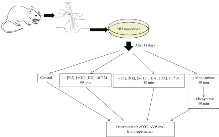
2.2. Cell Culture Treatment Protocol (Figure 1).The control samples were untreated and served as self-controls, which showed the basal release (60 min) of OT and AVP in NH.
The effects of PU, MU, and DU added to the NH for 60 minutes at concentration of 10−6M were examined one by one [36]: PU CAS registry number: 101-42-8, DU CAS registry number: 330-54-1, and MU CAS registry number:
150-68-5 (Sigma, Germany). In our earlier studies, dose-dependent kinetics of phenylurea agents were deter- mined, and the experimental doses were selected, because the saturation of receptor-binding sites depends on the affinity and number of receptor-binding molecules.
Monoamine-activated G-protein receptors in NH were treated with 10−6M (E, NE, 5-HT, HA, and DA) (Figures 2(a) and 2(b)) for 30 min [28–32] and the same concentration (10−6M) of phenylurea agents.
The combined treatment groups of the NH were treated firstly with monoaminergic compounds (for 30 min), then phenylurea compounds (for 60 min): (1) E + PU, NE + PU, 5-HT + PU, HA + PU, and DA + PU; (2) E + MU, NE + MU, 5-HT + MU, HA + MU, and DA + MU; and (3) E + DU, NE + DU, 5-HT + DU, HA + DU, and DA + DU.
The OT and AVP contents were detected in the superna- tant media. From the supernatant media, 500μL samples were removed by Gilson pipette at appropriate times and stored at−80°C until peptide radioimmunoassay (RIA) was performed to measure OT and AVP [10, 37].
A modified Lowry method [38] and Pierce BCA Protein Assay Kit (Thermo Fisher Scientific Inc., Rockford, USA) were used for the determination of total protein content.
2.3. Statistical Analysis.Pooled samples of neurohypophysis cell cultures (12 lots) were measured for OT and AVP
hormone release in different EDC groups (control, PU, MU, and DU) by monoamine regulation (basal, E, NE, 5-HT, HA, and DA) in rats (n= 10or 12 per group). Data were analyzed using mixed models [39–41]. The mono- amine regulation cycle was verified in mixed models for the comparison of 6 monoamine levels in the control groups for OT and for AVP. In the random intercept model, monoamine was used as the fixed factor and the lots as the intercept.
For the two investigated hormone data (OT, AVP), mixed models were applied with EDC and monoamine as fixed effects and random intercept for the lots. In the analy- sis models, the reference group was the control (no EDC treatment) basal (no monoamine) group.
Restricted maximum likelihood estimation and the Kenward-Roger method for adjusting the degrees of free- dom were applied in all models with unstructured covari- ance matrix. Pairwise comparisons were estimated by least squares means usingŠidákpvalue adjustment. Model resid- uals were checked for normality assumptions.
Statistical analyses were performed in SAS (Version 9.3 SAS Institute Inc., Cary, NC, USA), wherepvalues of<0.05 were considered to indicate statistical significance [42].
3. Results
Stability of homeostatic systems can be followed by examin- ing the endocrine regulation cycle. Therefore, the monoamine-activated OT/AVP release cycles were modelled by NHin vitro.
NH monolayer
Determination of OT/AVP level from supernatant + [PU], [MU], [DU]: 10−6 M
60 min
+ [E], [NE], [5-HT], [HA], [DA]: 10−6 M 30 min
+ Monoamines 30 min
+ Phenylureas 60 min After 14 days
Control
Figure1: Thein vitrotreatment protocol. PU: phenuron; DU: diuron; MU: monuron; E: epinephrine; NE: norepinephrine; 5-HT: serotonin;
HA: histamine; DA: dopamine; OT: oxytocin; AVP: arginine-vasopressin.
The result of PU, MU, and DU alone on neurohypophy- sis cell culture can be seen in Figures 3(a) and 3(b). OT release was not modified significantly by treatment with MU or DU, but PU (176 67 ± 1 25 ngOT/mg protein) can modulate it compared to the control (153 13 ± 0 63 ng OT/mg protein). AVP release did not show significant dif- ference in the case of phenylurea agents.
OT and AVP release is an indicator of the monoamine-activated receptor cycle of neurohypophysis cells. The changing of these mechanisms was investigated using phenylurea agents (Figures 4 and 5). E (292 81 ± 1 49 ng OT/mg protein) activated OT secretion (153 13 ± 0 63 ng OT/mg protein) (Figure 4(a)) was significantly decreased by PU (268 71 ± 1 07 ng OT/mg protein) and DU (255 ± 0 10 ngOT/mg protein). Figure 4(b) shows the effects of phenylureas on NE activation. MU (252 47 ± 4 75 ng OT/mg protein) and DU (277 09 ± 1 10 ng OT/mg protein) significantly reduced the effect of NE (316 55 ± 1 36 ng OT/mg protein). OT exocytosis induced by 5-HT is shown in Figure 4(c). The used phenylurea agents caused only
discrete changing of OT release in NH. In the case of HA (292 91 ± 3 16 ng OT/mg protein), only the combination with DU (264 89 ± 1 39 ng OT/mg protein) reduced the OT release significantly as seen in Figure 4(d). Figure 4(e) shows OT secretion for DA (342 06 ± 2 65 ng OT/mg pro- tein), and again, only the combination with DU (318 72 ± 1 74 ng OT/mg protein) decreased it significantly.
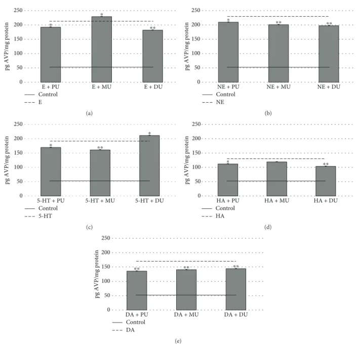
In the case of AVP hormone, the E (212 41 ± 0 59 pg AVP/mg protein) activation was modified significantly by PU (193 01 ± 0 54 pg AVP/mg protein), DU (182 94 ± 0 59 pg AVP/mg protein), and MU (230 18 ± 0 55 pg AVP/mg pro- tein) (Figure 5(a)). NE (231 33 ± 2 41 pg AVP/mg protein) induced AVP release (Figure 5(b)) was reduced by PU (211 96 ± 0 49 pg AVP/mg protein), MU (203 95 ± 0 64 pg AVP/mg protein), and DU (201 19 ± 0 52 pg AVP/mg pro- tein). The effects of 5-HT (194 11 ± 0 49 pgAVP/mg protein) on NH cell AVP release (Figure 5(c)) were decreased by PU (172 88 ± 0 43 pgAVP/mg protein) and MU (163 70 ± 0 54 pg AVP/mg protein) and increased by DU (214 44 ± 0 90 pg AVP/mg protein). In Figure 5(d), it can be seen that the effect
200 175 150 125 100 75 50 25 0
ng OT/mg protein
Monoamine groups Norepinephrine
Basal Epinephrine Serotonin Histamine Dopamine
(a)
125
100 75
50
25 0
pg AVP/mg protein
Basal Epinephrine Norepinephrine Serotonin Hitamine Dopamine Monoamine groups
(b)
Figure2: The effect of monoamines on OT (a) and AVP (b) release in neurohypophysis cell cultures. Pairwise comparisons verified the regulation cycle: all monoamine [10−6M] groups increased significantly (∗∗∗p< 0 0001) compared to the basal regulation in control OT (n= 12) and AVP (n= 10). All data presented asmean ± Std.
400 350 300 250 200 150 100 50
0 Control Phenuron Monuron Diuron
ng OT/mg protein
⁎
(a)
Control Phenuron Monuron Diuron 250
200
150
100
50
0
pg AVP/mg protein
(b)
Figure3: The effect of phenylureas [10−6M] on OT (a) and AVP (b) secretion in neurohypophysis cell cultures. (a)n= 12,mean ng OT/
mg protein ± Std;∗p< 0 01. (b)n= 10,mean pg AVP/AVP protein ± Std.
of HA (131 65 ± 0 53 pg AVP/mg protein) induced AVP release was diminished by PU (113 55 ± 0 72 pg AVP/mg protein) and DU (105 48 ± 0 89 pgAVP/mg protein). The DA (172 11 ± 0 94 pgAVP/mg protein) receptor-mediated AVP release (Figure 5(e)) in NH was significantly depressed by PU (138 72 ± 0 65 pg AVP/mg protein), MU (143 19 ± 0 71 pg AVP/mg protein), and DU (146 34 ± 1 24 pgAVP/mg protein).
4. Discussion and Conclusions
Bretaud et al. pointed out that exposures to sublethal con- centrations of carbofuran, DU, and nicosulfuron have remarkable effects on acetylcholinesterase activity in the brain of goldfish (Carassius auratus) [43]. It is described that the herbicide MU inhibits growth and heterocyst formation in the nitrogen-fixing cyanobacterium Nostoc muscorum
[44]. The results of Federico et al. showed direct genotoxic activity caused by the effects of PU, DU, and difenoxuron, as is demonstrated by the increasing number of chromo- somal aberrations and sister chromatid exchange in Chinese hamster ovary and epithelial cell lines [45]. However, there is a serious lack of information on the effects of these agents on the endocrine cells.
In neuroendocrine communication, monoamines are known to be relevant regulators for the hormone release.
Earlier studies have established that α1 receptors are involved in the E-induced increase in OT and AVP secre- tion, while the β2 receptor has a role in NE-mediated OT and AVP secretion. 5-HT1and 5-HT2receptors are involved in the 5-HT-induced increase in OT secretion while only 5-HT2 receptor has a role in the AVP secretion in NH.
Our results show that H1 and H2receptors are involved in
Control 400
350 300 250 200 150 100 50 0
E + PU E + MU E + DU
ng OT/mg protein
⁎ ⁎
E
(a)
Control NE 400 350 300 250 200 150 100 50 0
NE + PU NE + MU NE + DU
ng OT/mg protein
⁎⁎ ⁎
(b)
Control 5-HT 400
350 300 250 200 150 100 50 0
5-HT + PU 5-HT + MU 5-HT + DU
ng OT/mg protein
(c)
Control HA 400 350 300 250 200 150 100 50 0
HA + PU HA + MU HA + DU
ng OT/mg protein
⁎
(d)
Control DA 400 350 300 250 200 150 100 50 0
DA + PU DA + MU DA + DU
ng OT/mg protein
⁎
(e)
Figure4: The effect of 10−6M phenylureas on OT release in NH via 10−6M monoamine-activated receptor functions. n= 12; mean ng OT/mg protein ± Std; ∗p< 0 01 and ∗∗p< 0 001. OT: oxytocin; PU: phenuron; MU: monuron; DU: diuron; E: epinephrine; NE:
norepinephrine; 5-HT: serotonin; HA: histamine; DA: dopamine.
the HA-induced increase in OT and AVP release in neuro- hypophysis cell cultures. D1 receptors are involved in the DA-induced enhancement of OT and AVP secretion. All mentioned monoamine receptors are G-protein-coupled, and cAMP-mediated mechanisms are induced in cells. OT and AVP hormone secretion is realized through protein kinase-C mechanisms (Figures 2(a) and 2(b)) [20, 46, 47].
In our in vitro model system, the EDC effects were detected by following hormone release via the disturbance of monoamine receptor function. The phenylureas alone did not show effects on basal hormone secretion of NH (Figure 3(b)), expect for PU on OT release (Figure 3(a)). How- ever, the neurohypophysis cell function was modified by the combined treatment of monoamines and PU, MU, and DU.
With regard to OT secretion, PU has an effect in epinephrine
activation (Figure 4). MU has a stronger effect in NE activa- tion. E, NE, HA, and DA activation was decreased signifi- cantly by the effect of diuron. In the phenylurea homologue line, the used DU was twice chloro substituted which can explain the difference in the endocrine effect [48, 49].
With regard to AVP release, the effects of used phenylur- eas were more expressed in all monoamine activation (Figure 5). PU had a significant effect on E, NE, 5-HT, HA, and DA monoamine activation. E activation was enhanced, and NE, 5-HT, and DA activation was reduced significantly by the used dose of MU (with the exception of HA activation). DU increased serotonin activation and decreased E, NE, HA, and DA monoamine activation sig- nificantly. These results are interesting in the case of AVP (as neurotransmitter and neuromodulator) because this
pg AVP/mg protein 50
0 E + PU E + MU E + DU
Control E
(a)
Control NE pg AVP/mg protein 50
0 NE + PU NE + MU NE + DU
(b)
Control 5-HT
pg AVP/mg protein
250 200 150 100 50
0 5-HT + PU 5-HT + MU 5-HT + DU
⁎ ⁎⁎
⁎
(c)
Control HA
pg AVP/mg protein
250 200 150 100 50
0 HA + PU HA + MU HA + DU
⁎ ⁎⁎
(d)
Control DA
pg AVP/mg protein
250 200 150 100 50
0 DA + PU DA + MU DA + DU
⁎⁎ ⁎⁎ ⁎⁎
(e)
Figure5: The effect of 10−6M phenylureas on AVP release in NH via 10−6M monoamine-activated receptor functions.n= 10;mean pg AVP/mg protein ± Std;∗p< 0 01and∗∗p< 0 001; AVP: arginine-vasopressin; PU: phenuron; MU: monuron; DU: diuron; E: epinephrine;
NE: norepinephrine; 5-HT: serotonin; HA: histamine; DA: dopamine.
hormone has an essential role in the endocrine-regulating body of osmolality, blood volume, blood pressure, cell con- traction, and proliferation [23, 50]. The abovementioned hormones play a crucial role in horizontal homeostatic regu- lation; thus, altering the secretion of OT and AVP may influ- ence the adaptation processes of living organisms to the external environment.
Thesefindings confirm the classification of phenylureas as EDC. The environmental pollution by EDC may have chronic and/or acute effects on living organisms. Our results showed that the applied doses of phenylurea herbicides have direct and indirect effects on biological systems. Presumably, these phenylurea herbicides can disturb the association of monoamines to their receptors. The applied EDC per se play the role of possible modulator agents. In this context, neuro- hypophysis function is a crucial conditioning process.
E-, NE-, HA-, 5-HT-, and DA-activated hormone release mechanisms are mediated G-protein-coupled receptors [28, 29, 51]. These processes induce Gq/G11proteins, which, in turn, activate phospholipase-C, which catalyses the hydroly- sis of phosphatidylinositol 4,5-bisphosphate to yield inositol triphosphate and diacylglycerol [52–54]. These signal trans- ducers can enhance the intracellular [Ca2+] which is essen- tial for OT and AVP expression. A detailed understanding of the mechanisms of hormone expression and functions is required to fully understand the workings of EDC, particu- larly the possibility that low doses of PU, MU, and DU might interfere with monoaminergic receptors.
In conclusion, the effects of phenylurea herbicides on monoamine-induced OT and AVP release show an EDC character based our results of anin vitromodel system.
Data Availability
Requests for data, after initial publication, will be considered by the corresponding author.
Conflicts of Interest
The authors declare that they have no conflict of interest.
Authors’Contributions
Krisztián Sepp and Zsolt Molnár wish it to be known that, in their opinion, the first two authors should be regarded as jointfirst authors.
Acknowledgments
This research was supported by the European Union and the State of Hungary, co-financed by the European Social Fund in the framework of TÁMOP-4.2.4.A/2-11/1-2012-0001
“National Excellence Program” and EFOP-3.6.1-16- 2016-00008.
References
[1] H. Kitano, “Systems biology: a brief overview,” Science, vol. 295, no. 5560, pp. 1662–1664, 2002.
[2] E. Diamanti-Kandarikis, J. P. Bourguignon, L. C. Giudice et al.,
“Endocrine-disrupting chemicals: an Endocrine Society scien- tific statement,”Endocrine Reviews, vol. 30, no. 4, pp. 293–342, 2009.
[3] R. Melnick, G. Lucier, M. Wolfe et al., “Summary of the National Toxicology Program’s Report of the endocrine dis- ruptors low-dose peer review,”Environmental Health Perspec- tives, vol. 110, no. 4, pp. 427–431, 2002.
[4] J. R. Roy, S. Chakraborty, and T. Chakraborty,“Estrogen-like endocrine disrupting chemicals affecting puberty in humans – a review,” Medical Science Monitor, vol. 15, pp. 137–145, 2009.
[5] L. S. Birnbaum and S. Fenton, “Cancer and developmental exposure to endocrine disruptors,”Environmental Health Per- spectives, vol. 111, no. 4, pp. 389–394, 2003.
[6] M. A. Oturan, M. C. Edelahi, N. Oturan, K. El Kacemi, and J. J.
Aaron, “Kinetics of oxidative degradation/mineralization pathways of the phenylurea herbicides diuron, monuron and fenuron in water during application of the electro-Fenton pro- cess,” Applied Catalysis B: Environmental, vol. 97, no. 1-2, pp. 82–89, 2010.
[7] G. R. Scott and K. A. Sloman,“The effects of environmental pollutants on complexfish behaviour: integrating behavioural and physiological indicators of toxicity,”Aquatic Toxicology, vol. 68, no. 4, pp. 369–392, 2004.
[8] M. C. Gennaro, E. Marengo, V. Gianotti, and V. Maurino,
“New strategies for the determination of phenylurea pesticides by gas chromatography with hot splitless inlet systems,”Jour- nal of Chromatography A, vol. 910, no. 1, pp. 79–86, 2001.
[9] G. Pagano, M. Cipollaro, G. Corsale et al., “Comparative toxicities of benzene, chlorobenzene, and dichlorobenzenes to sea urchin embryos and sperm,”Bulletin of Environmen- tal Contamination and Toxicology, vol. 40, no. 4, pp. 481– 488, 1987.
[10] Z. Valkusz, G. Nagyéri, M. Radács et al.,“Further analysis of behavioral and endocrine consequences of chronic exposure of male Wistar rats to subtoxic doses of endocrine disruptor chlorobenzenes,” Physiology & Behavior, vol. 103, no. 5, pp. 421–430, 2011.
[11] Z. Molnár, R. Pálföldi, A. László et al.,“The effects of hypoka- laemia on the hormone exocytosis in adenohypophysis and prolactinoma cell culture model systems,”Experimental and Clinical Endocrinology & Diabetes, vol. 122, no. 10, pp. 575– 581, 2014.
[12] M. J. Brownstein, J. T. Russel, and H. Gainer,“Synthesis, trans- port, and release of posterior pituitary hormones,” Science, vol. 207, no. 4429, pp. 373–378, 1980.
[13] G. J. De Vries and R. M. Buijs,“The origin of the vasopressi- nergic and oxytocinergic innervation of the rat brain with spe- cial reference to the lateral septum,”Brain Research, vol. 273, no. 2, pp. 307–317, 1983.
[14] B. Olson, M. D. Drutarosky, M. S. Chow, V. J. Hruby, E. M.
Stricker, and J. G. Verbalis,“Oxytocin and an oxytocin agonist administered centrally decrease food intake in rats,”Peptides, vol. 12, no. 1, pp. 113–118, 1991.
[15] J. N. Ferguson, L. J. Young, and T. R. Insel,“The neuroendo- crine basis of social recognition,”Frontiers in Neuroendocri- nology, vol. 23, no. 2, pp. 200–224, 2002.
[16] J. A. Bartz, J. Zaki, N. Bolger, and K. N. Ochsner,“Social effects of oxytocin in humans: context and person matter,”Trends in Cognitive Sciences, vol. 15, no. 7, pp. 301–309, 2011.
pp. 1311–1315, 1996.
[19] Y. Ma, S. Shamay-Tsoory, S. Han, and C. F. Zink,“Oxytocin and social adaptation: insights from neuroimaging studies of healthy and clinical populations,”Trends in Cognitive Sciences, vol. 20, no. 2, pp. 133–145, 2016.
[20] G. Gimpl and F. Fahrenholz,“The oxytocin receptor system:
structure, function, and regulation,” Physiological Reviews, vol. 81, no. 2, pp. 629–683, 2001.
[21] K. Uvnas-Moberg,“Antistress pattern induced by oxytocin,”
Physiology, vol. 13, no. 1, pp. 22–25, 1998.
[22] M. W. Dünser, A. Mayr, H. Ulmer et al.,“The effects of vaso- pressin on systemic hemodynamics in catecholamine-resistant septic and postcardiotomy shock: a retrospective analysis,”
Anesthesia & Analgesia, vol. 93, no. 1, pp. 7–13, 2001.
[23] C. L. Holmes, K. R. Walley, D. R. Chittock, T. Lehman, and J. A. Russel, “The effects of vasopressin on hemody- namics and renal function in severe septic shock: a case series,”Intensive Care Medicine, vol. 27, no. 8, pp. 1416–1421, 2001.
[24] C. L. Holmes, D. W. Landry, and J. T. Granton, “Science review: vasopressin and the cardiovascular system part 1 – receptor physiology,”Critical Care, vol. 7, no. 6, pp. 427–434, 2003.
[25] M. Simard and M. Nedergaard,“The neurobiology of glia in the context of water and ion homeostasis,” Neuroscience, vol. 129, no. 4, pp. 877–896, 2004.
[26] O. G. Nilsson, P. Brundin, and A. Björklund,“Amelioration of spatial memory impairment by intrahippocampal grafts of mixed septal and raphe tissue in rats with combined choliner- gic and serotonergic denervation of the forebrain,” Brain Research, vol. 515, no. 1-2, pp. 193–206, 1990.
[27] K. Tully and V. Y. Bolshakow,“Emotional enhancement of memory: how norepinephrine enables synaptic plasticity,” Molecular Brain, vol. 3, no. 1, pp. 15–19, 2010.
[28] M. Radacs, M. Galfi, G. Nagyeri et al., “Significance of the adrenergic system in the regulation of vasopressin secretion in rat neurohypophyseal tissue cultures,”Regulatory Peptides, vol. 148, no. 1-3, pp. 1–5, 2008.
[29] M. Gálfi, M. Radács, A. Juhász, F. László, A. Molnár, and F. A. László, “Serotonin-induced enhancement of vasopres- sin and oxytocin secretion in rat neurohypophyseal tissue culture,” Regulatory Peptides, vol. 127, no. 1-3, pp. 225– 231, 2005.
[30] M. Gálfi, T. Janáky, R. Tóth et al.,“Effects of dopamine and dopamine-active compounds on oxytocin and vasopressin production in rat neurohypophyseal tissue cultures,”Regula- tory Peptides, vol. 98, no. 1-2, pp. 49–54, 2001.
[31] M. Radács, A. H. Molnár, F. A. László, C. Varga, F. László, and M. Gálfi, “Inhibitory effect of galanin on adrenaline- and noradrenaline-induced increased oxytocin secretion in rat neurohypophyseal cell cultures,”Journal of Molecular Neuro- science, vol. 42, no. 1, pp. 59–66, 2010.
[32] M. Radács, M. Gálfi, A. Juhász et al., “Histamine-induced enhancement of vasopressin and oxytocin secretion in rat
low frequency electromagneticfields on turkeys,”Poultry Sci- ence, vol. 97, no. 2, pp. 634–642, 2018.
[35] C. G. Langton,“Artificial Life an Overview,”inFifth Printing, a Bradford Book, The MIT Press, Cambridge, Massachusetts;
London, England, 2000.
[36] K. Sepp, A. M. Laszlo, Z. Molnar et al.,“The role of uron and chlorobenzene derivatives, as potential endocrine disrupting compounds, in the secretion of ACTH and PRL,” Interna- tional Journal of Endocrinology, vol. 2018, Article ID 7493418, 7 pages, 2018.
[37] G. Nagyeri, Z. Valkusz, M. Radacs et al.,“Behavioral and endo- crine effects of chronic exposure to low doses of chloroben- zenes in Wistar rats,” Neurotoxicology and Teratology, vol. 34, no. 1, pp. 9–19, 2012.
[38] O. H. Lowry, N. J. Rosebrough, A. L. Farr, and R. J. Randall,
“Protein measurement with the Folin phenol reagent,”Journal of Biological Chemistry, vol. 193, no. 1, pp. 265–275, 1951.
[39] J. Singer and J. Willett,Applied Longitudinal Data Analysis:
Modeling Change and Event Occurrence, Oxford University Press, New York, NY, USA, 2003.
[40] A. László,Longitudinal Studies: Repeated Measurements and Trends on Biomedical Data, [Ph.D. thesis], Department of Medical Physics and Informatics, University of Szeged, 2017.
[41] H. Brown and R. Prescott,Applied Mixed Models in Medicine, Second Edition, John Wiley & Sons Ltd., Edinburgh, UK, 2006.
[42] SAS,SAS/Stat 9.3 User's Guide, SAS Institute Inc., Cary, NC, USA, 2011.
[43] S. Bretaud, J. P. Toutant, and P. Saglio,“Effects of carbofuran, diuron, and nicosulfuron on acetylcholinesterase activity in goldfish (Carassius auratus),”Ecotoxicology and Environmen- tal Safety, vol. 47, no. 2, pp. 117–124, 2000.
[44] A. Vaishampayan,“Biological effects of a herbicide on a nitro- gen‐fixing Cyanobacterium (blue‐green alga): an attempt for introducing herbicide‐resistance,” New Phytologist, vol. 96, no. 1, pp. 7–11, 1984.
[45] C. Federico, S. Motta, C. Palmieri, M. Pappalardo, V. Librando, and S. Saccone, “Phenylurea herbicides induce cytogenetic effects in Chinese hamster cell lines,” Mutation Research, vol. 721, no. 1, pp. 89–94, 2011.
[46] E. V. Gurevich, R. R. Gainetdinov, and V. V. Gurevich, “G protein-coupled receptor kinases as regulators of dopamine receptor functions,” Pharmacological Research, vol. 111, pp. 1–16, 2016.
[47] K. M. Dumais and A. H. Veenema,“Vasopressin and oxytocin receptor systems in the brain: sex differences and sex-specific regulation of social behavior,” Frontiers in Neuroendocrinol- ogy, vol. 40, pp. 1–23, 2016.
[48] L. B. Moreira, G. Diamante, M. Giroux et al.,“Impacts of salin- ity and temperature on the thyroidogenic effects of the biocide diuron inMenidia beryllina,”Environemental science & tech- nology, vol. 52, no. 5, pp. 3146–3155, 2018.
[49] C. N. P. Boscolo, T. S. B. Pereira, I. G. Batalhão, P. L. R.
Dourado, D. Schlenk, and E. A. de Almeida, “Diuron
metabolites act as endocrine disruptors and alter aggressive behavior in Nile tilapia (Oreochromis niloticus),” Chemo- sphere, vol. 191, pp. 832–838, 2018.
[50] E. Kozniewska and K. Romaniuk, “Vasopressin in vascular regulation and water homeostasis in the brain,” Journal of Physiology and Pharmacology, vol. 59, pp. 109–116, 2008.
[51] S. Trumpp-Kallmeyer, J. Hoflack, A. Bruinvels, and M. Hibert,“Modeling of G-protein-coupled receptors: appli- cation to dopamine, adrenaline, serotonin, acetylcholine, and mammalian opsin receptors,”Journal Medicinal Chem- istry, vol. 35, no. 19, pp. 3448–3462, 1992.
[52] M. J. Shipston and D. L. Armstrong,“Activation of protein kinase C inhibits calcium-activated potassium channels in rat pituitary tumour cells,”Journal of Physiology, vol. 493, no. 3, pp. 665–672, 1996.
[53] S. R. Rawling,“Pituitary adenylate cyclase-activating polypep- tide regulates [Ca2+]iand electrical activity in pituitary cells through cell-type-specific mechanisms,”Trends in Endocrinol- ogy & Metabolism, vol. 7, no. 10, pp. 374–378, 1996.
[54] C. S. Bauer, R. J. Woolley, A. G. Teschemacher, and E. P.
Seward, “Potentiation of exocytosis by phospholipase C-coupled G-protein-coupled receptors requires the priming protein Munc13-1,” The Journal of Neuroscience, vol. 27, no. 1, pp. 212–219, 2007.
Stem Cells International
Hindawi
www.hindawi.com Volume 2018
Endocrinology
International Journal ofHindawi
www.hindawi.com Volume 2018
Hindawi
www.hindawi.com Volume 2018
BioMed
Research International
Oncology
Journal ofHindawi
www.hindawi.com Volume 2013
Hindawi
www.hindawi.com Volume 2018
Oxidative Medicine and Cellular Longevity
Hindawi
www.hindawi.com Volume 2018
PPAR Research
Immunology Research
Hindawi
www.hindawi.com Volume 2018
Journal of
Obesity
Journal ofHindawi
www.hindawi.com Volume 2018
Hindawi
www.hindawi.com Volume 2018
Computational and Mathematical Methods in Medicine
Hindawi
www.hindawi.com Volume 2018
Behavioural Neurology Ophthalmology
Journal ofHindawi
www.hindawi.com Volume 2018
Hindawi
www.hindawi.com Volume 2018
Research and Treatment
AIDS
Hindawi
www.hindawi.com Volume 2018
Parkinson’s Disease
Evidence-Based Complementary and Alternative Medicine
Volume 2018 Hindawi
www.hindawi.com