INVESTIGATION OF THE GENETIC BACKGROUND OF CHILDHOOD ASTHMA
AND ALLERGY
PhD Thesis
Lili Erika Készné Fodor
Doctoral School of Molecular Medicine Semmelweis University
Supervisor: Csaba Szalai, PhD, DSc.
Official reviewers: Krisztina Vellainé Takács, PhD Otília Menyhárt, PhD
Head of the Final Examination Committee: Péter Lakatos, MD, DSc
Members of the Final Examination Committee: Mária Sasvári, PhD, DSc Henriett Pikó, PhD
Budapest
2017
List of Contents
List of Abbreviations ... 3
1. Introduction ... 8
1.1. Asthma and Allergy ... 8
1.1.1. General Characteristics ... 8
1.1.2. Classification of Asthma and Allergy ... 9
1.1.3. Diagnostic Criteria ... 10
1.1.4. Pathogenesis ... 11
1.1.5. Medication ... 13
1.2. Epidemiology of Asthma ... 14
1.3. Genetic Background ... 16
1.3.1. Genetic Methods and Results in the Research of Asthma and Allergy ... 16
1.3.2. Apoptosis in Asthma ... 19
1.3.3. Angiogenesis in Asthma ... 22
1.4. Previous Results of Ovalbumin-Induced Mouse Model of Asthma ... 25
1.5. Current Shortcomings in the Research of Asthma and Allergy ... 26
2. Objectives ... 27
3. Methods ... 29
3.1. Subjects ... 29
3.1.1. Characteristics of Participants of Sputum Induction and Gene Expression Measurements ... 29
3.1.2. Characteristics of Participants of Genotyping Analysis ... 30
3.2. Sputum Induction ... 33
3.3. DNA Isolation ... 34
3.4. RNA Isolation and cDNA transcription ... 34
3.5. Gene Expression with TaqMan ... 35
3.6. SNP Selection and Genotyping with Competitive Allele-Specific PCR ... 35
3.7. SNP Selection and Genotyping with Sequenom iPLEX Gold MassARRAY of Previous Mouse Model Based Asthma Investigation, and Statistical Analyses ... 38
3.8. Cell Culturing ... 38
3.9. Western Blot Analysis ... 39
3.10. Bioinformatics ... 39
4. Results ... 46
4.1. Results of the Investigation of the Role of the Hippo Signalling Pathway in Asthma ... 46
4.1.1. Results of Gene Expression Analysis of the Hippo Signalling Pathway ... 46
4.1.2. Results of Genotyping Analysis of YAP1 Gene ... 47
4.1.3. Bayesian Results of Genotyping Analysis of YAP1 Gene ... 50
4.1.4 Haplotype Analysis of YAP1 SNPs ... 53
4.1.5. Results of Western Blot Analysis ... 54
4.1.6. Results of HeLa Cell Treatment with YM155... 55
4.2. Results of Investigation of the Role of Angiopoietin Receptor Tie-2 in Asthma and its Phenotypes ... 56
4.2.1. Results of Genotyping Analysis of TEK Gene ... 56
4.2.1.1. Prevalence of Co-morbidities Among the Asthmatic Patients ... 56
4.2.1.2. Comparison of Gene Expressions in a Mouse Model of Asthma ... 56
4.2.1.3. SNP Association Study ... 56
4.3. Results of SNP Association Study Based on Previous Results from Mouse Model of Asthma ... 59
4.3.1. Genotyping Results of 90 SNPs ... 59
5. Discussion ... 62
6. Conclusions ... 70
7. Summary ... 72
8. Összefoglaló ... 73
9. References... 74
10. Publications ... 91
11. Acknowledgements ... 92
List of Abbreviations
ACT Actin-like protein
ADAM33 A disintegrin and metalloproteinase domain-containing protein 33 ADAMTS12 ADAM metallopeptidase with thrombospondin type 1 motif 12 ADRB2 2 adrenergic receptor
Ang1/2 Angiopoietin 1/2
ANKRD5 Ankyrin repeat domain 5 BCA Bicinchoninic acid assay BDP Beclomethasone dipropionate BIRC5 Baculoviral IAP repeat containing 5
BN-BMLA Bayesian network based Bayesian multilevel analysis of relevance CD14 Cluster of differentiation 14
CHML Choroideremia-like protein
CI Confidence interval
CNV Copy number variation
CO2 Carbon dioxide
COPD Chronic obstructive pulmonary disease CTLA4 Cytotoxic T lymphocyte associated protein 4 CYFIP2 Cytoplasmic FMR1 interacting protein 2 CYSLTR2 Cysteinyl leukotriene receptor 2
DAG Directed acyclic graph
DMEM Dulbecco’s Modified Eagle Medium
DMSO Dimethyl sulfoxide
DNA Deoxyribonucleic acid
DPP10 Dipeptidyl peptidase 10
DZ Dizygotic twins
ECL Electrochemiluminescence ECM Extra cellular matrix
ELISA Enzyme-linked immunosorbent assay eQTL Expression quantitative trait locus
EQTN Equatorin
ESR1 Estrogen receptor 1 FAT4 FAT atypical cadherin 4
FBS Foetal bovine serum
FCERIA Gene for high affinity IgE receptor I FcRII Low-affinity IgE receptor II
FDA Food and Drug Administration FDR False discovery rate
FENO Fractional exhaled nitric oxide
FEV1 Forced expiratory volume in 1 second FRMD6 FERM-domain containing 6
FVC Forced vital capacity
GAPDH Glyceraldehyde 3-phosphate dehydrogenase GBD Global Burden of Disease Study
GINA Global Initiative for Asthma Guidelines
GM-CSF Granulocyte-macrophage colony-stimulating factor
GP General practitioner
GPR154 G-protein coupled receptor PGR14
GPRA G-protein coupled receptor for asthma susceptibility GSTP1 Glutathione S-transferase pi 1
GWAS Genome-wide association study
H1 Histamine receptor 1
H2 Histamine receptor 2
HDAC Histone deacetylase
HLA-DQB1 Human leukocyte antigen DQ 1 HLA-G Human leukocyte antigen G HRP Horseradish peroxidase HWE Hardy-Weinberg Equilibrium
IAP Inhibitor of apoptosis
ICS Inhaled corticosteroid
IFN Interferon-
IFNA Gene for interferon alpha IFNG Gene for interferon gamma
Ig Immunoglobulin
IL Interleukin
IL18R1 Interleukin 18 receptor 1
IL7R Interleukin 7 receptor
IRAK3 Interleukin 1 receptor associated kinase 3
ISAAC International Study of Asthma and Allergies in Childhood ITLN1 Intelectin-1
JAG1 Jagged 1
JNK Janus kinase
KLF15 Kruppel Like Factor 15
KASP KBioscience Competitive Allele-Specific PCR LABA Long-acting beta agonist
LATS1/2 Large tumour suppressor kinase 1/2
LD Linkage Equilibrium
LIFR Leukaemia inhibitory factor receptor
LTA Lymphotoxin
MAF Minor Allele Frequency MOB1 MOB kinase activator 1
mRNA Messenger RNA
MS4A2 Membrane spanning 4-domains A2
MST1/2 Mammalian STE20-like protein kinase 1/2
MZ Monozygotic twins
NAT2 N-acetyltransferase 2 NGS Next-generation sequencing
NRP1/2 Neurophilin-1/2
NSAID Non-steroid anti-inflammatory drug
OPN3 Opsin 3
OR Odds ratio
ORMDL3 Orosomucoid-like 3
OVA Ovalbumin
PBS Phosphate buffered saline
PCR Polymerase chain reaction
PDZ Post synaptic density protein, Drosophila disc large tumour suppressor, Zonula occludens-1 protein
PGF Placental growth factor
PGF2 Prostaglandin F2
PHF11 PHD Finger Protein 11
PPARGC1B Peroxisome proliferator-activated receptor gamma coactivator 1- beta
PTGER4 Prostaglandin E receptor 4 PVDF Polyvinylidene fluoride
Ras Ras superfamily
Rho Rho family of GTPases
RIPA buffer Radioimmunoprecipitation assay buffer RLT buffer Guanidine thiocyanate buffer
RNA Ribonucleic acid
ROS Reactive oxygen species
rs SNP reference number
RT-PCR Reverse transcriptase PCR SABA Short-acting beta agonist
SAV1 Salvador family WW domain containing protein 1
SCIN Scinderin
SD Standard deviation
SFRS8 Splicing factor, Arginine/Serine-rich 8
SH3 SRC homology 3
SMAD SMAD family members
SNP Single-nucleotide polymorphism TAD Transcriptional activation domain
TAZ Tafazzin
TB Transcriptional enhancer factor-binding
TCR T cell receptor
TEAD TEA domain containing protein
TEK Gene for tyrosine-protein kinase receptor 2 TFF1 Trefoil Factor 1
Th1 T helper type 1 lymphocyte
Th2 T helper type 2 lymphocyte
Tie2 Tyrosine-protein kinase receptor 2 TLE4 Transducin like enhancer of split 4 TLR9 Toll-like receptor 9
TNF Tumour necrosis factor
TP73 Tumour protein p73
UTR Untranslated region
VDR Vitamin D receptor
VEGF Vascular endothelial growth factor
VEGFR Vascular endothelial growth factor receptor
WGS Whole genome sequencing
YAP1 Yes-associated protein 1
YM155 Sepantronium Bromide
ZFR3 Zinc finger RNA binding protein 3
1. Introduction
1.1. Asthma and Allergy 1.1.1. General Characteristics
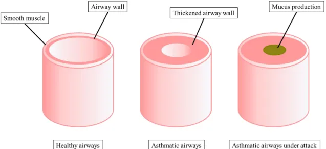
Asthma is a chronic inflammatory respiratory disease influenced by a wide range of environmental and genetic factors (Chen, Wong, and Li 2016). It is characterized by airflow obstruction due to smooth muscle constrictions and airway inflammation with symptoms such as coughing, wheezing, tightness in the chest, bronchoconstriction and airway hyperresponsiveness that may remit spontaneously or upon treatment. Further, long term inflammation leads to mostly irreversible structural and functional changes in the airway smooth muscles called airway remodelling that is characterized by bronchial wall thickening and increased vascularity, sub-mucosal gland hyperplasia and hypertrophy as well as extracellular matrix (ECM) deposition and angiogenesis (‘New NHLBI guidelines for the diagnosis and management of asthma. National Heart, Lung and Blood Institute’, 1997) (Figure 1).
Figure 1. Schematic diagram of healthy and asthmatic airways.
Asthma exacerbations may be caused by different environmental triggers. These factors, among others, may be grouped into indoor and outdoor stimuli, where indoor factors include allergens of dust mites, cockroaches, mice and pets, indoor burning of tobacco, wood and biomass, indoor endotoxins or products from Gram-positive bacteria.
Outdoor factors include viral and microbial pathogens, airborne particles, ozone, diesel exhaust particles, pollens, outdoor moulds, tobacco smoke, cold air or humidity.
Environmental stimuli also include exercise, occupation or even diet (Ho 2010; Diette et al. 2008).
1.1.2. Classification of Asthma and Allergy
Based on which cause initiated asthma or the exacerbation, asthma phenotypes can be distinguished. These include allergic-, non-allergic asthma, viral-induced asthma, exercise-induced asthma. Within the limits of this thesis, we have included these four groups in our analyses and have created subgroups for these asthma phenotypes with the help of respiratory specialists (Figure 2). Other asthma types can also be characterized for example aspirin-exacerbated respiratory disease that is a combination of asthma, chronic rhinosinusitis with nasal polyps and a sensitivity to aspirin or other types of non-steroid anti-inflammatory drugs (NSAIDs), pre-asthma wheezing in infants where recurrent episodes of the abnormality is likely due to asthma (Martinez et al. 1995), but other reasons may exist such as allergies, infection or obstructive sleep apnea. Furthermore, there is exacerbation-prone asthma with more frequent visits to the hospital due to recurrent asthma attacks, and asthma associated with apparent irreversible airflow limitation, where irreversibility may only be defined based on longitudinal studies, a progressive development of airway obstruction and treatment irresponsiveness (Pascual and Peters 2009), as well as eosinophilic and neutrophilic asthma (Bruijnzeel, Uddin, and Koenderman 2015; Patterson, Borish, and Kennedy 2015; Pelaia et al. 2015).
We have added subgroups to the pre-existing ones included in our analyses, for example asthma comorbidities of allergic rhinitis and allergic conjunctivitis, as these are the most frequent asthma associated allergic diseases which often occur together (Shaker and Salcone 2016; Rosario and Bielory 2011; Lee et al. 2013). Conjunctivitis is an inflammatory disease of the eye characterized by flushing, swelling, itching, and watering of the eyes whereas rhinitis is an inflammation of the nasal mucosal surface indicated by sneezing, a runny and/or stuffy nose, and post-nasal dripping. In different studies, between 50-65% of patients with rhinitis also had conjunctivitis, but conjunctivitis could also exist without rhinitis (Rosario and Bielory 2011). Our subgroups also include clinical parameters of asthma such as total IgE level and absolute eosinophil concentrations, which have been found to correlate with asthma severity. In children, the most frequent phenotype is the IgE mediated allergic asthma which can also have heterogeneous
Figure 2. Subgroups of asthma phenotypes included in our analyses. Asthma phenotypes may overlap.
1.1.3. Diagnostic Criteria
It is well-known that asthma is not a single disease but rather a series of overlapping individual diseases or phenotypes, each defined by its unique interaction between genetic and environmental factors (Lötvall et al. 2011; Borish and Culp 2008). Moreover, non-asthmatic disease symptoms may also overlap with asthma.
Diagnosis of the disorder may therefore be difficult, but crucial in terms of the therapy applied, morbidity and mortality. There are guidelines, such as The Global Strategy for Asthma Management and Prevention 2015 report update or the National Institutes of Health Guidelines for the Diagnosis and Management of Asthma Expert Panel Report-3, for an easier diagnosis (Global Initiative for Asthma; National Asthma Education and Prevention Program 2007). The detailed history of symptoms and a physical exam aids diagnosis of asthma subtypes. Measurements of forced expiratory volume in 1s (FEV1) and forced vital capacity (FVC) and especially their ratio, FEV1/FVC are good indicators of airflow obstruction. Specialists also examine diffusing capacity or lung volumes and may apply Broncho provocation (Global Initiative for Asthma; National Asthma Education and Prevention Program 2007).
Cause of asthma
Asthma
Clinical parameters
Comorbidities of asthma
Allergic asthma
Non-allergic asthma
Exercise-induced asthma
Viral-induced asthma Outdoor allergy
Indoor allergy Inhalative allergy
Allergic rhinitis Allergic Conjunctivitis
Total IgE concentration Absolute eosinophil number
1.1.4. Pathogenesis
It is important to understand the pathophysiology of asthma which has still not been fully elucidated. The description of the course of the disease goes beyond the scope of this paper, therefore, here I only summarize the main aspects of asthma pathogenesis.
Asthma is a chronic inflammatory disorder, where many cells and elements of the immune response play a role in its pathogenesis. Once the body encounters an allergen, virus or a noxious agent the immune system will be activated and in genetically susceptible individuals will over-react. In allergic asthma, dendritic cells, that are antigen- presenting cells, encounter the allergen and migrate to lymph nodes to present the peptide to naïve T lymphocytes that will be activated to mature into T helper 2 (Th2) cells with the aid of other regulatory cells (Kuipers and Lambrecht 2004). T lymphocyte subpopulations among others, include Th1 and Th2 cells with distinctive cytokine profiles that include interleukin-12 (IL-12), interferon- (IFN) and IL-4, -5, -9 and -13, respectively. There are several factors that determine the Th1/Th2 balance. According to the ‘hygiene hypothesis’ the Th1/Th2 balance may be skewed towards the cytokine profile of Th2 cells in newborns. This imbalance is usually lifted by infections, the presence of older siblings, rural environment or daycare attendance at an early age, that all entail a Th1 response. On the other hand, urban environment, the use of antibiotics or sensitization to diverse allergens do not involve Th1 cytokines, hence the early imbalance remains making the individual more susceptible to allergies, asthma or other chronic inflammatory diseases (Sears et al. 2003; Horwood, Fergusson, and Shannon 1985; Gern, Lemanske, and Busse 1999; Gern and Busse 2002; Eder, Ege, and von Mutius 2006).
The release of Th2 cytokines activates a cascade of events that lead to airway inflammation and in the long run, airway remodelling. IL-4 aids the differentiation of Th2 cells and along with IL-13 they play a role in the formation of IgE immunoglobulins through the induction of class-switching of B-lymphocytes, hence IgE receptors will be produced once they have become plasma cells. IgE receptors are important actors in hypersensitivity type I and diseases such as allergic asthma, atopic diseases or allergic conjunctivitis. IL-5 and granulocyte-macrophage colony-stimulating factor (GM-CSF) help the maturation of eosinophil granulocytes in the bone marrow and after infiltration to the inflamed airways their prolonged survival, respectively. Furthermore, tumour necrosis factor- (TNF-) further enhances the inflammatory processes in the lungs
Beside eosinophil infiltration, other immune cells, such as neutrophils, macrophages or mast cells also transmigrate into the airways. Eosinophils have increased numbers in asthmatic airways. By releasing pro-inflammatory mediators and cytokines, they contribute to the inflammatory response. It has been shown that higher numbers of eosinophils correlate with asthma severity. Mast cells play a critical role in the pathogenesis of allergic diseases, as having many IgE receptors on their surface allows these immunoglobulins to be physically cross-linked by allergens, hence degranulation of the mast cells begin, which then empty bronchoconstrictors, such as histamine, leukotrienes or prostaglandins into the surrounding tissues (Boyce 2003; Robinson 2004).
Histamine mediates oedema and mucus secretion as well via its histamine receptors 1 (H1) and 2 (H2), respectively (White 1990). Leukotrienes not only influence airway smooth muscle, but also recruit neutrophils (Gelfand and Dakhama 2006). Among several types of prostaglandins, PGF2 causes direct constriction of airway smooth muscles. It has been shown that upon PGF2 treatment asthmatics had an 8000-fold increase in sensitivity to it compared to healthy subjects (Mathé et al. 1973). It has been suggested that airway hyperresponsiveness also has a relation to the increased numbers of mast cells found in the airway smooth muscle. Further, mast cells not only release cytokines upon allergen contact, but in exercise-induced asthma they may also be activated by osmotic changes (Brightling et al. 2002). Macrophages may be activated by IgE receptors as well, releasing more inflammatory mediators and other cytokines enhancing the inflammatory response (Peters-Golden 2004). The role of neutrophils remains unclear in the pathogenesis of allergic diseases, but elevated numbers have been found in the airways of more severe asthmatics (Fahy et al. 1995; Wenzel 2006; Wenzel et al. 1997).
Epithelial cells of the airway also play a role in asthma. These cells lining the airways have a barrier function and they also maintain tissue homeostasis (Moheimani et al. 2016). By releasing more pro-inflammatory mediators during the inflammatory processes in asthma, epithelial cells may also suffer injury. Repair mechanisms in asthmatic patients are impaired, further worsening the controlled state of asthma.
Oxidative stress also has an effect on the bronchial epithelium in asthma.
Oxidative stress is the imbalance between the production of increased oxidative sources and the impaired mechanisms of detoxifying the reactive intermediates and repairing the caused damage (Holguin 2013). Reactive oxygen species (ROS) are produced either upon environmental exposure to air pollution of gases and particulate matter or the local inflammation will secondarily induce the production of ROS (Bowler 2004; Ghio,
Carraway, and Madden 2012). Oxidative stress is associated with inflammatory cell activation and hence the production of pro-inflammatory mediators (Paredi, Kharitonov, and Barnes 2002; Wood, Gibson, and Garg 2003). The increased amount of ROS results in oxidative lipid peroxidation and DNA damage, further aggravating inflammation and the severity of asthma.
1.1.5. Medication
To date there is no cure for asthma. It is a very complex disease with many factors, pathways, mechanisms that play a role in the pathophysiology of asthma. Which medication an asthmatic individual will take depends on age, what triggered their asthma, symptoms and whether the drug is effective. It is important for patients to have controlled asthma regardless of the severity of their disease. There are several types of long-term and short-term asthmatic medications. Long-term medications help to maintain a controlled asthmatic state on an everyday basis so that the incidence of an asthma attack is lower, while short-term drugs are a quick relief in case of such an attack.
The most essential drug among long-term medications is inhaled corticosteroids (ICS), which are anti-inflammatory medicines (e.g. Budesonide (Pulmicort), Fluticasone (Flovent)). ICSs reduce airway inflammation by down-regulating pro-inflammatory proteins (Adcock, Ito, and Barnes 2004; De Bosscher, Vanden Berghe, and Haegeman 2003), reversing components of airway remodelling, such as increased vascularity of the bronchial wall (Chanez et al. 2004), suppressing the production of chemotactic mediators and adhesion molecules that attract immune cells to the site of inflammation (eosinophils, dendritic cells, mast cells, lymphocytes) and also by inhibiting their survival (Schwiebert, Stellato, and Schleimer 1996). ICSs are better than orally taken corticosteroids, because they locally treat inflammation, rather than causing side-effects. Leukotriene modifiers are also effective oral anti-inflammatory drugs (e.g. Zafirlukast (Accolate), Montelukast (Singulair)), but in some cases, may cause side-effects of depression, aggression or agitation. Long-acting beta agonists (LABAs) (e.g. Salmeterol (Serevent), Formoterol (Foradil)) are inhaled drugs taken with ICSs enhancing their effects by suppressing inflammatory genes (Korn et al. 1998) and increasing the localization of glucocorticoid receptors in the nucleus for a better uptake of the medication (Eickelberg et al. 1999). On the other hand, taken alone, LABAs may increase the risk of severe asthma attacks.
to ICSs. Theophylline increases the activity of histone deacetylase (HDAC), which in turn reduces the number of eosinophils. Because ICSs activate HDAC through a different mechanism, it has been suggested that the low dose of theophylline enhances the anti- inflammatory effect of ICSs both in asthma and chronic obstructive pulmonary disease (COPD) (Cosio et al. 2004; Hossny et al. 2016).
Short-term asthma medications include short-acting beta agonists (SABAs) which offer ease of symptoms within minutes once inhaled through a nebulizer directly to the airways (e.g. Metaproterenol, Levalbuterol (Xopenex)). Orally or intravenously taken corticosteroids upon an asthma attack are also very effective in treating episodes of asthma attacks. In such case, the most used corticosteroids are prednisone, prednisolone or methylprednisolone, which should not be taken for long periods of time as may cause side-effects of weakness, weight gain, mood or behaviour changes, etc.
There are of course many approaches to target different factors in asthma that lead to a decrease in the inflammation in the airways. For instance, omalizumab is a humanized antibody (IgG1k) against IgE antibodies, one of the key players in asthma pathogenesis.
It has been approved in the 2000s in the United States by the Food and Drug Administration (FDA), as well as in the European Union to treat patients 12 years-of-age or older (Allergic Asthma and CIU Treatment | XOLAIR® (Omalizumab)). It is used in cases of corticosteroid resistance, but due to its higher price and only a few long-term trial studies, it is not yet frequently used nor it is administered for longer time-periods (Chang et al. 2007; Humbert et al. 2014; Normansell et al. 2014; Schulman 2001).
Furthermore, several drugs have been developed to target cytokines IL-5, IL-4 or IL-13 with antibody therapy. All of these medications are only effective in eosinophilic asthma phenotypes, but unfortunately minute non-eosinophilic asthma biomarkers are available to use in the search for potent therapies (Guilleminault et al. 2017).
1.2. Epidemiology of Asthma
It is well known that there is an increasing number of persons with asthma and/or allergies. The higher numbers of patients burden the countries both financially and socially. Many factors must be included in order to gain a complete knowledge of the burden of the disease. These include the number of adult and child patients in a country, the number of hospital visits, the cost of their treatment, morbidity (e.g. number of days missed from school or place of work/year) or mortality.
According to the Global Asthma Report 2014 that is based on the Global Burden of Disease Study (GBD) between 2008 and 2010 it has been estimated that around 334 million people suffer from asthma world-wide. Furthermore, there was an increase in the number of asthmatics according to the 2011 report based on 2000-2002 population data, where 235 million people had asthma globally (The Global Asthma Report 2014). This increase may perhaps be due to better and more precise diagnosis of asthma phenotypes (Carr and Bleecker 2016). Moreover, it is estimated that due to incomplete data, these numbers may be higher. The International Study of Asthma and Allergies in Childhood (ISAAC) estimated that around 14% of all the children in the world have asthma each year (Lai et al. 2009). The GBD study found that asthma is the fourteenth in number of years lost to asthma-associated morbidity and mortality world-wide (Walter et al. 2015).
In the USA in 2013 it was predicted that 16.5 million adults (8.3% of population) and 6.1 million children (7% of population) have asthma. The disease is more prevalent in women compared to men, also more common in children, than in adults, furthermore, it is more frequent in boys compared to girls. Asthma was the leading cause of absenteeism in children, where 50% of children missed school for at least 1 day every year, in 2013 this meant 13.8 million missed school days. Asthma morbidity is similar in adults, as 14 million days of work is missed each year. Although in 2014 3651 people died from asthma in the USA, death rates have decreased by 26% since 1999 (https://www.cdc.gov/asthma/pdfs/asthma_facts_program_grantees.pdf,
https://www.cdc.gov/asthma/most_recent_data.htm).
The cost of asthma in the USA is $56 billion/year, which means a $3259 per person. The yearly cost of the disease in Europe is €19.3 billion (https://www.cdc.gov/asthma/impacts_nation/), whereas in Asia-Pacific the cost varies between $184 and $1189 per person (www.globalasthmareport.org/burden/burden.php).
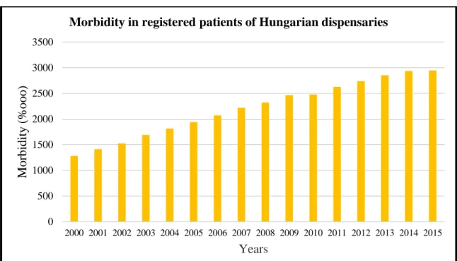
The Bulletin of the Hungarian Korányi Pulmonology Institute summarized the epidemiology of several pulmonology related diseases including asthma. According to the study, in which information of registered asthma patients were collected from all Hungarian dispensaries, in 2013, 282 754 people were affected by asthma, which means a 2.8% prevalence in Hungary (Csoma et al. 2016). The yearly increase in number of asthmatic adults is 16 000 - 19 000 (Figure 3).
Figure 3. Diagram showing morbidity rates of asthma patients collected from all Hungarian dispensaries. (Csoma et al. 2016)
1.3. Genetic Background
1.3.1. Genetic Methods and Results in the Research of Asthma and Allergy
Asthma is a multifactorial disorder, considering that apart from several small effect genes and variations that may also be inherited in an additive fashion, environmental factors also play a key role in the development of such diseases. With the rapid advances in genetics and genetic technologies, the amount of research of complex disorders have become substantial, however, our understanding of the mechanisms of their inheritance is very limited.
The heritability of asthma is estimated by the comparisons of correlations and concordance rates of monozygotic (MZ) and dizygotic (DZ) twins. According to twin studies, 36-77% of asthma is heritable (Duffy et al. 1990; Harris et al. 1997; Koppelman, Los, and Postma 1999; Nieminen, Kaprio, and Koskenvuo 1991), where the concordance of MZ twins is 0.74 and 0.35 of DZ twins, presenting a significant difference (Liu, Spahn, and Leung 2011). Furthermore, when neither of the parents have asthma, the likelihood of the children having the disease is 11-13%, but when both parents have asthma, this rate is 50-70% (Barnes and Marsh 1998), also showing the importance of the genetic component of asthma.
0 500 1000 1500 2000 2500 3000 3500
2000 2001 2002 2003 2004 2005 2006 2007 2008 2009 2010 2011 2012 2013 2014 2015
Morbidity (%ooo)
Years
Morbidity in registered patients of Hungarian dispensaries
Essentially, there are three different types of genetic studies to elucidate the genetic background of complex diseases. These include candidate gene association studies, genome-wide association studies (GWASs) and whole genome sequencing (WGS).
Candidate gene association studies analyse the relationship of a prespecified gene or genetic region and a given disease or phenotype by comparing variation frequencies between cases and controls. The previously determined genes are chosen based on the protein’s function or role in the disease. There are more than 1000 candidate gene association studies of asthma, from which 120 genes have been identified to have a relationship with the disorder (March, Sleiman, and Hakonarson 2011). There are 54 genes found to be associated with asthma that were reproduced in 2-5 independent studies. Fifteen genes were found to be associated with the disease in 6-10 independent studies, whereas only 10 were found in more than 10 independent investigations. These ten, most likely to indeed have a functional role in asthma, include IL-4, IL-13, 2 adrenergic receptor (ADRB2), major histocompatibility complex, class II, DQ 1 (HLA- DQB1), TNF, lymphotoxin (LTA), high affinity IgE receptor (FCERIA), IL-4 receptor (IL4R), CD14, and a disintegrin and metalloproteinase domain-containing protein 33 (ADAM33) (Basehore et al. 2004; Haller et al. 2009; Howard et al. 2002; Kabesch et al.
2006; Liggett 1995; Munthe-Kaas et al. 2008; Potaczek, Okumura, and Nishiyama 2009;
Potter et al. 1993; Pykäläinen et al. 2005; Randolph et al. 2004; Suttner et al. 2009; Van Eerdewegh et al. 2002; Vladich et al. 2005; Wu et al. 2010; Zhou et al. 2009).
Unfortunately, candidate gene association studies cannot give a full picture of the complex genetic background of diseases such as asthma, because multifactorial diseases develop through many inherited genetic variations that also influence each other, while environmental factors also contribute to the formation of the disorder. This problem was partially solved by WGS, which is a hypothesis-free research tool examining hundreds of microsatellite markers in affected siblings. Thus, many 10-20 million base pair regions have been identified to contain candidate genes for asthma and atopy. These regions need to be restricted to find the disease susceptibility genes or variations, which is done by positional cloning. This technique begins with taking a linked marker in a region and depicting the area in its proximity by several levels of recombination. This task was daunting until the results of the Human Genome Project were available to search on a computer, for a much easier identification of variations in a given region on the DNA
(Jorde, Carey, and Bamshad 2010). WGS and positional cloning have identified the following among others. ADAM33, dipeptidyl peptidase 10 (DPP10), G-protein coupled receptor for asthma susceptibility (GPRA), human leukocyte antigen G (HLA-G), cytoplasmic FMR1 interacting protein 2 (CYFIP2) or orosomucoid-like 3 (ORMDL3) (Allen et al. 2003; Laitinen et al. 2004; Moffatt et al. 2007; Nicolae et al. 2005; Noguchi et al. 2005; Van Eerdewegh et al. 2002).
Technical advances in genetics, such as microarrays or next-generation sequencing (NGS) have led to the evolution of GWASs. These case-control studies allow millions of markers (single nucleotide polymorphisms (SNPs), or copy number variations, (CNVs)) to be investigated in large populations with a given phenotype (Jorde, Carey, and Bamshad 2010). Many genes and regions have been identified with high- throughput GWASs (Table 1). On the other hand, GWASs also faced a problem of
’missing heritability’. Researchers expected previously identified susceptibility genes with a strong cumulative effect to be replicated during the association study, however the results could not be reproduced. In this regard, the analysis of GWAS results is essential, as well as the development of new techniques for the identification of genetics regions, candidate genes and variations.
Table 1. Asthma or asthma-associated phenotype susceptibility genes and regions.
Adapted from Gu and Zhao, 2011 (Gu and Zhao. 2011).
Chromo-
some Locus Candidate genes
1 1p36,1qter,1q23 FCER1A, OPN3, CHML, IL10
2 2q14,2q32,2q33,2p DPP10, IL18R1, CTLA4, CD28
3 3q21-q22,3q21.3,3p TLR9
5 5q31-q33,5q31,5p13,5p15,5q23.3 IL4, IL9, ZFR3, LIFR, PTGER4, ADAMTS12, IL7R
6 6p21,6q24-q25,6q25.3 HLAG, ESR1, TNF
7 7p14-p15,7q GPR154
8 8p23.3-23.2 NAT2
9 9p1,9p21,9p22 TLE4, IFNA
11 11q13,11q21,11q,11p14 MS4A2, GSTP1
12 12q13.12-q23.3,12q13-
12q24,12q21,12q24.31,12q24.33
SFRS8, CD45, IFNG, IRAK3, VDR
13 13q14,13q PHF11, CYSLTR2
14 14q11.2,14q13-q23,14q24,14q23 TCR, ACT
17 17q21 ORMDL3
19 19q13,19q13.3 FCER2
20 20q13,20p12 ADAM33, JAG1, ANKRD5
21 21p21 -
x Xp, Xq -
1.3.2. Apoptosis in Asthma
It is well-known, that apoptosis is a key feature in the pathomechanism of asthma (Vignola et al. 1999). The most reviewed process is eosinophil-clearance, which is impaired in asthmatic patients, hence the high numbers of eosinophils accumulated in the bronchial tissues will neither go through apoptosis, nor be cleared by phagocytosis of macrophages (Kankaanranta et al. 2000; Walsh 2000; Woolley et al. 1996). Additionally, it has been shown that the lack of eosinophil apoptosis in asthmatics correlates with disease severity (Duncan et al. 2003).
The balance between cell apoptosis and survival depends on the control and maintenance of different regulatory elements and pathways. For instance, the members of the inhibitor of apoptosis protein (IAP) family not only inhibit apoptotic pathways in a caspase-dependent manner, they also play a role in the regulation of cell cycle and cell
initially thought. Furthermore, the aberrant expression of the members of this protein family results in pathologic cell functioning and uncontrolled cell division (Altieri 2010).
Baculoviral IAP repeat containing 5 (BIRC5), also called survivin is an important anti-apoptotic member of the IAP family. BIRC5 has been previously thought to be only expressed in foetal tissues during growth. Moreover, it is abnormally expressed in cancerous tissues, hence being a featured target of therapeutic research (Altieri 2010).
Recently, it has been shown that BIRC5 has additional roles in inflammatory mechanisms and disorders, such as asthma (Altznauer et al. 2004; Valentin et al. 2009; Vassina et al.
2006). Furthermore, our research group has found several important aspects of BIRC5 in asthma. Namely, that the mRNA level of Birc5 in ovalbumin (OVA) induced asthmatic mouse model was significantly increased compared to normal mice (fold-change of 5.94, p=0.001) (Tölgyesi et al. 2009). This result was replicated by Tumes et al, who also found that in mice, the mRNA and protein expression of Birc5 found in the bronchoalveolar fluid correlated with the number of eosinophils (Tumes, Connolly, and Dent 2009). Our research group has further shown, that the gene expression level of BIRC5 was significantly higher in asthmatic patients compared to healthy controls, and both the gene expression level and one of the studied variations, rs9904341, were significantly correlated with the eosinophil ratio found in the induced sputum of asthmatics (Ungvári et al. 2012a).
Furthermore, our group’s previous results have shown that the gene expression of FERM-domain containing 6 (FRMD6) is significantly decreased in both the OVA- induced mouse model, as well as asthmatic patients compared to controls. Additionally, a gene polymorphism has been shown to be associated with asthma verified by both frequentist and Bayesian statistical approaches (Ungvári et al. 2012b). FRMD6 is the upstream activator of the Hippo signalling pathway, which also regulates the expression of several proteins, such as BIRC5 (Ungvári et al. 2012a; Ungvári et al. 2012b).
The Hippo pathway is highly conserved from Drosophila melanogaster to mammals and regulates organ size via promoting apoptosis and inhibiting cell proliferation in the embryonic stages of development (Huang et al. 2016; Yu, Zhao, and Guan 2015). Its name originates from the Drosophila Hippo protein kinase (Hpo), which produces tissue overgrowth or „hippopotamus-like” phenotype upon mutations in its coding gene. It is still not exactly known whether the pathway is regulated by determinants of cell polarity and cell-cell junctions, mechanical cues of the cell, soluble factors regulating Hippo members or metabolic status, like cellular energy and oxygen
stress (Yu, Zhao, and Guan 2015) (Figure 4). However, it has been proposed that FRMD6 (also known as Willin) influences the activity of the Hippo pathway by turning on its central kinase cascade (Angus et al. 2012). The members of this signalling cascade, mammalian STE20-like protein kinase 1 and 2 (MST1/2) and large tumour suppressor kinase 1 and 2 (LATS1/2) with scaffold proteins salvador family WW domain containing protein 1 (SAV1) and MOB kinase activator 1 (MOB1), respectively, phosphorylate one another to inhibit yes-associated protein 1 and tafazzin (YAP1/TAZ), the main effectors of the pathway (Harvey and Tapon 2007; Harvey, Pfleger, and Hariharan 2003; Huang et al. 2005; Jia et al. 2003; Justice et al. 1995; Lai et al. 2005; Lange et al. 2015; Pan 2007;
Wu et al. 2003; Xu et al. 1995). YAP1/TAZ, upon phosphorylation on several serine sites by its upstream regulators, are sequestered in the cytoplasm by 14-3-3 proteins, unable to enter the nucleus, then, they may also be degraded by proteasomes (Piccolo, Dupont, and Cordenonsi 2014). YAP1 and TAZ are transcriptional coactivators that bind to transcription factors when active, such as TEA domain containing proteins (TEAD), SMAD family members (SMAD) or tumour protein P73 (TP73), to regulate the expression of anti-apoptotic, (e.g. BIRC5) or apoptotic genes that play a role in cell differentiation, survival and migration (Alarcón et al. 2009; Strano et al. 2001; Vassilev et al. 2001).
Figure 4. Examples of signals and pathways regulating YAP1 activity, including the Hippo signalling pathway.
The gene, YAP1, that codes for the main effector of the Hippo pathway, is located on the long arm of chromosome 11. It is a 123 kb gene comprising 10 exons and 9 introns that will be a 54 kDa protein after translation. YAP1 contains a transcriptional enhancer factor-binding domain (TB), a 14-3-3 binding site, two WW domains that aid the binding and interaction with LATS kinases, as well as playing a role in the regulation of transcription, cell transformation and tissue growth (Sudol and Harvey 2010; Zhang et al.
2010). Furthermore, YAP1 has an SRC homology 3 domain (SH3) binding motif, a transcriptional activation domain (TAD), a PDZ binding domain and several serine phosphorylation sites throughout its sequence (Iglesias-Bexiga et al. 2015) (Figure 5).
Figure 5. Simplified schematic diagram of YAP1 protein structure. N: N-terminus, C: C terminus
YAP1 behaves as an oncogene, that has been investigated and applied as a therapeutic target in different types of cancers, such as liver, prostate, thyroid, gastric, or lung cancer.
Besides embryonic tissues, where YAP1 plays an important role in, for example, lung maturation and postnatal airway homeostasis, it is widely expressed in respiratory epithelial cells of the embryonic and mature lung, where the Hippo/YAP1 signalling regulates epithelial cell proliferation and differentiation (Mahoney et al. 2014).
Furthermore, in mice it has been demonstrated that YAP is dynamically regulated during regeneration of the airway epithelium following lung injury suggesting a possible role of Hippo/YAP1 signalling in the pathogenesis of acute and chronic lung diseases (Lange et al. 2015).
1.3.3. Angiogenesis in Asthma
Processes such as cytokine production, inflammatory cell infiltration to the lungs, injury to epithelial cells or apoptosis all play an important role in the development and severity of asthma. Structural changes in the airway walls due to both neovascularization
N C
TEAD binding domain
14-3-3 binding domain
WW2 domain
SH3 binding domain
TAD PDZ
binding domain YAP1
Serine 127
WW1 domain
and angiogenesis are also key aspects of asthma (Bischof et al. 2009). Increased vascularity and angiogenesis in asthmatic patients may cause an increase in airway wall thickness, and hence, airway occlusion (Makinde and Agrawal 2011). Moreover, Salvato has found that the number of vessels as well as vascularity have a positive correlation with asthma severity (Salvato 2001).
Angiogenesis is the process where new blood vessels form from pre-existing ones (Madeddu 2005). It takes place in embryonic development, as well as adults, where angiogenesis is an important feature of both many physiological and pathological processes. Physiological processes include hair growth, the female reproductive cycle, wound healing, or bone repair (Carmeliet 2005). On the other hand, uncontrolled angiogenesis is present in cancer, rheumatoid arthritis, diabetes or psoriasis, but poor angiogenesis results in myocardial or brain ischemia or non-healing ulcers (Bellou et al.
2013; Costa and Soares 2013; Zachary and Morgan 2011). Angiogenesis is regulated by several molecules, such as vascular endothelial growth factor (VEGF), which is the most distinct growth factor for the vascular endothelium (Breier et al. 1992; Ferrara 2002;
Shweiki et al. 1993). There are several growth factors in this family that either primarily regulate the growth of blood vessels or lymphangiogenesis, furthermore, placental growth factor (PGF) is expressed in the placenta or certain types of tumours (De Falco, Gigante, and Persico 2002; Ferrara 2002; Maglione et al. 1991). The VEFG family function through VEGF receptors (VEGFR), VEGFR-1, -2 and -3 (Ferrara 2002), that also have coreceptors, Neurophilin-1 and -2 (NRP1 and -2) that increase VEGF affinity for VEGFR (Becker et al. 2005; Gluzman-Poltorak et al. 2000; Soker et al. 1998) (Figure 6). Other regulators of angiogenesis are angiopoietins, like Ang1 and Ang2 in humans. These are glycoproteins that have both been characterized by acting as ligands for tyrosine-protein kinase receptor, Tie2 (Saharinen et al. 2008; Yuan et al. 2009). Ang1 and Ang2 have a similar affinity for binding to Tie2. Ang1 stimulates kinase activity by binding to Tie2;
on the other hand, Ang2 may act as an agonist or an antagonist for Tie2 in a tissue- and cell-dependent manner (Augustin et al. 2009). Ang1 provides an anti-inflammatory effect on blood vessels during angiogenesis, whereas Ang2 and vascular endothelial growth factor (Vegf) contribute to a pro-inflammatory effect. (Gamble et al. 2000; Makinde and Agrawal 2011)
Figure 6. Schematic diagram of VEGFs, VEGF receptors and their primary functions.
Adapted from Detoraki et al, 2010. (Detoraki et al. 2010)
Tie2 is encoded by the TEK gene in humans and is found on the short arm of chromosome 9. It plays a role in angiogenesis, endothelial cell survival, proliferation, migration, adhesion and cell spreading, as well as the reorganization of the actin cytoskeleton or the maintenance of vascular quiescence (Audero et al. 2004; Cascone et al. 2003; Fukuhara et al. 2008; Martin et al. 2008; Saharinen et al. 2008; Yuan et al. 2009;
Lee et al. 2004). Tie2 also has an anti-inflammatory effect by averting the leakage of pro- inflammatory elements from the blood vessels into the surrounding tissues. It has been found that Tie2 receptor mRNA and protein are abundantly expressed in the lungs (Kanazawa 2007). In a recent genome wide association study in European American populations the strongest signals were identified at the 9p21.2 locus consisting of four SNPs in weak LD with each other and spanning three genes (EQTN (Equatorin), MOB3B (MOB kinase activator 3B), TEK) (Almoguera et al. 2016). It was hypothesized that, based on its function, variations in the TEK gene were responsible for the association. In another study, three non-linked expression quantitative trait locus (eQTL) SNPs were identified in the introns of the TEK gene, which all associated with lower TEK gene expression in a HapMap3 cohort and increased risk for developing acute respiratory distress syndrome (Ghosh et al. 2016).
The Tie2 pathway also has an important role in the development and function of the eye and the TEK gene is highly expressed in the Schlemm’s canal endothelium in the eye (Souma et al. 2016). Mutation in the TEK gene is associated with primary congenital
glaucoma and Tie2 is a highly-investigated target in different eye diseases like subretinal and choroidal neovascularization, macular oedema or diabetic retinopathy (Campochiaro and Peters 2016; Campochiaro 2015; Tan et al. 2016).
1.4. Previous Results of Ovalbumin-Induced Mouse Model of Asthma
Our research group has previously developed an ovalbumin-induced mouse model of asthma and have carried out a whole genome gene expression microarray analysis on different healthy and asthmatic mouse groups. Using a high-throughput microarray technology may lead to greater insights into new genes and pathways regulating the development of asthma. This study has been carried out by Tölgyesi et al and the details can be found in the GEO database with the record number GSE11911. Therefore, here, I only summarize the experimental setup and findings that are relevant for our following analyses.
Six- to eight-week-old, female, pathogen-free BALB/c mice were used in the experiment that were kept on an OVA-free diet in order to senzitize them to the compound. All together, 4 groups of mice have been created, where one of them received placebo (PBS) during the allergen challenge which comprised the control group. The other asthmatic groups received OVA during the allergen challenge, where groups 1 and 2 comprised the mice from which BALF and lung tissue was collected 4 hours after the first and third allergen challenge on days 28 and 30, respectively. From group 3 mice, we have collected BALF and lung tissue 24 hours after the third allergen challenge on day 31. From lung tissues, RNA was isolated for the whole genome gene expression analysis and mRNA levels have been compared between control and asthmatic mice. As a result, 533, 1554 and 1134 genes showed a larger than 2-fold expression change that were statistically significant in groups 1, 2 and 3 compared to the control group, respectively.
Furthermore, 861 transcripts showed a statistically significant, larger than 2-fold difference in gene expression between the asthmatic groups (Tölgyesi et al. 2009). These data have been used for further analysis in order to find orthologous genes in humans that may influence the development of asthma by being differentially regulated on an mRNA or DNA level.
We have chosen 60 genes based on the results of this study. Our main reasons for considering a gene cover several aspects. Firstly, a gene having a larger than 2-fold
expression change between asthmatic and control groups. Secondly, a gene (protein) having a potential role in the pathogenesis of asthma. Thirdly, finding previous scientific results for a given gene. And last, having scientific novelty (Temesi et al.
2014). These criteria led to the selection of a variety of genes that may be found in Table S2.
1.5. Current Shortcomings in the Research of Asthma and Allergy
Based on the above, it is clear that asthma is a prevalent, often severe, complex disorder that is yet impossible to cure. Understanding the mechanisms and pathways of the disease, its pathogenesis, its genetic background will bring solutions to asthma one step closer.
Our research is the very first to identify and hypothesize the Hippo pathway to play a role in a phenotype of asthma, as well as a variation of the TEK gene to act in allergic conjunctivitis. Although research of apoptosis and angiogenesis in asthma is a small piece in a bigger picture, nonetheless, such scientific contributions may advance further research in this field.
2. Objectives
Our goal was to study the genetic and genomic background and the pathogenesis of childhood asthma and its associated phenotypes. The main objectives can be summarised as follows.
1. Investigation of the role of the Hippo signalling pathway in asthma
• Evaluating the differences in gene expressions of seven Hippo pathway genes between asthmatics and healthy subjects based on previous results of Ungvári et al. (Ungvári et al. 2012a; Ungvári et al.
2012b)
• Assess relationship of polymorphisms (tagSNPs) spanning the whole of YAP1 gene (based on above results) and asthma and its phenotypes by estimation of allele frequencies between asthma patients and healthy controls. Moreover, gaining further associations through the haplotype analysis of our data and a more extensive Bayesian statistical analysis.
• Comparing FRMD6, BIRC5 and YAP1 protein levels in induced sputum samples from asthmatics and controls in order to evaluate role of Hippo signalling pathway in asthma through protein expression.
• Investigating HeLa cells in vitro upon BIRC5 antagonist, YM155 treatment in order to find functional roles for Hippo signalling pathway components, BIRC5, YAP1 and FRMD6.
2. Investigation of the role of angiopoietin receptor Tie-2 in asthma and its phenotypes
• Assessing the incidence of different comorbidities of asthma within our study population.
• Evaluating tagSNPs of Tie2, encoded by the TEK gene, in asthmatic and control subjects to find biomarkers for asthma susceptibility.
3. Investigation of associations between 60 previously identified genes and asthma
• Sixty genes have been previously identified by gene expression microarray on an ovalbumin-induced mouse model of asthma by our research group (Tölgyesi et al. 2009). Following variation selection, identification of associations by estimation of allele frequencies between asthma patients and healthy controls.
3. Methods 3.1. Subjects
3.1.1. Characteristics of Participants of Sputum Induction and Gene Expression Measurements
The gene expression analysis was done using the induced sputum of 18 asthmatic patients and 10 healthy controls. All subjects completed a detailed, pre-edited questionnaire based on the ISAAC Phase Three Questionnaire. The recent Global Initiative for Asthma guidelines (www.ginasthma.org) were used to diagnose asthma by a respiratory medicine specialist. The evaluation of asthma severity was done at the time of acquisition of induced sputum samples from the patients based on patient history, including number of exacerbations per year, lung function test results, medical treatment applied and response to medication. Asthmatics were divided into four severity groups, but due to low number of patients GINA 1,2 (mild) and GINA 3,4 (moderate-severe) were aggregated. GINA groups of severity from 1-4 are summarised as follows. GINA 1 is
‘intermittent’ asthma, with symptoms less than once a week and brief exacerbations.
Nocturnal symptoms do not occur more than twice a month. FEV1 is more than 80%, where their variability is less than 20%. GINA 2 is ‘mild persistent’ with symptoms more than once a week but less than once a day and exacerbations may affect the patient’s activity and sleep. Nocturnal symptoms occur more than twice a month. FEV1 is more than 80% with variability of less than 20-30%. GINA 3 is ‘moderate persistent’ with daily symptoms and exacerbations affecting activity and sleep. Nocturnal symptoms occur more than once a week and there is a need for daily use of inhaled short-acting beta- agonist. FEV1 is between 60-80% with a variability of more than 30%. GINA 4 is ‘severe persistent’ with daily symptoms and frequent exacerbations. There are also frequent nocturnal symptoms of asthma. The patients suffer from the limitation of physical activites. FEV1 is less than 60% with a variability of more than 30%. Out of the 18 asthmatic patients, 14 regularly used inhaled corticosteroids (ICS): <500 μg/day beclomethasone dipropionate (BDP) or equivalent (n = 5), 500–1000 μg/day BDP or equivalent (n = 7) and >1000 μg/day BDP or equivalent (n = 2); while four were considered steroid naive. Healthy volunteers with no previous history of asthma or any airway conditions comprised the control group. Everyone participated in a lung function test (PDD-301/S, Piston Inc., Budapest, Hungary) and were assessed for fractional
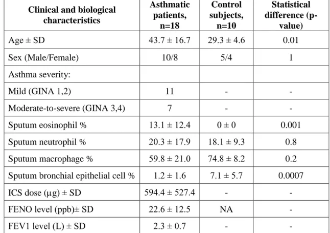
healthy subjects had a normal lung function and had no respiratory tract infection four weeks prior to the analysis. A skin prick test was also performed for common allergens to test the presence of atopy, which is a genetic predisposal for developing allergic rhinitis, atopic dermatitis and/or asthma. The participants’ sex, age, smoking habits and allergic statuses were compared between cases and controls and between severity groups, but no statistical significance was found (Ungvári et al. 2012a). Table 1 shows a summary of this study population.
Table 1. Detailed characteristics of subjects participating in sputum and gene expression analysis.
Clinical and biological characteristics
Asthmatic patients,
n=18
Control subjects,
n=10
Statistical difference (p-
value)
Age ± SD 43.7 ± 16.7 29.3 ± 4.6 0.01
Sex (Male/Female) 10/8 5/4 1
Asthma severity:
Mild (GINA 1,2) 11 - -
Moderate-to-severe (GINA 3,4) 7 - -
Sputum eosinophil % 13.1 ± 12.4 0 ± 0 0.001
Sputum neutrophil % 20.3 ± 17.9 18.1 ± 9.3 0.8
Sputum macrophage % 59.8 ± 21.0 74.8 ± 8.2 0.2
Sputum bronchial epithelial cell % 1.2 ± 1.6 7.1 ± 5.7 0.0007
ICS dose (g) ± SD 594.4 ± 527.4 - -
FENO level (ppb)± SD 22.6 ± 12.5 NA -
FEV1 level (L) ± SD 2.3 ± 0.7 - -
GINA: Global Initiative for Asthma; ICS: Inhaled corticosteroid in g; FENO:
Fractional exhaled nitric oxide in parts per billion; FEV1: Forced expiratory volume in 1 second in litres.
3.1.2. Characteristics of Participants of Genotyping Analysis
The genotyping analysis of YAP1 SNPs included 1233 unrelated individuals, out of which 522 were asthmatic children (mean age±SD: 10.20±5.31 years; 328 males and 194 females) and 711 healthy controls (mean age±SD: 14.0±11.2; 429 males and 282
females). Further, the genotyping analysis of TEK SNPs included 1189 unrelated individuals, out of which 435 were asthmatic children (mean age±SD: 10.3 ± 4.4 years;
269 males and 166 females) and 754 healthy controls (mean age±SD: 13.8±11.8 years;
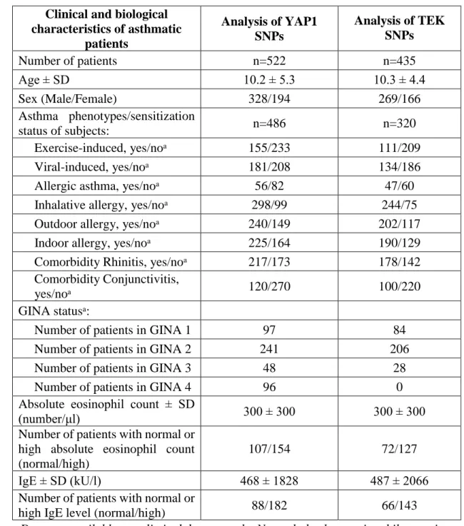
453 males and 297 females). Moreover, the genotyping analysis of 60 previously identified genes (list of genes found in Temesi et al. 2014) included 671 individuals, out of which 311 were asthmatic children (mean age±SD: 10.6 ± 4.8 years; 203 males and 18 females) and 360 healthy controls (mean age±SD: 21.7 ± 13.9 years;181 males and 179 females). The patients were all Hungarian (Caucasian), from which about 5% were of Gypsy origin based on state population data. Asthma was diagnosed based on GINA guidelines, as before (Ungvári et al. 2012a). Atopy was identified by a positive skin prick test and/or positive total or specified serum IgE levels. The total and specified serum IgE levels were evaluated with the 3gAllergy blood tests in the Immulite 2000 Immunoassay system (Tarrytown, NY, USA). The resulting serum IgE levels were either normal or high based on age-specific ranges (kU/litre).
Different types of asthma were defined in patients. A number of participants were excluded from the analysis, where insufficient information was available on asthma phenotypes, hence altogether, 391 and 320 asthmatic children were involved in the phenotype analysis of YAP1 and TEK genes, respectively. Asthma was divided into allergic and non-allergic asthma subtypes. If asthma was not provoked by allergens, but allergy was also present, allergy types were, nonetheless, marked. In allergic patients depending on the types and quantities of allergens, subgroups of indoor, outdoor, or inhalative allergic phenotypes have been designated. Asthma was categorized as exercise- induced asthma when the asthma exacerbation was provoked by exercise in the medical history of the patients. If the onset of asthma or the asthma exacerbations have been associated with an infection related acute respiratory illness the asthma was classified as viral-induced asthma. Non-atopic patients with viral-induced asthma phenotype composed the non-allergic asthma subgroup (Wenzel 2006) (Figure 2). Indoor allergens included dust mites, mould, pet dander and cockroaches, whereas outdoor allergens consisted of different types of pollen. Eosinophil cell counts from blood were measured with the Coulter AXM analyser, where the normal relative range was between 1-6%, and the normal range of absolute eosinophil count was between 50-200/μl. None of the patients had a change of therapy before the blood samples were taken. Neither had they exacerbations or respiratory infections for at least four weeks prior to the blood test. Table
2 shows the detailed characteristics of the asthmatic patients participating in the gene association study.
The samples from the control children were collected at the Orthopaedic Department in the Budai Children’s Hospital or at the Urological Department in the Heim Pál Hospital, both in Budapest. None of the controls had any symptoms of asthma or airway conditions, nor any need for medication.
A written informed consent was signed by all patients or by their parent/guardian.
The study was carried out according to the Declaration of Helsinki and was approved by the Ethics Committee of the Hungarian Medical Research Council.
Table 2. Detailed characteristics of asthmatic patients participating in SNP analysis of YAP1 and TEK genes.
Clinical and biological characteristics of asthmatic
patients
Analysis of YAP1 SNPs
Analysis of TEK SNPs
Number of patients n=522 n=435
Age ± SD 10.2 ± 5.3 10.3 ± 4.4
Sex (Male/Female) 328/194 269/166
Asthma phenotypes/sensitization
status of subjects: n=486 n=320
Exercise-induced, yes/noᵃ 155/233 111/209
Viral-induced, yes/noᵃ 181/208 134/186
Allergic asthma, yes/noᵃ 56/82 47/60
Inhalative allergy, yes/noᵃ 298/99 244/75
Outdoor allergy, yes/noᵃ 240/149 202/117
Indoor allergy, yes/noᵃ 225/164 190/129
Comorbidity Rhinitis, yes/noᵃ 217/173 178/142 Comorbidity Conjunctivitis,
yes/noᵃ 120/270 100/220
GINA statusᵃ:
Number of patients in GINA 1 97 84
Number of patients in GINA 2 241 206
Number of patients in GINA 3 48 28
Number of patients in GINA 4 96 0
Absolute eosinophil count ± SD
(number/μl) 300 ± 300 300 ± 300
Number of patients with normal or high absolute eosinophil count (normal/high)
107/154 72/127
IgE ± SD (kU/l) 468 ± 1828 487 ± 2066
Number of patients with normal or
high IgE level (normal/high) 88/182 66/143
ᵃData are available on a limited data set only. Normal absolute eosinophil count is 200 /μl and high absolute eosinophil count is 200 /μl. Normal IgE level is 200 kU/l,
high IgE level is 200 kU/l.
3.2. Sputum Induction
Induced sputum was used for gene expression assays and Western blot analysis.
Participants were first treated with 400μg of inhaled salbutamol, then, they inhaled 4.5%
saline solution generated by a De Vilbiss Nebulizer (Ultra-NebTm 2000 model 200 HI, Somerset, PA, USA) for 5 minutes. This procedure was repeated two more times, where after each sputum induction, the subjects’ pulmonary function was assessed. All samples were examined for salivary contamination and only those were used in the study, which were macroscopically free of such contamination. These samples were, then, diluted with phosphate buffered saline (PBS) that contained 0.1% dithiothreitol from Sigma (St Louis, MO, USA). The samples were thoroughly mixed with a vortex and placed on a bench rocker for 30 minutes at room temperature. Then, the samples were filtered with a 40μm Falcon cell strainer and centrifuged at 1500rpm for 10 minutes at room temperature. The cell pellet was resuspended in 1ml PBS. The Trypan Blue Exclusion test was used to determine cell viability in a Burker chamber. After differential cell count by a specialist, cells were stored on lysis buffer at -80°C until use (Ungvári et al. 2012a).
3.3. DNA Isolation
Genomic DNA was isolated from whole blood samples of 1233 individuals using the QIAamp blood DNA midi kit (Qiagen, Maryland, USA) or the iPrep PureLink gDNA Blood Kit on iPrep Purification Instrument (Invitrogen, Carslbad, CA, USA) starting out from 1 ml whole blood. The average DNA concentration of samples was between 30- 60ng/l measured by Nanodrop ND-1000 spectrophotometer (NanoDrop Technologies, Wilmington, DE).
3.4. RNA Isolation and cDNA transcription
RNA was isolated successfully from the induced sputum samples of 18 patients and 10 control subjects with the Qiagen Mini RNeasy Kit according to the manufacturer’s instruction (Qiagen, Maryland, USA). The total RNA quantity was measured by Nanodrop ND-1000 spectrophotometer (NanoDrop Technologies, Wilmington, DE). The cDNA used in the gene expression analysis, was produced with a High Capacity cDNA Reverse Transcription Kit from Thermo Fisher Scientific (Thermo Fisher Scientific, Waltham, MA USA).
3.5. Gene Expression with TaqMan
Real-time quantitative PCR was performed on LATS1, LATS2, MST1, MST2, SAV1, YAP1, TAZ and β-actin genes using 7900HT Fast Real-Time PCR System (Thermo Fisher Scientific, Waltham, MA USA) according to the manufacturer’s instructions with 1.5 μl cDNA/well and final volume of 25μl. β-Actin was used as an endogenous control and all results were normalized to it using the delta delta Ct method.
3.6. SNP Selection and Genotyping with Competitive Allele-Specific PCR
SNPs were selected from YAP1 gene using UCSC Genome browser. Preferably, promoter, missense or UTR SNPs were chosen where MAF in the Caucasian population was higher than 0.1, which was confirmed by HapMap. We also checked for linkage between SNPs and have chosen tagSNPs with Haploview 4.2 program (Broad Institute of MIT, Cambridge, MA, USA). TEK gene SNPs were chosen based on the paper of Ghosh et al. (Ghosh et al. 2016).
KBioscience Competitive Allele-Specific PCR (KASP) version 4.0 genotyping assays were used (LGC Genomics, Berlin, Germany) to genotype fourteen SNPs on the YAP1 gene and three SNPs on the TEK gene (Table 3) according to the manufacturer’s instructions. PCR reactions were carried out using a 7900HT Fast Real-Time PCR System (Thermo Fisher Scientific, Waltham, MA USA).
36
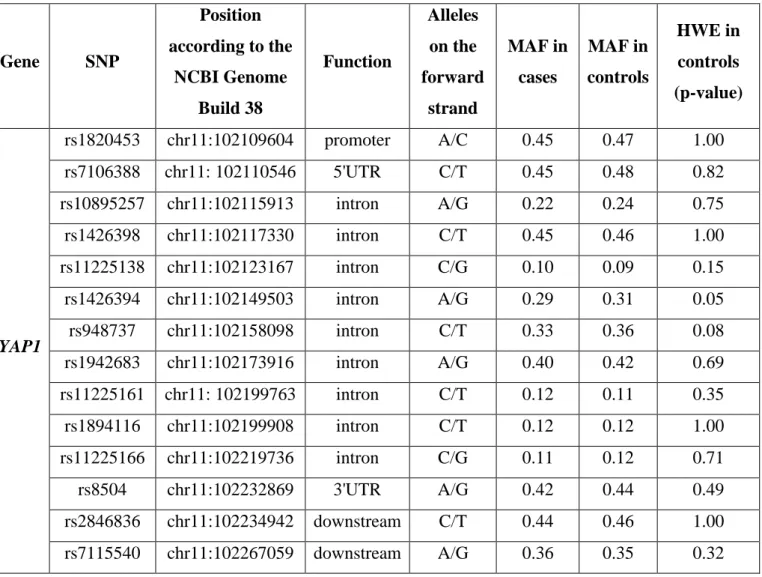
Table 3. Description of selected SNPs on YAP1 and TEK and results of the genotyping.
Gene SNP
Position according to the
NCBI Genome Build 38
Function
Alleles on the forward
strand
MAF in cases
MAF in controls
HWE in controls (p-value)
YAP1
rs1820453 chr11:102109604 promoter A/C 0.45 0.47 1.00
rs7106388 chr11: 102110546 5'UTR C/T 0.45 0.48 0.82
rs10895257 chr11:102115913 intron A/G 0.22 0.24 0.75
rs1426398 chr11:102117330 intron C/T 0.45 0.46 1.00
rs11225138 chr11:102123167 intron C/G 0.10 0.09 0.15
rs1426394 chr11:102149503 intron A/G 0.29 0.31 0.05
rs948737 chr11:102158098 intron C/T 0.33 0.36 0.08
rs1942683 chr11:102173916 intron A/G 0.40 0.42 0.69
rs11225161 chr11: 102199763 intron C/T 0.12 0.11 0.35
rs1894116 chr11:102199908 intron C/T 0.12 0.12 1.00
rs11225166 chr11:102219736 intron C/G 0.11 0.12 0.71
rs8504 chr11:102232869 3'UTR A/G 0.42 0.44 0.49
rs2846836 chr11:102234942 downstream C/T 0.44 0.46 1.00 rs7115540 chr11:102267059 downstream A/G 0.36 0.35 0.32
37
MAF: minor allele frequency; HWE: Hardy-Weinberg Equilibrium Gene SNP
Position according to the
NCBI Genome Build 38
Function
Alleles on the forward
strand
MAF in cases
MAF in controls
HWE in controls (p-value)
TEK
rs581724 chr9:27187424 intron T/G 0.39 0.39 0.99
rs3780315 chr9:27196292 intron G/A 0.47 0.44 0.000062
rs7876024 chr9:27202665 intron A/G 0.24 0.24 0.02