PREPARATION AND SURFACE MODIFICATION OF MAGNETIC PLGA NANOPARTICLES FOR SUSTAINING NATURAL INTERFERON RELEASE
Ph.D. THESIS
By:
QUAZI TANMINUL HAQUE SHUBHRA
SUPERVISORS:
PROF. JÁNOS GYENIS DR. TIVADAR FECZKÓ
UNIVERSITY OF PANNONIA
DOCTORAL SCHOOL OF MOLECULAR- AND NANOTECHNOLOGIES RESEARCH INSTITUTE OF CHEMICAL AND PROCESS ENGINEERING
HUNGARY
2014
DOI: 10.18136/PE.2014.553
PREPARATION AND SURFACE MODIFICATION OF MAGNETIC PLGA NANOPARTICLES FOR SUSTAINING NATURAL INTERFERON RELEASE
Értekezés doktori (PhD) fokozat elnyerése érdekében Írta: Quazi Tanminul Haque Shubhra
Készült a Pannon Egyetem Molekuláris- és Nanotechnológiák Doktori Iskolája keretében
Témavezetők: Dr. Gyenis János, professor emeritus
Dr. Feczkó Tivadar, tudományos főmunkatárs
Elfogadásra javaslom (igen / nem)
……….
(aláírás) A jelölt a doktori szigorlaton ...%-ot ért el,
Az értekezést bírálóként elfogadásra javaslom:
Bíráló neve: …... …...) igen /nem
……….
(aláírás) Bíráló neve: …... …...) igen /nem
……….
(aláírás) A jelölt az értekezés nyilvános vitáján …...%-ot ért el.
Veszprém, ……….
a Bíráló Bizottság elnöke A doktori (PhD) oklevél minősítése…...
………
Az EDHT elnöke
TABLE OF CONTENT List of abbreviations ABSTRACT
6 8
KIVONAT 9
AUSZUG 10
1 INTRODUCTION 11
1.1 Objectives 12
2 REVIEW OF THE LITERATURE 13
2.1 Interferon (IFN) 13
2.1.1 Natural interferon alfa (IFN-α) 13
2.1.2 The mechanisms of action 14
2.2 Model drug 15
2.3 Nanoparticles in drug delivery 16
2.3.1 Characteristics of NPs for drug delivery 17
2.3.2 Toxicological hazards of nanoparticles 18
2.3.3 Forces acting on nanoparticle systems 18
2.4 Microencapsulation methods 19
2.4.1 Double emulsion method 21
2.4.2 W1/O/W2 double emulsion solvent evaporation techniques 23
2.5 Polymers used for drug delivery 24
2.5.1 Poly(lactic-co-glycolic acid) 24
2.6 Magnetic nanoparticles (MNPs) 26
2.6.1 Surface modification of magnetic nanoparticles 28
2.6.2 Applications of MNPs 29
2.6.3 Toxicity of MNPs 30
2.7 Drug Release 31
2.8 Microencapsulation of interferon alpha by PLGA 32 2.9 Surface modification of PLGA particles 34
3. MATERIALS AND METHODS 35
3.1 Materials 35
3.2 Methods 35 3.2.1 Synthesis of oleic acid-coated superparamagnetic iron oxide
nanoparticles
35 3.2.2 Preparation of IFN-α (or model drug HSA) loaded magnetic PLGA
NPs
36
3.2.3 Process parameters 37
3.2.4 Experimental design 37
3.2.5 Redispersion of PLGA NPs 39
3.2.6 Surface modification of PLGA NPs 39
3.2.7 Hydrodynamic size, electrophoretic mobility and zeta (ζ) potential measurement
40 3.2.8 Determination of encapsulation efficiency (EE%) 40
3.2.9 Morphology of NPs 41
3.2.10 Process Optimization 41
3.2.11 Protein adsorption studies 42
3.2.12 In vitro IFN-α release 43
4. RESULTS AND DISCUSSIONS 44
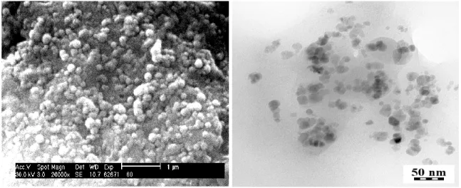
4.1 Morphology of HSA loaded magnetic PLGA NPs 44 4.2 Hydrodynamic size and size distribution 45
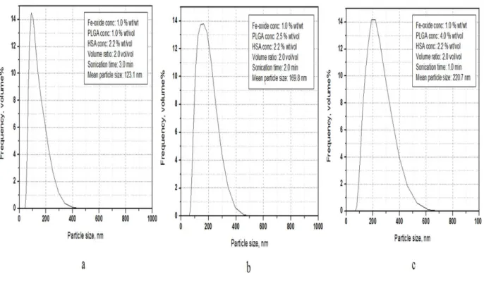
4.2.1 Effect of Fe3O4/PLGA weight ratio 48
4.2.2 Effect of PLGA concentration 49
4.2.3 Effect of HSA concentration 51
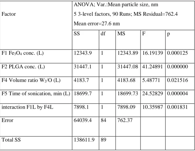
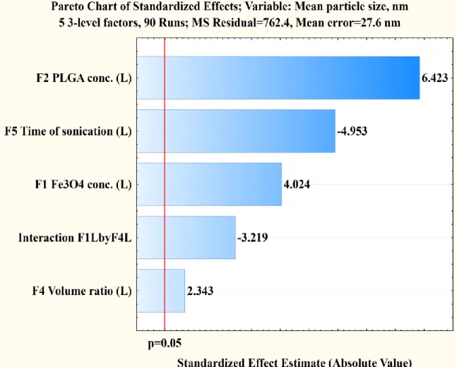
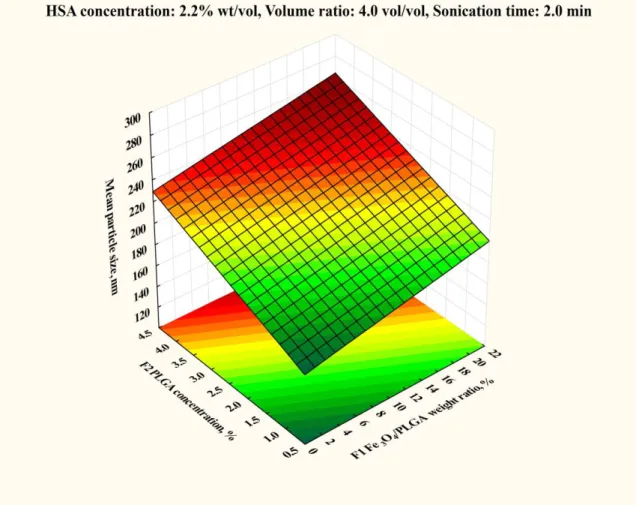
4.2.4 Effect of volume ratio of the W2 and O phases 51 4.2.5 Effect of sonication time during second emulsification 53 4.2.6 Prediction of the expected mean particle size 54 4.2.7 Optimization of the process variables to achieve smaller sized NPs 55 4.3 Encapsulation efficiency of model drug 57
4.3.1 Effect of PLGA and HSA concentrations 59
4.3.2 Effect of the magnetite/PLGA mass ratio 62
4.3.3 The interaction of the PLGA concentration and the volume ratio 63 4.3.4 The interaction of HSA concentration and volume ratio 65
4.3.5 Effect of sonication duration on the second emulsification 66
4.3.6 HSA loading in the particles 68
4.4 Optimization of the process variables 69
4.5 Surface modification 73
4.5.1 The surface charge of PLGA NPs 74
4.5.2 Surface attachment of poloxamer 75
4.5.3 Protein adsorption studies 77
4.5.4 Isothermal calorimetric analysis 80
4.6 In vitro IFN-α release 83
4.6.1 IFN-α decomposition kinetics 85
4.6.2 Modelling IFN-α release from NPs 86
5 CONCLUSION 89
6 NEW SCIENTIFIC RESULTS 90
7 ÚJ TUDOMÁNYOS EREDMÉNYEK 92
8 REFERENCES 94
List of publications Acknowledgements
101 103
Appendix 105
List of abbreviations BBB
BCA BSA Dmean
DCM DOE Dox
Blood brain barrier Bicinchoninic acid Bovine serum albumin
Mean hydrodynamic particle size Dichloromethane
Experimental design Doxorubicin
EE ELISA GA HSA IFN- LA
Encapsulation efficiency
Enzyme-linked immunosorbent assay Glycolic acid
Human serum albumin Interferon alfa
Lactic acid MNPs
MPs MPS MRI Ms NPs PBS PEG PLA PLGA PVA RES SEM SPIO TEM
Magnetic nanoparticles Microparticles
Mononuclear phagocyte system Magnetic resonance imaging Saturation magnetization Nanoparticles
Phosphate-buffered saline Poly(ethylene glycol) Poly(lactic acid)
Poly(lactic-co-glycolic acid) Poly(vinyl alcohol)
Reticuloendothelial system Scanning electron microscope Superparamagnetic iron oxide Transmission electron microscopy
VHSA Volume of the introduced HSA solution (internal phase)
VPLGA Volume of the introduced PLGA/magnetite solution (intermediate organic phase)
VPVA Volume of the outer aqueous phase (PVA solution) W/O/W
O/W/O XFe3O4
Water-oil-water Oil-water-oil
Relative mass of the introduced magnetite compared to the mass of PLGA
XPLGA Concentration of PLGA in the intermediate oily phase XHSA Concentration of HSA in the inner aqueous phase
XVOLR Volume ratio of the outer aqueous phase to the intermediate oily phase Xtime Time of the second sonication
ABSTRACT
The aim of this Ph.D. work was to create sustained and targeted drug delivery device for natural interferon alfa (IFN- ) which is an effective medicine mainly applied against hepatitis viruses and viral hepatocellular carcinoma. Thus, IFN- and superparamagnetic iron oxide (Fe3O4) nanoparticles (NPs) were co-encapsulated inside poly(lactic-co-glycolic acids) (PLGA) matrix. PLGA NPs were prepared using double emulsion solvent evaporation method, and the relation between the process variables, in order to obtain small sized NPs sterilizable by ultrafiltration with high encapsulation efficiency; the surface modification to evade macrophages in the bloodstream; and the in vitro IFN-α release were investigated. Process optimization and functionalization were carried out using human serum albumin model drug. By experimental design and statistical analysis with STATISTICA® software, the number of experiments was reduced to 90 instead of 243 for five important process variables and the results were analyzed. The mean hydrodynamic size and encapsulation efficiency of NPs ranged from 115-329 nm and 18 to 97%, respectively depending on the process conditions. The optimization process, carried out by exact mathematical tools using GAMSTM/MINOS software, enabled to find out optimum process conditions to achieve high encapsulation efficiency (92.3%) for relatively small sized PLGA NPs (155 nm). NPs prepared with selected optimum condition were subjected to surface modification to prolong their lifetime in the bloodstream by using triblock copolymer poloxamer. Surface functionalization was verified by size, zeta potential and isothermal titration calorimetry investigations. In blood serum protein adsorption measurements, bovine serum albumin tended to be adsorbed 50% less on the surface modified NPs in comparison to the unmodified ones. In vitro IFN- release study carried out using enzyme linked immunosorbent assay test showed that prepared PLGA NPs are capable to sustain the release of interferon, and protect it from the quick degradation. Interferon release from poloxamer modified PLGA NPs was much slower than that for unmodified ones.
KIVONAT
A PhD munka célja késleltetett és célzott hatóanyag leadási rendszer létrehozása természetes interferon alfa (IFN- ) számára, mely a hepatitisz vírusok és a virális hepatocelluláris karcinóma egyik hatékony gyógyszere. E célból poli(tejsav-glikolsav) (PLGA) mátrixban IFN- és szuperparamágneses vas-oxid nanorészecskéket együttesen mikrokapszuláztak. A PLGA nanorészecskéket összetett emulziós oldószer elpárologtatásos módszerrel állították elő, és vizsgálták az eljárás változói közötti összefüggést annak érdekében, hogy ultraszűréssel sterilizálható kisméretű, nagy kapszulázási hatékonyságú részecskéket nyerjenek; a felületi módosítást, hogy a véráramban elkerüljék a makrofágokat; és a nanorészecskék in vitro IFN- kibocsátását.
Az eljárás optimalizálását és a részecskék funkcionalizálását humán szérum albumin modell gyógyszerrel végezték. A STATISTICA® kísérlettervező és statisztikai analízis szoftverével a kísérletek számát 243-ról 90-re csökkentették az öt legfontosabb folyamatváltozó figyelembevételével, és az eredményeket elemezték. A nanorészecskék átlagos hidrodinamikai mérete és a kapszulázási hatékonysága az eljárási körülmények függvényében 115-329 nm és 18-97 % tartományban változott. Az optimalizálás egzakt matematikai eszköze a GAMSTM/MINOS szoftver lehetővé tette a legjobb eljárási körülmények megtalálását magas kapszulázási hatékonyság (92,3 %) és viszonylag kisméretű (155 nm) PLGA nanorészecskék eléréséhez. Az optimális körülmények között előállított nanorészecskék felszínét poloxamer triblokk kopolimerrel módosították, hogy megnöveljék a véráramban az élettartamukat. A felszíni funkcionalizálást méret, zeta potenciál és izotermális titrációs kalorimetriás analízissel támasztották alá. Vér szérum fehérje adszorpciós vizsgálata során a felületmódosított nanorészecskék 50 %-kal kevesebb bovine szérum albumint adszorbeáltak, mint a nem módosított nanorészecskék. In vitro IFN- leadási vizsgálat enzimhez kapcsolt immunszorbens meghatározással kimutatta, hogy a PLGA nanorészecskék képesek az interferont késleltetetten kibocsátani, és megvédeni a gyors lebomlástól. A poloxamerrel módosított PLGA nanorészecskék lényegesen lassabban adták le az interferont, mint a nem módosított részecskék.
AUSZUG
Das Ziel der Ph.D These war Herstellung eine Vorrichtung für nachhaltige und gezielte Arzneimittelabgabe welche naturgemäß Interferon Alpha (IFN-α) enthält. Interferon Alpha ist eine effektive Medizin vorzugsweise verwendet gegen Hepatitis-Viren und virales Leberzellkarzinom. So IFN-α und superparamagnetischen Eisenoxid (Fe3O4) Nanopartikeln (NPs) waren innerhalb einer Matrix von Poly(Milchsäure-co- Glykolsäure) (PLGA) co-gekapselt. PLGA NPs waren mit der Hilfe einer Doppelemulsion Methode präpariert. Die Relation zwischen Die Prozessvariablen und die in vitro IFN-α Freigabe war untersucht um feinen NPs zu beschaffen, die für Sterilisation durch Zentrifugation geeignet sind, und eine hohe Verkapselung Leistungsfähigkeit haben, und mit Oberflächenmodifizierung Makrofagen in den Blutstrom zu vermeiden können. Die Optimierung des Verfahrens und die Funktionalisierung waren Aufwendung von Humanes Serumalbumin als Modell Arzneimittel durchgeführt. Die Anzahl von Experimenten war von 243 bis 90 Prozessvariablen Aufwendung das Versuchsdesign und statistische Analyse von Statistica Software reduziert. Der hydrodynamischen Durchschnittskerngröße und Verkapselung Leistungsfähigkeit von NPs waren 115-239 nm und 18-97 %, beziehungsweise abhängig von den Prozessbedingungen. Die Optimierung des Verfahrens, die mit exaktem mathematischem Instrument, der GAMSTM/MINOS Software durchgeführt war, hat ermöglicht zu finden optimal Verfahren Bedingungen zu erreichen eine hohe Verkapselung Leistungsfähigkeit (92,3 %) von relativen kleinen PLGA NPs (155 nm). NPs, die mit ausgewählten optimalen Verfahren Bedingungen hergestellt waren zu prolongieren ihre Lebensdauer in den Blutstrom, waren mit Hilfe dreiblock copolymer Poloxamer Oberflächenmodifizierung untergezogen.
Oberflächenfunktionalisierung war mit Korngröβe- und Zetapotenzialanalyse, und isometrische Kalorimetrie Titrieren geprüft. In der Blutserum Absorption Messungen Bovines Serum Albumin was tendieren zu absorbieren 50 % weniger auf die oberflächenmodifizierte NPs als auf die Unmodifizierten. Die in vitro IFN-α Freigabenuntersuchung zeigte, was mit der Enzyme verbundenen immunosorbens Probe durchgeführt war, daβ die präparierten PLGA NPs fähig sind zu retardieren die Freigabe von Interferon, und die Oberflächenmodifizierung bewährt die NPs von einer schnellen Degradierung. Freigabe von Interferon mit Poloxamer modifizierten PLGA NPs war viel langsamer als von Unmodifizierten.
1 INTRODUCTION
Last two decades have witnessed tremendous efforts by researchers for developing potent drugs to treat cancer and viral diseases. Cancer has overtaken heart disease as the number one killer and as a single entity, cancer takes highest number of human lives worldwide with an estimated 8.2 million deaths in 2012. World cancer report 2014 forecasted 75% increase in cancer cases worldwide over next two decades which is going to result in 25 million cancer cases. The estimated total annual economic cost of cancer was approximately US$ 1.16 trillion in 2010. This amount is equivalent to 2% of total global GDP (gross domestic product). According to recent report released by Centers for Disease Control and Prevention on World Hepatitis Day (July 28, 2013), viral hepatitis is a leading cause of infectious disease mortality globally, each year causing approximately 1.4 million deaths. Most of these deaths occur among the approximately 400 million persons living with chronic hepatitis B virus or hepatitis C virus infection.
Nanoparticles (NPs) are solid colloidal particles with diameters ranging from 1- 150 nm, nevertheless, regarding microencapsulation, 1-1000 nm is generally termed as nano size range. Numerous NP-based drug delivery and drug targeting systems are currently under development and many of them are already developed [1]. Especially since 1980s, scientists have been trying to develop potent drug carriers and deliver medicines to targeted sites. The primary cause behind tremendous development of medical industry is recognizing essentiality for developing current therapies for drugs already marketed and yet to be marketed. Drug loaded magnetic nanoparticles/nanocapsules (NPs) are very promising tools to realize targeted and sustained drug delivery, especially in fighting against cancer. Several disease related bioactive molecules/drugs are successfully encapsulated by appropriate matrices to improve bioactivity, bioavailability and applied for controlled delivery. Nanomedicines of the diseases such as AIDS, cancer, tuberculosis, diabetes, malaria, prion disease, etc.
are in different trial phase for the testing and some of them are commercialized [2].
Probably that’s why recent years have witnessed not only unprecedented growth of research, but also vast applications in the area of nanoscience and nanotechnology.
Therapeutic NP technologies are able to revolutionize the drug development process and capable of changing the landscape of the pharmaceutical industry. Encapsulation of
medicinal drugs by nanocarriers increases specificity, drug efficacy, therapeutic index of corresponding drugs and tolerability.
1.1 Objectives
The aim of this Ph.D. work was to create sustained and targeted drug delivery device for natural interferon alfa (IFN- ). IFN- and superparamagnetic iron oxide (Fe3O4) NPs were co-encapsulated inside poly(lactic-co-glycolic acids) (PLGA) matrix.
PLGA NPs were prepared using suitable method of production and the relation between the process variables to obtain smaller sized NPs with higher encapsulation efficiencies, surface modification to evade macrophages in the bloodstream and in vitro IFN-α release were investigated. Due to high cost of IFN-α, process optimization and functionalization were carried out using human serum albumin (HSA) model drug instead of IFN-α. IFNs exhibit antiviral and antitumor activities. Magnetic NPs can target IFN- loaded PLGA NPs to liver and spleen and can be used to cure cancers of respective body parts. On the other hand, due to the presence of magnetic NPs, targeting any body part with the help of magnetic field (by external or internal magnet) may be possible and drug uptake process might also be visualized by MRI (Magnetic resonance imaging).
2 REVIEW OF THE LITERATURE 2.1 Interferon (IFN)
Interferon (IFN) was first discovered in 1957 by Alick Isaacs and Jean Lindenmann [3]. Interferon (IFN) is a class of cytokines (cell signaling proteins) with immune stimulating activity carrying out important physiological functions in higher vertebrates. IFNs are a family of small proteins and glycoproteins. They are produced and secreted in vivo by cells mainly in response to infections caused by viruses and also in response to synthetic or biological inducers. IFNs are being used widely for the biologic therapy of infectious viral diseases such as hepatitis C and B and for the treatment of several cancers.
IFNs are divided into type 1 and type 2 based on common biochemical features and/or amino acid sequence similarities. IFN-γ is the only type 2 member having dissimilar structure than type 1 IFNs. Type 1 IFNs share a common cell surface receptor. Type 1 IFN is subdivided into several types depending on the basis of nucleotide and amino acid sequence identities, species-restricted distributions or unique biological functions. Among different subtypes, two most ancient and best- characterized type 1 IFNs are ‘IFN-α’ and ‘IFN-β’ which are the most broadly distributed type 1 IFNs among mammals.
2.1.1 Natural interferon alfa (IFN- )
Natural IFN-α proteins are partially glycosylated, whereas recombinant IFNs are not. Natural IFN-α has certain advantages over recombinant products. Natural IFNs show comparatively lower side effects than recombinant or synthetic IFNs, which is their main advantage and the reason behind the use of natural IFN-α in this study. Cell culture-derived "natural" IFN products contain a multiplicity of IFN types or species, thus, are expected to provide potentially better therapeutic efficacy, than single-species recombinant IFN products. Since each subtype has its own specific biological activity, it is expected that natural IFN-α has more balanced effect against a variety of diseases.
Therefore, it can be considered more advantageous for patients to be treated with natural product that potentially elicits an effective immune response, and is tolerated to a higher extent by the treated patients.
IFN- is produced by leukocytes (T cells, B cells, macrophages and null cells) upon exposure to viruses, B cell mitogens, tumor cells or foreign cells [4]. IFN- is the
most extensively studied interferon species. Each subtypes of IFN-α showed different growth inhibitory, antiviral and other biologic activities. Regarding antiviral activity, IFN-α1 was the least, while IFN-α8 was the most potent.
IFN-α is used in more than 40 countries for treating more than 14 types of cancers, including some hematological malignancies (chronic myeloid leukemia, hairy cell leukemia, some B and T cell lymphomas) and certain solid tumors, such as melanoma, renal carcinoma and Kaposi’s sarcoma [5].
2.1.2 The mechanisms of action
IFNs show numerous and diverse actions, functioning at the physiological and molecular level. They bind to specific membrane receptors on the cell surface to show their cellular activities. After being bound to the cell membrane, IFNs initiate a complex sequence of intracellular events, including the up-regulation of certain other cytokines, suppression of cell proliferation, induction of certain enzymes, inhibition of virus replication in virus-infected cells, immunomodulating activities such as enhancement of the phagocytic activity of macrophages and augmentation of the specific cytotoxicity of lymphocytes for target cells.
In spite of years of intense works in animal tumor models and considerable experiences in the clinical use of IFNs, the exact mechanisms underlying the antitumor response are not fully understood. Likewise, not too much is known about why some tumors are highly responsive to IFN treatments, while others show little or no response.
IFN-α plays very important role in the antiviral defense by regulating the immune system or by direct inhibition of the intracellular viral lifecycle [6]. IFN-α has the ability to stimulate the host cell to resist viral infection and can inhibit many steps of viral replication. The major antiviral action is the affecting of translation of the viral genome.
IFN-α inhibit viral transcription, enzymatically degrade viral RNA, reduce the translation of viral proteins by interfering with the normal host cell translation mechanism, modify glycosylation patterns of viral proteins (which could influence virus packaging, release, or virulence) and can alter the cell membrane (e.g. increasing cell membrane fluidity) which may influence virion maturation and release. IFNs inhibit cell growth and division by increasing the length of the multiplication cycle, enhancing cell lysis through cytotoxic mechanisms, depleting essential metabolites, inhibiting the
expression of oncogenes, inducing the expression of tumor suppressor genes and inducing apoptosis.
IFN-α is eliminated from the body mainly by the kidney, where they are totally filtered through the glomeruli. In the kidney, during tubular reabsorption, they undergo rapid proteolytic degradation resulting in negligible reappearance of intact IFN-α in the systemic circulation. Minor pathways of elimination for IFN-α involve liver metabolism and subsequent biliary excretion [7]. For healthy people, IFN-α exhibited an elimination half-life of 3.7 to 8.5 hours (mean 5.1 hours).
Primary adverse effect is "flu-like" symptoms (headache, fever, myalgias, etc.) which are in most cases mild or moderate, and transient, and does not affect the treatment. Alfa IFNs have suppressive effect on the bone marrow, which may lead to a fall in the white blood cell count, the platelet count and less commonly the hemoglobin concentration. Moreover, abnormalities in the blood clotting mechanism have been observed. These effects can enhance the risk of infection, thrombosis and hemorrhage.
2.2 Model drug
To make the research more economical, researchers frequently use model drug during the development of drug loaded NPs/microparticles (MPs) since in many cases drugs are quite expensive. In this study, HSA was selected as a model drug, since natural interferon accompanied with a number of other cytokines and proteins is secreted as a protein mixture from the human blood, and HSA is the most abundant protein in this mixture. HSA is not removed from the mixture, since it was found that it assures appropriate medium to cytokines, and preserves their activities and prevents them from environmental damages. HSA concentration is higher than 95% in the natural interferon mixture (Trigon Biotechnological Zrt.), therefore in the encapsulation process it affects strongly the properties of the obtained NPs. HSA is a monomeric multi-domain macromolecule, which is the most abundant plasma protein in the human body with a plasma concentration of 0.6 mM [8]. HSA consists of 585 amino acids that form three structurally similar α-helical domains.
2.3 Nanoparticles in drug delivery
The use of nanotechnology in medicine, specifically for drug delivery has been increasing day by day. From a positive viewpoint, NPs have the ability to access into the cell and various cellular compartments including the nucleus, and they have the potential to cross the blood brain barrier also. Currently a multitude of substances are being investigated for the preparation of NPs for drug delivery. Not too much is known regarding all aspects of nanoparticle toxicology in the field of drug delivery, however, vast number of research is being carried out to explore that.
Many studies have demonstrated that NPs possess several advantages over MPs [9]. For example, NPs are capable of travelling through the bloodstream without blockage or sedimentation of the microvasculature. Small NPs can circulate throughout the body and penetrate tissues e.g. tumors. NPs having unique small size can strongly influence drug loading, drug release, stability of the therapeutic agents, etc. Because of their small size, unique qualities are shown by NPs that are not found in the same material at larger size [10]. Drug release is strongly affected by the size of particles.
Smaller particles possess larger surface-area-to-volume ratio, and most of the drugs associated with small particles normally remain at or near the particle surface, which leads to a faster drug release [11]. NPs have relatively high cell uptake in comparison to MPs. They are useful for a wider range of cellular and intracellular targets because of their mobility and small size. NPs can be taken up by cells through natural routes e.g.
endocytosis. These vehicles can be designed in a way so that they will represent biophysical characteristics that are unique to the target cells, and are capable of minimizing drug loss and toxicity that can occur due to the delivery to the non-desired tissues. Similar study showed that NPs penetrated throughout the submucosal layers of a rat intestinal loop model, whereas MPs were predominantly localized in the epithelial lining [12]. This indicates that particle distribution can be tuned by controlling size of particles. In recent years, significant researches have been done on NPs as oral drug delivery vehicles, and the major interest for this purpose was lymphatic uptake of the NPs by the Peyer’s patches in the gut associated lymphoid tissue. There are lots of reports on the optimum size for Peyer’s patch uptake ranging from < 1 μm to < 5 μm [13]. Results show that MPs remain in the Peyer’s patches, while NPs are systemically disseminated. Another advantageous feature of NPs smaller than 220 nm is that they
could be easily sterilized by ultrafiltration, since the sizes of bacteria and viruses are larger [14].
One big challenge for nanomedicinal formulations is scaling up. Nanodrug formulation to a large-scale is not always easy. It is much easier to maintain the size or composition of NPs at the laboratory scale than at a large scale. A number of technologies for the production of nanodrug may not be compatible with large-scale production due to the type of preparation method need to be used and high cost of materials employed. Although there are a good number of patents published on nanodrug delivery technologies, commercialization is still in its early stage [15].
2.3.1 Characteristics of NPs for drug delivery
Particle size is one of the most important characteristics of NPs intended for drug delivery applications. Size plays vital roles in particle functions, in vivo distribution, targeting, drug loading, drug release, toxicity, degradation, clearance and uptake mechanisms, etc. [16]. Depending on size, particles show different diffusion characteristics, velocities and adhesion properties which result in different uptake efficiencies. The size of NPs must be large enough to prevent rapid leakage in blood capillaries and at the same time small enough to escape the capture of macrophages in the reticuloendothelial system (RES).
The surface properties of NPs also control the fate of nanoparticles in the circulation. Opsonization and mononuclear phagocyte system (MPS) clearance are the two most important factors that are strongly affected by NP surface properties.
Generally, positively charged NPs are internalized by cells due to electrostatic interaction with the negatively charged cell membrane by non-specific endocytosis [17].
On the other hand, negatively charged NPs show more selective internalization due to slight charge repulsion effect. Higher surface charge (either positive or negative) lead to an increase in macrophage surveillance, followed by MPS clearance. The in vivo biodistribution studies showed that highly negatively or highly positively charged NPs accumulated in the liver due to MPS cells uptake, whereas slightly negatively charged NPs accumulated in the tumor tissue and were excluded by the MPS system [17].
NPs used for drug delivery purpose should have desirable release profile. Both drug release and biodegradation of nanocarriers need to be considered significantly during the development of nanoparticulate delivery systems. Drug release is a complex
solubility, desorption of the adsorbed or surface-bound drug, NP matrix degradation or erosion, the combination of diffusion and erosion processes, etc. [13]. Polymeric coating of nanocarriers can also affect the release of drug.
2.3.2 Toxicological hazards of nanoparticles
Safety and toxicological issues need to be strongly considered for the use of nanotechnology in nanomedicines. NPs have significant negative impact on human body due to the interactions between NPs and biological systems, which is termed as
“nanotoxicology”. Preliminary nanotoxicological studies have revealed that NPs can contribute to free radicals formation, undesirable penetration through the epidermis or other physiological barriers to enter different areas of the body that are more susceptible to toxic effects and damage of brain cells [15]. The cationic NPs (e.g. gold, polystyrene, etc.) may cause blood clotting and hemolysis, whereas anionic NPs are nontoxic in comparison to cationic ones. However, scientists have been working on the surface coatings and modifications of NPs to make them comparatively safer and more effective [18].
2.3.3 Forces acting on nanoparticle systems
The primary attractive force among NPs dispersed in a liquid is van der Waals force. For two nanospheres of radius ‘R1’and ‘R2’ separated by a distance ‘D’ that is much smaller than ‘R’, the van der Waals interaction energy can be written: V = - {AR1R2/6D(R1+R2)} where A is the effective Hamaker constant of the system [19]. If this attraction is strong enough, the NPs will hit each other and coalesce to form large particle. If only van der Waals forces would operate in a particle system, we could expect all dissolved particles to coagulate immediately and precipitate out of solution as a mass of solid material. Fortunately this does not happen due to the existence of repulsive forces between particles suspended in water or any liquid of high dielectric constant. Particles suspended in liquid are usually charged and can be prevented from coalescing by repulsive electrostatic forces. Other repulsive forces capable of preventing coalescence are solvation and steric forces.
Electrostatic repulsion arises when electric charge exists at the surface of the NPs. These charges may originate from the adsorption of charged ions at the surface (e.g. the adsorption of –OH- groups to the water-air or water-polymer interfaces that
ionization of carboxyl group to carboxylate ion). In immediate vicinity of the NP surface, solution composition is in fact relatively complex. It is often described by a double-layer model: the inner layer (also known as the Stern layer), where the water molecules are highly structured by solvation of surface charges, and the diffuse layer involving counterions.
In case of very small separations between the surfaces or particles (e.g. few nanometers), the non-DLVO (Derjaguin and Landau, Verwey and Overbeek) forces such as the solvation forces and hydrophobic interaction forces become important.
Solvation forces may arise from the ordering of the solvent molecules into discrete layers between particle surfaces in a highly restricted space. When NPs are immersed in a liquid, particle surfaces may be solvated. If two solvated surfaces come close enough together so that the solvation layers overlap, repulsive force originates. The repulsion may be caused because of the hydrated groups at the surfaces when they approach each other. Sometimes, solvation forces are also called structural force. Hydrophobic interaction occurs due to the water-fearing property of hydrocarbons by releasing water from the surface or hydrophobic particles resulting in interaction between hydrophobic groups.
Steric repulsion helps to stabilize colloidal particles and provides a more general method, exploiting the fact that the strength of van der Waals forces falls off very quickly with the distance. Colloidal particle surfaces can be coated with neutral and hydrophilic molecules that are long enough to maintain a certain distance between the particles, thereby rendering the van der Waals attraction negligible. In a physiological medium, extremely hydrophilic macromolecules like dextran or poly(ethylene glycol) (PEG) are often used to stabilize colloidal particles, since they interact strongly with the particle surface, and that they cannot form ‘bridges’ between different particles.
2.4 Microencapsulation methods
Microencapsulation is a process of enclosing submicron/micron sized particles of solids/droplets of liquids/gasses in an inert shell, which in turn protects and isolates the particles from the external environment. Microcapsules consist of two parts: the core and shell. The core is the intrinsic part (contains the active ingredient) and is protected by the shell (the extrinsic part). The obtained products of microencapsulation process are termed as microparticles/microcapsules/microspheres depending on the internal
structure and the morphology. If the particle size goes below 1 m, they are termed as nanoparticles, nanocapsules, nanospheres, respectively.
The morphology of microcapsules mainly depends on the arrangement of core material inside the shell and deposition process for shell material. Microcapsules may be regular and irregular in shapes. Based on their morphology, microcapsules can be classified as matrix, mononuclear and polynuclear type (Fig. 1).
Fig. 1: Morphology of microcapsules.
Matrix type microcapsules exhibit homogeneous distribution of the core material inside the shell material. Mononuclear (also known as core-shell) type microcapsules contain a shell around the core. Many cores are encapsulated within the shell in polynuclear microcapsules. Beside these three primary morphologies, multiple shell mononuclear microcapsules are also observed.
The goals of the microencapsulation are diverse, however, most important directions are: (i) protection of the sensitive substances from the external environment, (ii) masking of organoleptic properties such as odor or taste, (iii) enzyme and microorganism immobilization, (iv) controlled, sustained or timed release of drug, (v) for safe handling of the toxic materials, (vi) better processability (improving flowability, solubility, dispersibility), etc.
Several techniques are used for the encapsulation of core materials. Broadly these methods can be divided into three types [20]:
(i) Chemical methods: In-situ processes like suspension, emulsion, precipitation or dispersion polymerization, interfacial polycondensations, etc. are the most widely used chemical techniques for microencapsulation.
(ii) Physico-chemical methods: Among physico-chemical methods, most common microencapsulation process is coacervation. Coacervation is based on aggregation of macromolecules by partially desolvating fully solvated macromolecules. Coacervation can be divided into two types: simple coacervation and complex coacervation. In simple coacervation processes, addition of salt or alcohol, change in temperature or pH issues in phase separation. On the other hand, oppositely charged polymer, when added to the polymer solution, forms a coacervate phase via anion–cation interactions in complex coacervation process. Sol-gel encapsulation, layer-by-layer (L-B-L) assembly, supercritical CO2-assisted microencapsulation, etc. are also physico-chemical methods of microencapsulation used for specific applications.
(iii) Physico-mechanical methods: There are several physico-mechanical methods which are being used widely. Solvent evaporation technique finds widespread applications in different pharmaceutical industries. It facilitates the release of drug in a controlled way, which has a lot of clinical benefits. The core material to be encapsulated is dissolved or dispersed in the solution of coating polymer. The coating polymer is dissolved in a volatile solvent. The forming solution is added to a stirred solution containing a suitable stabilizer to form small polymer droplets containing encapsulated material. From the mixture, solvent is evaporated. As a result, coating material shrinks around the core material encapsulating the core.
2.4.1 Double emulsion method
A double emulsion is a complex liquid dispersion system in which the droplets of a dispersed liquid are further dispersed in another liquid. In a double emulsion, the inner dispersed droplets/globules are separated (compartmentalized) from the outer liquid phase by a layer of another phase. So far, different types of double emulsions have been documented. Some may have single internal compartment, while others may contain many internal droplets (known as “multiple-compartment emulsions”). Two common types are the water-oil-water (W/O/W) and oil-water-oil (W/O/W) double
emulsions. The most common and widely used double emulsion is W/O/W type, although O/W/O emulsions are needed for some specific applications.
Potential applications for double emulsions are well documented and a large number have been patented [21]. The important applications are in pharmaceuticals, agriculture, foods and cosmetics. In most cases, double emulsions are used to achieve slow and sustained release of active matter from an internal reservoir into the continuous phase (mostly water).
Usually a two-step emulsification process is used to form double emulsions using high pressure valve homogenizers or conventional rotor-stator. The primary emulsion of either type (W/O or O/W) is prepared under high-shear conditions to obtain inner droplets of small size, while less shear is applied during the secondary emulsification step to avoid rupture of the liquid membrane between the innermost and outermost phase. However, the second step can affect encapsulation efficiency and might reduce it, if homogenization is too intensive. It can also result in highly polydisperse outer drops, if homogenizing conditions are too mild.
Double emulsions can alternatively be produced by other methods e.g. by forcing a primary emulsion through a microporous membrane or microfabricated channel arrays into a continuous phase liquid. Much less shear is needed in this case, than in the conventional emulsification processes. As a result, the droplets are intact and both monodispersity and high entrapment efficiency can be achieved.
Double emulsions possess two different interfaces and require two sets of different types of emulsifiers. For O/W/O type emulsions, the first set of emulsifiers needed for the internal interface must be hydrophilic. For the same type of emulsion, the second set of emulsifiers needed for the external interface must be hydrophobic. In case of W/O/W type of double emulsions, the order of the emulsifiers is just opposite. The inner emulsifiers must be hydrophobic, while the outer ones need to be hydrophilic. In many cases a mixture of two or more emulsifiers in each set is used for better stabilization results. Too little emulsifier might fail to give a stable emulsion, whereas too much emulsifier may lead to toxic effects and can even cause destabilization.
By double emulsion, active materials are generally entrapped in the inner aqueous/organic phase. It is well documented in the literature that, due to the osmotic pressure difference, the active matter tends to diffuse and migrate from the internal phase to the external interface; in most cases through a mechanism known as “reverse
applications. Flocculation, aggregation, coalescence are the major factors affecting the instability of the double emulsions, resulting in the separation of the phases and the rupture of the droplets. Double emulsions with low molecular weight emulsifiers (so called monomeric emulsifiers) are mostly thermodynamically unstable. This is because in the second stage of the emulsification severe homogenization or shear takes place forming large droplets. Proper and more suitable combinations of emulsifiers can improve the emulsion stability and reduce the droplet sizes. Among different surfactants, partially hydrolyzed poly(vinyl alcohol) (PVA) is mostly used since it is capable of producing smallest particles [22].
2.4.2 W1/O/W2 double emulsion solvent evaporation techniques
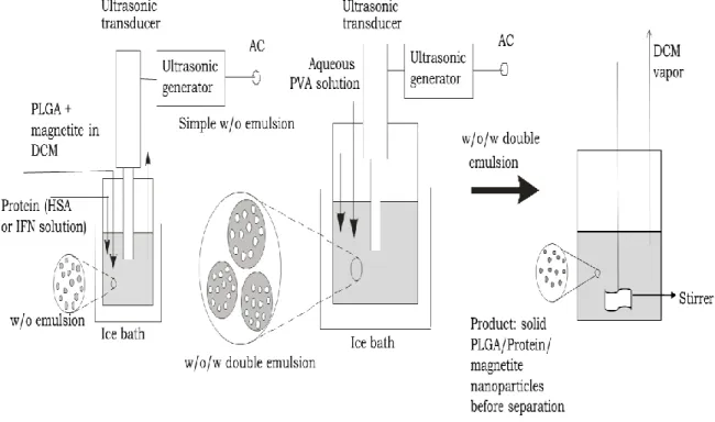
W1/O/W2 double emulsion solvent evaporation technique is normally used to prepare hydrophobic biodegradable micro/nanospheres containing hydrophilic proteins, pharmaceuticals and polypeptides for sustained release applications.
For the generation of transient W1/O/W2 emulsions, a two-stage emulsification process is used. The basic principle of the encapsulation in this process is based upon inducing phase separation of the polymer dissolved in a volatile organic solvent (O phase) due to partial extraction of the solvent in a large volume of the outer water phase (W2 phase) and finally evaporation of the volatile solvent. The polymer then forms a coacervate which encloses the internal aqueous phase (W1) that contains the active compound. MPs/NPs are hardened upon the removal of the residual solvent. Suitable solvents for this technique need to be volatile and immiscible with water.
Dichloromethane is being employed frequently due to its low boiling point which facilitates the removal of residual solvent from the finished product. The other aspects include high dissolving power for a wide range of polymers, its partial solubility in water, and toxicity profile. The hydrophilic active substance (e.g. drug) is dissolved either in buffer or water and represents the internal water phase (W1). Stabilizing excipients can be added for the protection of drug in the core of the microspheres. W1/O emulsion is resulted upon the homogenization of W1 phase with the O phase. The resulted primary emulsion is then rapidly added to a large volume of an external aqueous solution (W2) that usually contains emulsifier(s) (e.g. PVA), and then emulsified to obtain a double W1/O/W2 emulsion.
Particles are formed based on coacervation. Because of its initial diffusion into
inducing phase separation of the polymer. The organic solvent elimination occurs by two steps: firstly by extraction and secondly by evaporation. For further removal of the organic solvent and particle hardening, the system is continuously stirred at a constant rate for few hours. After complete solidification, the MPs/NPs are isolated either by filtration or centrifugation and washed several times with water to remove residual PVA and non-encapsulated drug. Depending on the glass transition temperature of the polymer, the MPs/NPs are lyophilized or dried in a vacuum at room temperature.
The main advantage of the W1/O/W2 double emulsion technique is that hydrophilic drug substances can be encapsulated under very gentle process conditions.
Additionally, high yields and encapsulation efficiencies can be obtained.
2.5 Polymers used for drug delivery
Polymeric materials used for formulating NPs and MPs include both natural and synthetic polymers. Natural polymers in most cases are less expensive and have aqueous solubility. They have not been widely employed for biomedical purpose, since they vary in purity, show variability from batch-to-batch, and often require crosslinking that could denature the embedded drug. Synthetic polymers have a number of advantages including high control of polymer properties, such as molecular weight and functionality, and are feasible for commercial-scale production. Synthetic polymers show more reproducibility and can be prepared with the desired copolymer compositions, molecular weights and degradation rates. Synthetic polymers such as PLGA, poly(lactic acids) (PLA), poly(ε-caprolactone) (PCL), poly(methyl methacrylates) and poly(alkyl cyanoacrylates) or natural polymers such as gelatin, chitosan, albumin, collagen or alginate, etc. are commonly used as drug carriers. It is reported in the literature that polyesters alone and in combination with other polymers are most commonly used for the formulation of NPs and MPs. PLGA and PLA are highly biocompatible and biodegradable. They have been employed since the 1980’s for numerous in vivo applications (biodegradable implants, controlled drug release, etc.).
2.5.1 Poly(lactic-co-glycolic acid)
PLGA is one of the most successfully used biodegradable polymers. PLGA is approved by the US Food and Drug Administration (FDA) and European Medicine Agency (EMA) in various drug delivery systems in humans.
This unique carrier system has the potential to change the current scenario of cancer research and diagnosis in real time. Encapsulation or conjugation of drugs in PLGA carriers can reduce several undesirable shortcomings. PLGA particles loaded with drugs can prolong the in vivo circulation time of the therapeutics from minutes to hours and are also able to reduce the cellular uptake along the endocytic route [23]. A very significant property of PLGA is the capability to manipulate its physico-chemical properties in order to tailor its degradation rate, which subsequently affects the release profile of the encapsulated molecules. Tailoring the release of encapsulated drugs from the polymeric particles reduces the use of frequent doses in the treatment regimen. The degradation rate of PLGA depends mainly on the hydrophilicity, molecular weight and crystallinity of the polymer. More hydrophilic and lower molecular weight PLGA exhibits an increased rate of polymer degradation. Ratio of PLA and PGA monomers present in the copolymer mainly determines the hydrophilicity of PLGA. Glycolic acid (GA) is more hydrophilic than LA (lactic acid). If the PLGA contains higher proportions of GA, increased degradation rate of the NP formulations is observed due to higher hydrophilicity of PLGA. The only exception to this rule found in the literature is the co-polymer having 50:50 GA:LA ratio which shows fastest degradation rate (half- life (t1/2) about 2 weeks) among PLGA polymers of different GA:LA ratios even those with higher GA content. This is because of the amorphous nature of PLGA 50:50. At 50:50 GA:LA ratio the PLGA polymer possesses least crystallinity in its structure and more prone to hydrolysis and degradation than other PLGA polymers with higher or lower GA:LA ratios.
PLGA particles are biodegradable in the body since in the presence of water they undergo hydrolysis of their ester linkages leading to two metabolite monomers: LA and GA, which, under normal physiological conditions, are byproducts of several metabolic pathways in human body [24]. The degradation rate for PLGA polymer depends on the monomer ratio. The degradation products (LA and GA) are endogenous and easily metabolized by the body via the Krebs cycle, and are eliminated. It leads to very minimal systemic toxicity related with using PLGA for drug delivery or biomaterial applications. According to a review by Athanasiou et al., many in vitro and in vivo studies associated with the toxicity/biocompatibility demonstrated that PLGA biomaterials show absence of significant toxicity and satisfactory biocompatibility [25].
In vivo studies investigated applications in articular cartilage, bone and meniscus. A
muscle. The results of all those studies supported the in vivo application of PLA-PGA biomaterials, although some cases showed inflammatory responses [24,25].
Biodistribution studies demonstrate that delivery by PLGA NP enhances accumulation of therapeutic/diagnostic agents by the enhanced permeability and retention effect [24].
PLGA is soluble in a wide range of common solvents including acetone, chlorinated solvents, tetrahydofuran, ethyl acetate, etc. The glass transition temperature (Tg) of PLGA copolymers are higher than the physiological temperature, hence they are glassy in nature. Thus, their chain structure is fairly rigid, which gives them good mechanical strength to be used for the formulation of drug delivery devices. PLGA polymer goes through bulk erosion during its degradation. The degradation bulk erosion model of release is considered advantageous in vaccine delivery, since it hinders the early release of large adjuvants or antigens from the delivery system before the particle is taken up by dendritic cells and reduces the systemic distribution of encapsulated molecules.
2.6 Magnetic nanoparticles (MNPs)
NPs show physical and chemical properties that differ from the characteristic of their atom and the bulk counterparts. MNPs have large surface area that dramatically changes some of the magnetic properties. Each MNP can be considered as a single domain, which is the reason why MNPs can exhibit superparamagnetic property and quantum tunneling of magnetization. Superparamagnetism is a type of magnetism observed in small ferromagnetic or ferrimagnetic NPs. Superparamagnetism is especially important in applications like MRI or drug delivery, where NPs exhibit no magnetic properties upon removal of the external field and therefore possess no attraction for each other eliminating the major driving force for aggregation. More importantly, superparamagnetic NPs show better control over the application of their magnetic properties since they are capable to provide strong response to an external magnetic field.
To date, iron oxide particles such as magnetite (Fe3O4) or maghemite (γ -Fe2O3) are by far the most commonly employed for clinical use, although nickel, cobalt, neodymium–iron–boron, etc. are also magnetically responsive materials. This is because magnetite and maghemite may be nontoxic, show good chemical stability, biological compatibility, and relatively ease of manufacture. Nickel and cobalt are
highly magnetic materials, but they are both toxic and susceptible to oxidation and hence are of little interest [26].
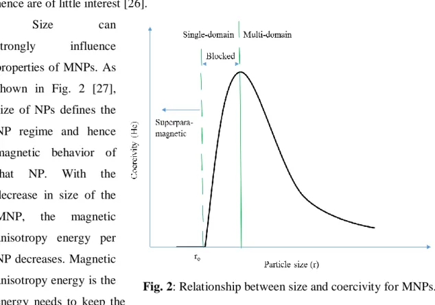
Size can
strongly influence properties of MNPs. As shown in Fig. 2 [27], size of NPs defines the NP regime and hence magnetic behavior of that NP. With the decrease in size of the MNP, the magnetic anisotropy energy per NP decreases. Magnetic anisotropy energy is the energy needs to keep the
magnetic moment in a particular orientation. The anisotropy energy becomes equal to the thermal energy at a characteristic size for each type of MNP allowing random flipping of the magnetic moment. The flipping is observed at sizes below ro (transition point from superparamagnetic to single domain), and the nanoparticle is then defined as being superparamagnetic [28].
Generally saturation magnetization (Ms) increases linearly with size until it reaches the bulk value. Above a certain size (typically 5-40 nm) known as critical particle diameter (Dc), nanoparticles become multi-domain instead of single domain.
Multi-domain nanoparticles show bulk magnetism, become either ferro/ferri or antiferromagnetic. Iron nanoparticles in the size range below 20 nm are superparamagnetic (range of 10–20 nm).
Above a temperature called blocking temperature (TB), both ferromagnetic and ferrimagnetic NPs exhibit superparamagnetic behavior. A blocking temperature TB can be defined as the temperature between the blocked and the superparamagnetic state.
Blocking temperatures rapidly increase with particle size as can be seen from the following equation [28]:
TB = KV/25kB = K(4πro3/3)/25kB
Fig. 2: Relationship between size and coercivity for MNPs.
where kB is the Boltzmann constant, K is an anisotropy constant, V is the volume of one MNP and ro is the MNP radius.
During nanoparticle synthesis, if the MNP size is maintained below a critical volume/size, the MNPs tend to develop as single magnetic domain structures and at the smallest sizes, NPs exhibit superparamagnetic behavior under standard conditions.
Increasing size of MNPs would undoubtedly aid attraction to external magnets.
Generally, larger particles possess shorter plasma half-life and cleared from the body quickly, whereas smaller nanoparticles (smaller than 10 nm) are subjected to rapid renal elimination. Though the smaller the magnetic carrier, the higher the efficiency of cell uptake. MNPs smaller than 4 μm are removed by the RES, mainly in the liver (60–90%) and spleen (3–10%) [29]. Spleen usually filters MNPs larger than 200 nm. MNPs up to 100 nm are mainly phagocytosed by liver cells [29].
Drug loaded magnetic nanoparticles target the liver cells, since iron oxide is accumulated in the liver. Jia et al. found higher antitumor activity for doxorubicin (DOX) drugs in comparison to free DOX when co-encapsulated with magnetite inside PLGA [30]. In the presence of magnetic field, the antitumor activity of the DOX-MNPs was higher also. Akbarzadeh et al., studied cytotoxicity of DOX loaded magnetic PLGA-poly (ethylene glycol) (PEG) using magnetite and found from the in vitro cytotoxicity test that the Fe3O4 had no cytotoxicity and were biocompatible [31].
2.6.1 Surface modification of magnetic nanoparticles
In the absence of any surface coating, magnetic iron oxide NPs have a large surface-to-volume ratio possessing high surface energies. As a result, they tend to aggregate so as to minimize the surface energies. Additionally, uncoated iron oxide NPs show high chemical activity, and oxidized easily in air (especially magnetite), generally causing loss of magnetism and dispersibility [32].
Magnetic iron oxide NPs have hydrophobic surfaces and agglomerate to form large clusters resulting in increased particle size due to hydrophobic interactions between the NPs. Clusters of MNPs possess strong magnetic dipole–dipole attractions between them and exhibit ferromagnetic behavior. Type and geometric arrangement of surface coatings on the NPs determine the overall size of the colloid and play a significant role in the biokinetics and the biodistribution of NPs in the body [32].
Generally, the types of specific coatings for these MNPs depend on the end application, and should be chosen by keeping the intended application in mind.
The coated surfaces are suitable for further functionalization by the attachment of various bioactive molecules. Although MNPs are considered biodegradable, the iron in MNPs can be reused/recycled by cells using normal biochemical pathways for iron metabolism. Such recycling of iron may evoke adverse effects on homeostasis, causing damage to critical cells in the heart, liver and other metabolically active organs [33].
Modification of surface can also decrease these side effects by avoiding exposure and preventing the leaching of magnetic cores and facilitating intact excretion of MNPs through the kidney [33].
Surface modification with polymers can significantly increase the half-life of MNPs by retarding RES clearance. Opsonization involving opsonin binding is a major step in the phagocytosis process of MNPs. To avoid opsonization of MNPs, both non- biodegradable and biodegradable inorganic and organic coatings are used to retard detection and uptake by macrophages. MNPs can be coated with organic stabilizers (e.g.
oleic acid), polymeric stabilizers (e.g. PVA, PEG) and with inorganic molecules (e.g.
silica, gold).
2.6.2 Applications of MNPs
Most promising use of colloidal MNPs is in drug delivery to carry drugs to specific site for site-specific delivery of drugs. Ideally, MNPs are capable to bear pharmaceutical drug on their surface or in their bulk that can be driven to organ of target and released there. They can also be co-encapsulated with drug molecules inside a matrix and can be administered for targeting specific sites. For drug delivery application, the charge, the surface chemistry of the MNPs and most importantly the size strongly affect both the bioavailability of the NPs within the body and the blood circulation time. Both larger and smaller particles are removed from the circulation system very quickly and not useful for drug delivery purpose. Particles in the range of ca. 10 to 100 nm are optimal for intravenous administration and exhibits most prolonged blood circulation time. NPs in this range of size are small enough not only to evade RES of the body, but also capable of penetrating very small capillaries within the body tissues. As a result, they can offer the most effective distribution in specific tissues. Ex vivo experiments on the toxicity of magnetite-loaded polymeric particles or magnetite have demonstrated that
the former one has rather low cytotoxicity, and magnetite itself has many adverse effects. Nevertheless, particle size must be considered very strongly, because any fraction greater than 5 µm can cause capillary blockade, and must be avoided [34].
Superparamagnetic iron oxide (SPIO) NPs are extensively used as MRI contrast agents, to better differentiate pathological and healthy tissues. These contrast agents find particular application for imaging organs associated with RES (e.g. spleen, liver), which is where SPIO NPs tend to be amassed quickly after intravenous administration.
The ultra small SPIO NPs show the tendency to accumulate in the lymph nodes and are used as contrast agent for MR-based lymphography. Cell tracking by iron oxide NP based MRI is now getting popular, and is very useful tool in the field of biomedicine.
2.6.3 Toxicity of MNPs
There are several reports indicating the potential toxicity of MNPs. Iron is an innate metal and essential for life, mainly because of its ability to donate and accept electrons readily by switching between ferrous (Fe2+) and ferric (Fe3+) ions. This oxidation-reduction reaction plays crucial role in energy production and in many important metabolic pathways. The total amount of iron in the body is strictly regulated, because excess iron can be very toxic.
High levels of free iron ions resulted from MNPs will cause an imbalance in body homeostasis leading to aberrant cellular responses including oxidative stress, DNA damage, epigenetic events, and inflammatory processes [33]. Many researchers have reported probable toxicity because of overloaded iron and several conditions that affect toxicity of MNPs. One of them is mitochondrial activity, because the toxic mechanism of iron is evident from the generation of reactive oxygen species (ROS). As a result, organs having highly active mitochondria such as liver, heart, and pancreatic beta cells are vulnerable to iron toxicity. Gender and age can also affect the degree of iron toxicity due to different normal serum ferritin ranges (6–155 mg/ml for women and 15–320 mg/ml for men). Age-related macular degeneration (AMD), Alzheimer’s disease and atherosclerosis are accelerated by excess iron. Hence, for practical application of MNPs, strategies to avoid possible toxic effects of therapeutics involving MNPs should be developed and focus can be given to the use of lower quantity of MNPs.
2.7 Drug Release
The release profile of drugs from NPs depends on both the physicochemical nature of the encapsulated drug molecules and the type of matrix material. Mode of the drug attachment and/or encapsulation (e.g. covalent conjugation, surface adsorption, dispersion homogeneity of drug molecules in the polymer matrix), matrix hydration and/or degradation controlling parameters, and the physical state of the drug within the matrix (such as crystal form) are the factors that affect the release profile.
Drug release from biodegradable polymers occurs in one of the two ways:
erosion and diffusion. In vivo release from biodegradable polymers is generally governed by a combination of both erosion and diffusion mechanisms, which depend on the relative rates of them. Erosion is the physical dissolution of a polymeric material due to its degradation. Most of the biodegradable polymers used for drug delivery are degraded by hydrolysis (reaction between typically ester bonds in the polymer backbone and water molecules, which repeatedly cuts the polymer chain until it is returned to monomers). Other biodegradable polymers degrade enzymatically, which also takes place by chain scission. Upon the breakage of chemical bonds along the polymer chain by water molecules, the physical integrity of the polymer degrades allowing the release of the drug. There are two possible mechanisms of erosion. First one is the confinement of water to the matrix surface, as in the case of a hydrophobic polymer chain scission is limited only on the surface. Hence, the drug will be released due to the erosion of the polymer matrix. On the other hand, if the water penetrates the polymer matrix faster than it hydrolyzes the bonds on the surface, erosion will take place throughout the entire material (also known as bulk erosion). In many cases, in vivo erosion of a polymer matrix is some combination of these mechanisms. Degradation by surface erosion alone is preferred in many cases, since the rate of degradation can be controlled through the surface area of the matrix.
For diffusion-controlled release, concentration gradient of drug inside the polymer matrix is the driving force for drug molecules to diffuse out into the surrounding medium. The diffusion of any drug molecule through the polymer matrix depends on several factors like the molecular weight of the drug, the solubility of the drug in the polymer matrix and the surrounding medium, the diffusion coefficient of the drug molecule, the distance necessary for diffusion, its concentration throughout the polymer matrix, etc. Drugs can be either evenly distributed throughout the polymer
matrix or they can be encapsulated as a reservoir. For the reservoir system, the release rate also depends on the membrane area and thickness. Practically, a lag period is often observed for reservoir system after placement in vivo, and burst release is present in most other systems. When a drug is dissolved in the matrix and released by diffusion mechanism, the driving force for the release is the concentration gradient and the release can be predicted based on Fick’s laws of diffusion. Cumulative release from diffusion- controlled matrix devices is inversely proportional to the square root of time [35]. This presents an engineering challenge since smaller surface area resulted from the degradation decreases the release rate.
2.8 Microencapsulation of interferon alpha by PLGA
Development of drug carriers for the delivery of drug in a bio-available and safe manner to targeted sites is now becoming an exceedingly important area of biopharmaceutical researches. As a result of vast number of researches carried out by researchers all over the world, various effective drug delivery technologies with significant outcomes have been reported. Among various available biodegradable polymers, PLGA is most popular due to its favorable degradation characteristics, long clinical experience and possibilities for sustained drug delivery [36]. PLGA has been extensively studied in the literature to illustrate the delivery of large number of various drugs, proteins, peptides, gene, etc. Among various drugs, different types of interferons (natural, synthetic, interferon- , interferon-γ, etc.) have been investigated by many researchers. To the best of our knowledge, until now no study has been carried out to prepare nanoparticles co-encapsulating natural interferon-α and magnetite for targeted delivery using PLGA. Below, results of similar studies are shown by emphasizing that none of them investigated the entrapment of natural interferon-α along with magnetite inside NPs.
Zhou et al. encapsulated IFN-α loaded magnetic PLA and PLGA microspheres (MPs) with mean size of 2.5 µm [37]. The microparticles were prepared by modified solvent evaporation method.
Yang et al., studied IFN-α loaded PLGA MPs varying the concentrations (5%, 10%, 15%, 20% and 25%) and the viscosities (0.39, 0.6, 0.89 and 1.13 dL/g) of PLGA using double emulsion solvent evaporation method without magnetic particles [3]. Nine groups of rats were injected intramuscularly with three doses (0.5, 1 and 2 MIU) of


![– ].Recentstudiesrevealedtheevidencethatinsulinplaysacriticalroleinmaintainingthemitochondrial inthenormalbrainfunctionhasbeenconfirmedbyevidencethatinsulindysregulationplaysaroleinthepathophysiologyofAlzheimer’sdisease(AD)[ therapiesareconcernedwithdelive](data:image/gif;base64,R0lGODlhAQABAIAAAP///wAAACH5BAEAAAAALAAAAAABAAEAAAICRAEAOw==)