Hypothermia in preclinical models of neonatal hypoxic-ischemic encephalopathy
Doctoral Dissertation
Dr. Áron Kerényi
Semmelweis University
Doctoral School of Clinical Medicine
Consulent: Dr. Miklós Szabó, MD, Ph.D
Official reviewers: Dr. Ferenc Domoki, MD, Ph.D, Dr. Krisztián Szigeti, Ph.D
Head of the
Final Examination Committee: Dr. András Szabó, MD, D.Sc
Members of the
Final Examination Committee: Dr. Tibor Ertl, MD, D.Sc, Dr. Alán Alpár, MD, D.Sc
Budapest
2016
1
1. TABLE OF CONTENTS
1. TABLE OF CONTENTS ... 1
2. LIST OF ABBREVIATIONS ... 3
3. INTRODUCTION ... 6
3.1 Neonatal hypoxic-ischemic encephalopathy (HIE) ... 6
3.2 Animal models of neonatal HIE ... 8
3.3 Neonatal HIE pathomechanisms ... 12
3.4 Experimental neuroprotective interventions ... 21
3.5 Therapeutic hypothermia ... 22
3.6 Mechanisms of action in therapeutic hypothermia ... 25
3.7 Application of therapeutic hypothermia ... 26
3.8 Hypothermia as an endogenous protective mechanism ... 28
3.9 Endogenous hypothermia in preclinical research ... 31
4. OBJECTIVES ... 33
5. METHODS ... 34
5.1 Piglet experiments ... 34
5.1.1 Animal preparations ... 34
5.1.2 Induction of cerebral hypoxia-ischemia ... 35
5.1.3 Experimental groups ... 35
5.1.4 Serum cortisol & troponin measurements, pathological examinations .... 36
5.1.5 In Situ Hybridization, mRNA Quantification and immunocytochemistry 36 5.1.6 Data analysis ... 36
5.2 Rodent experiments ... 37
5.2.1 Animal preparations ... 37
5.2.2 Blood biochemistry and tissue cytokine measurements ... 38
5.2.3 Histology and immunohistochemistry ... 38
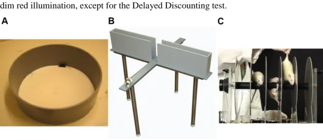
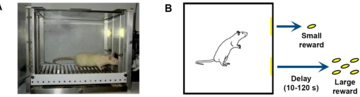
5.2.4 Behavioral testing ... 39
5.2.5 Statistical analysis ... 43
6. RESULTS ... 44
6.1 Cooling to different target temperatures in a piglet model of HIE ... 44
6.1.1 General characteristics ... 44
2
6.1.2 Cardiovascular parameters ... 45
6.1.3 Changes in blood biochemistry parameters ... 47
6.1.4 Ex vivo investigations ... 53
6.2 Investigating endogenous hypothermia in a rodent model of HIE ... 56
6.2.1 Effect of ambient temperature on neonatal asphyxia tolerance ... 56
6.2.2 Role of endogenous hypothermia ... 57
6.3 Characterization of a novel rodent model of HIE ... 58
6.3.1 Preliminary experiments ... 58
6.3.2 Randomized experiments ... 58
6.3.3 Early brain histology ... 59
6.3.4 Behavioral tests ... 62
7. DISCUSSION ... 65
7.1 Deep hypothermia in the piglet model of HIE ... 65
7.2 Hypothermia and hypoxic hypometabolism ... 68
7.3 Creating translational models of HIE ... 69
7.4 A novel rodent model of birth asphyxia ... 70
7.5 Relevance and limitations of these findings ... 72
8. CONCLUSIONS ... 75
9. SUMMARY ... 76
10. ÖSSZEFOGLALÁS ... 77
11. BIBLIOGRAPHY ... 79
12. PUBLICATION LIST ... 92
13. ACKNOWLEDGEMENTS ... 93
3
2. LIST OF ABBREVIATIONS
5CSRTT – Five-Choice Serial Reaction Time Test ADH – Antidiuretic hormone
ADHD – Attention-deficit/hyperactivity disorder ADP – Adenosine diphosphate
AED – Acute energy depletion aEEG – Amplitude-integrated EEG AIF – Apoptosis inducing factor AMP – Adenosine monophosphate
Apaf-1 – Apoptotic protease-activating factor−1 ATP – Adenosine triphosphate
AVP – Arginine vasopressin β-NTP – β-nucleotide triphosphate BAK – Bcl2-antagonist/killer1 BAT – Brown adipose tissue BAX – Bcl2-associated × protein
Bcl2 – B-cell lymphoma 2 protein family Bcl-XL – B-cell lymphoma-extra-large BID – BH3 interacting-domain death agonist bpm – Beats per minute
CA – Cornu ammonis CBF – Cerebral blood flow CNS – Central nervous system CI – Confidence interval CP – Cerebral palsy Cx - Cortex
DAB - 3,3'-Diaminobenzidine DD – Delayed Discounting DG – Dentate gyrus
Diablo – Direct inhibitor of apoptosis binding protein with low Pi
4 EAA – Excitatory amino acid
EPM – Elevated Plus Maze
EPP – Exchangeable phosphate pool FiO2 – Fraction of inspired oxygen HC - Hippoxampus
HI – Hypoxia-ischemia
HIE – Hypoxic-ischemic encephalopathy HR – Heart rate
IBA-1 – Ionized calcium-binding adapter molecule 1 ICAM – Intercellular adhesion molecule
IL – Infralimbic cortex ITI – Intertrial interval LH – Limited hold period
MABP – Mean arterial blood pressure MRI – Magnetic resonance imaging MRS – Magnetic resonance spectroscopy MWM – Morris Water Maze test
NAA – N-acetyl-aspartate NE – Neonatal encephalopathy
NF-κB – Nuclear factor kappa-light-chain-enhancer of activated B cells NO – Nitric oxide
nNOS – Neuronal nitric oxide synthase NTP – Nucleoside triphosphate
OF – Open Field
OFR – Oxygen free radical
31P-MRS – Phosphorus-31 Magnetic Resonance Spectroscopy PaO2 – Arterial partial pressure of oxygen
PaCO2 – Arterial partial pressure of carbon dioxide PBS – Phosphate-buffered saline
PCr - Phosphocreatine PFA – Paraformaldehyde PFK – Phosphofructokinase
5 Pi – Inorganic phosphate
PrL – Prelimbic cortex RI – Resident-Intruder test RNS – Reactive nitrogen species
RSG Cx – Granular part of the Retrosplenial Cortex SD – Standard Deviation
SEF – Secondary energy failure SEM – Standard Error of Mean SI – Social Interaction test
Smac – Second mitochondria-derived activator of caspase tBID – Truncated BH3 interacting-domain death agonist TNF – Tumor necrosis factor
TO – Timeout period
TRAIL – TNF-related apoptosis-inducing ligand receptor Trec – Rectal temperature
VGAT – Vesicular GABA transporter VGLUT – Vesicular glutamate transporter
6
3. INTRODUCTION
3.1 Neonatal hypoxic-ischemic encephalopathy (HIE)
Neonatal hypoxic-ischemic encephalopathy (HIE) is one of the most devastating diseases of the perinatal period. Approximately 1-2 newborns per 1000 live births are affected in the developed countries while its prevalence in the developing world is much higher, altogether amounting to around 700.000 neonatal deaths worldwide annually.1 An additional 1.15 million newborns are estimated to develop neonatal encephalopathy and almost half a million will suffer lifelong neurodevelopmental impairments due to HIE.1 These children present an enormous socio-economic burden to the families and to the whole of society as well. Looking at only cerebral palsy (CP) which is traditionally considered one of the most pervasive neurodevelopmental disorders related to perinatal hypoxic events, it can be estimated that HIE introduces a lifelong economic burden of
$1.9 billion every year in the US alone.2 These statistics unambiguously show that neonatal HIE presents one of the most severe problems in perinatal care.
Our definitions of “birth asphyxia” and neonatal HIE have evolved in parallel with our understanding of these conditions over the past few decades. Originally “birth asphyxia” was the umbrella term used for all cases of depressed birth, whether a mild and transitory respiratory problem or genuine neonatal stillbirth.3 Additionally, from the middle of the 19th century such a broad diagnosis was thought to be causally linked to the development of CP in later childhood.4 This one-dimensional causal association have been called into question repeatedly5 and it has become clear that in order to efficiently design therapeutic interventions, the diagnostic criteria need to be more specific. In order to avoid confusion, it might be desirable to retain the term “birth asphyxia” for its literal meaning, i.e. a combination of (arterial) hypoxemia and hypercapnia present at birth. For the neonatal condition associated with certain cases of birth asphyxia, there is an ongoing debate whether hypoxic-ischemic encephalopathy (HIE), neonatal encephalopathy (NE), or some other term would be most adequate.6 Due to its widespread use, I will use the term HIE to indicate the clinical condition,
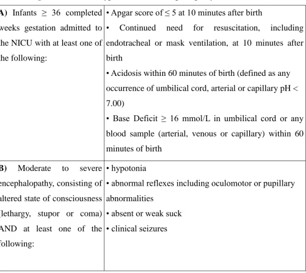
7 which is outlined in Table 1.
Table 1: Diagnostic criteria of hypoxic-ischemic encephalopathy.7 A) Infants ≥ 36 completed
weeks gestation admitted to the NICU with at least one of the following:
• Apgar score of ≤ 5 at 10 minutes after birth
• Continued need for resuscitation, including endotracheal or mask ventilation, at 10 minutes after birth
• Acidosis within 60 minutes of birth (defined as any occurrence of umbilical cord, arterial or capillary pH <
7.00)
• Base Deficit ≥ 16 mmol/L in umbilical cord or any blood sample (arterial, venous or capillary) within 60 minutes of birth
B) Moderate to severe encephalopathy, consisting of altered state of consciousness (lethargy, stupor or coma) AND at least one of the following:
• hypotonia
• abnormal reflexes including oculomotor or pupillary abnormalities
• absent or weak suck
• clinical seizures
HIE indicates infants, who fulfil two sets of criteria. The first category is related to the newborn's condition at birth (asphyxia, A criteria), including various markers of intrauterine or intrapartum hypoxia and a need for cardiorespiratory support. The second set of criteria is related to the infant's condition in the first hours after birth (encephalopathy, B criteria), i.e. whether neurological impairment can be observed following resuscitation and stabilization. Some clinical trials have employed a third set of criteria, which included abnormalities on amplitude-integrated EEG (aEEG),7 but in current clinical practice the diagnosis of HIE can be given without aEEG recording.8
Thus the definition of HIE is now rather narrow, compared to the historical use of birth asphyxia. On the one hand, infants who develop neonatal encephalopathy or CP in later life, but do not show signs of severe hypoxia at birth (thus they do not fulfil the
8
A criteria) are excluded, as the underlying cause of their impairment is likely to have been present prior to birth, e.g. genetic causes or placental insufficiency.5 On the other hand, approximately 10% of all newborns require some form of assistance at birth and 1% need vigorous cardiopulmonary resuscitation, but the majority of these infants do not develop encephalopathy or other neurological impairments later on.9 Thus the term HIE is used for infants, who demonstrably suffer from some level of neurological impairment in the first few hours of life and this is likely related to peripartum hypoxic events.
Historically, infants with HIE have had a highly variable prognosis.
Approximately 35% of these infants died, 35% suffered some form of neurodevelopmental impairment and 30% survived with normal neurological outcome.10 Neurodevelopmental impairment is generally measured at 18 to 24 months of age, when mental and psychomotor development can already be assessed adequately.11 However, the majority of children showing impaired neurological function at 2 years also display lower IQ-scores in school age.12
It is important to note that while infants with moderate to severe HIE have a highly variable prognosis, there is an even larger group of babies, who require some assistance at birth, but do not develop acute encephalopathy.9 These newborns have a highly favorable prognosis in terms of the severe neurodevelopmental impairments of asphyxic babies. However, a number of cohort studies have attempted to follow these newborns and some have suggested an increased risk for subtle neurodevelopmental impairments in later life.13-17
3.2 Animal models of neonatal HIE
In order to design rational therapeutic interventions for neonatal HIE, it was necessary to develop a working understanding of its pathomechanisms.18 Due to the imprecise timing of hypoxia, ischemia and hypercapnia in the peripartum period, as well as to the difficulties of indirectly measuring the severity of encephalopathy in newborns, most of the mechanistic understanding of HIE emerged from animal models.19 From 1955 to 1994 there were almost 300 preclinical papers published related to neonatal HIE.20 The
9
earliest recorded experiments of neonatal asphyxia were conducted 1813 by LeGallois, who noted that respiratory efforts in newborn rabbits subjected to asphyxia persisted for 27 minutes compared to only 2 minutes in adult animals.21 In 1870 Bert et al. made similar observations when comparing newborn pups with 20-day-old juvenile rats.22 In addition to the inverse correlation between maturity and tolerance to asphyxia, some of the earliest observations on various animal species concluded that males are less tolerant to asphyxia than females and high temperature, thyroxin, insulin injection or adrenalectomy all reduce resistance to hypoxia.23
In order to draw adequate conclusions from animal experiments, one needs to take into account a wide range of considerations regarding animal species, post- conceptual age, level of maturity and the method of inducing hypoxia or asphyxia.
Some of the most frequently used animal species in investigations of HIE have traditionally been the immature rat24 or mouse,25 the newborn piglet,26 the fetal sheep27 and various species of non-human primates.28 The optimal age of the animal at the time of hypoxia depends on the developmental dynamics of the specific species as well as the focus of the investigation. The developmental timing of particular markers of brain maturity (eg. brain growth spurt, neuronal migration, myelination, etc.) can be rather diverse in different species.29 This makes it impossible – in principle – to develop animal models which reflect all or most aspects of human HIE and therefore requires researchers to take a holistic view of the strengths and limitations of various animal models.
Historically, the foundations of perinatal asphyxia research were laid down by the work of Myers and Brann on the rhesus monkey.30 Throughout the 1970s these studies employed both fetal and neonatal models of HIE with various methods of achieving hypoxia and/or ischemia and investigated cardiovascular as well as neurological outcomes in both acute and chronic settings.31-33 The major contributions of these studies to our understanding of perinatal HIE have been summarized by Raju as follows:34 (1) the immature brain has a greater degree of tolerance to an asphyxic insult than the mature brain; (2) in addition to a reduction in PaO2 (hypoxemia alone), ischemia is also required to cause measurable brain damage; (3) depending on the type of insult, two distinct patterns of neuropathological damage can be observed: acute nuclear damage in the brain stem in case of global ischemia (together with anoxia), and
10
oedema with neuronal necrosis in the cerebral hemispheres in case of prolonged partial asphyxia. Limitations of these primate models have also been pointed out, in particular that more severe maternal hypoxia is necessary to generate fetal CNS injury compared to humans, and also that the prevalent pattern of oedema with neuronal necrosis in the primate can rarely be seen in humans, while the human pathology of intra- and periventricular hemorrhage was scarcely observed in these animals. Additionally, by the 1990s primate experiments have become increasingly difficult to conduct due to strict regulations and prohibitive costs.
The fetal or neonatal sheep has been one of the most important experimental large animal models in the investigations of cerebral blood flow (CBF) and brain metabolism during and after HIE. A large number of these studies were published in the 1980s and they generally involved a Cesarean-section performed at varying periods of gestation and the instrumentation of the fetal lamb for later monitoring and manipulation.35-37 The uterus and the abdomen were closed and after 2-3 days of recovery either the mother or the fetus were subjected to hypoxia, hypercapnia, acidosis, anemia, polycythemia or other procedures. These studies played a major role in elucidating the specific effects of different components of asphyxia (hypoxia, acidosis, ischemia, etc) on CBF and brain energy metabolism, understanding the developmental aspects of CBF control and describing the extent and limitations of CBF autoregulation in the fetal lamb.34 The major limitations of these models include the fact that most of the studies focused on acute changes in CBF and brain metabolism and less so on the middle to long term outcomes of HIE, and also that cortical neuronal maturation before birth is much more rapid in the sheep than in the human. While a fetal lamb at 120 days (86%) of gestation can be considered similar to a term human infant regarding brain maturity, the neonatal lamb already possesses an adult pattern of cortical maturity.38 Additionally, maintaining this model requires resources in laboratory space, personnel and funding which only a few centers can provide.
Parallel to the development of ovine preparations, the newborn piglet has also emerged in the 1980s as an affordable and ‘workable’ model in numerous laboratories.34 These studies also focused primarily on the acute effects of HIE on CBF and brain metabolism, and thus confirmed and elaborated on many of the findings of lamb studies.39,40 More recently, however, the piglet model has gained increasing popularity
11
among researchers investigating diseases of the neonatal period.41 This is partially due to the emergence of magnetic resonance imaging and spectroscopy (MRI and MRS) as invaluable tools for the in vivo investigation of the brain as well as potential bridging biomarker modalities, which could possibly provide direct links between animal studies and the human clinical condition.42 The neonatal piglet is ideally suited for such investigations, owing to its similarity to human neonates in terms of brain development, as well as its optimal size for imaging.41 In the clinical translation of therapeutic hypothermia, piglet studies have been invaluable in providing guidelines about the timing and the optimal depth of hypothermia,43,44 a work which is still ongoing, as will be highlighted later on.
With more than 1300 citations to the original 1981 paper, the single most widely used preclinical animal model of HIE is the Rice-Vannucci rodent model.24 In this preparation 7-days-old rat pups are subjected to permanent unilateral carotid artery ligation followed by a transient period of hypoxia. The authors of the original article cite Levine, who conducted experiments regarding the tolerance of adult rats to anoxia and ischemia.45 His 1960 paper reported that anoxia alone was unsuited for producing significant histological brain damage, as most of the animals either died or survived without lesions. Therefore, he introduced permanent unilateral carotid artery ligation to sensitize the forebrain to subsequent anoxia. Since these findings were in accordance with those of Myers et al. on rhesus monkeys,30 Vannucci and colleagues adopted this preparation to the immature rat, successfully producing significant unilateral brain injury without acute signs of neuromotor dysfunction.24 While numerous modifications to the original setup have been introduced since, including the occlusion of both carotid arteries46 or using different degrees of hypoxia for various durations, the core of this model has been instrumental in uncovering many pathophysiological features of HIE, including the depletion of high-energy phosphate metabolites as well as the role of excessive excitatory amino acid release in the development of brain injury.47
The advantages of this preparation – its relative cost-efficiency and ease of use – are well reflected in its widespread adaptation and usage even today. However, if one considers the fact that more than 500 pharmacological agents were found to be neuroprotective in preclinical models of neonatal HIE in the past decades, while none of these could be successfully translated to clinical care, the low predictive power of these
12
models becomes obvious.48 The reasons for this are probably several fold, but likely include: (1) the difficulty of determining the optimal age for comparison with the term human newborn; (2) the major anatomical differences between the rodent and the human brain (eg. archicortex volume, axonal myelination, grey/white matter ratio, developmental velocity); (3) the absence of particular patterns of injury in the rodent model, which are regularly seen in the human (eg. injury to the parasagittal cortex, subcortical white matter and brain stem); (4) the absence of systemic level multi-organ involvement, which is also commonly observed in severe cases of human HIE;49 (5) the invasive and permanent surgical ligation of one common carotid artery which has virtually no translational equivalent in human HIE.50 Additionally, a number of investigators have noted lately that while this preparation reliably produces some level of injury on most surviving animals, the variability of injury is still relatively large, which consequently requires a great number of animals to be sacrificed for each study.51 While some researchers have tried to argue that this is in fact an advantage of the model, as such a variability is also seen in human infants with HIE,52 this difficulty of early patient stratification into meaningful prognostic groups is one of the reasons why human clinical trials in HIE require so prohibitively high number of subjects.48
3.3 Neonatal HIE pathomechanisms
Despite their limitations, animals models have enabled us to gain a mechanistic understanding of HIE pathology.30 An excellent paper by Michel J. Painter summarized the state-of-the-art of perinatal HIE research in 1995, which I will attempt to further expand using data from the last 20 years since that review was published.19
One of the earliest observations was that in addition to the direct detrimental effects of hypoxia and ischemia, neuronal death continued upon reperfusion.53 It was recognized early on that, in principle, this late neuronal death could be ameliorated via post-insult neuroprotective interventions and hence it has been the focus of numerous investigations. More recent studies showed that while a number of neurons indeed die during the primary phase of the injury, the hypoxia-induced impairment of cerebral oxidative metabolism, cytotoxic edema and the accumulation of excitatory amino acids
13
(EAAs) typically recover, at least partially over approximately 30-60 minutes upon resuscitation.54 This period is usually followed by a ‘latent phase’, when the EEG is still suppressed, but high-energy phosphates have recovered to almost baseline levels.55 During this phase cerebral metabolism is believed to be actively suppressed, since tissue oxygenation is increased while cerebral perfusion is reduced.56 In the case of moderate to severe HIE, this latent phase is generally followed by a ‘secondary energy failure’
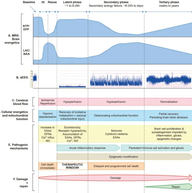
(SEF), which is characterized by the accumulation of EAAs, cytotoxic edema, mitochondrial failure and spreading neuronal death. 57,58 More severe HIE appears to produce higher levels of neuronal death during the primary phase and also an earlier and more severe SEF with more extensive neuronal loss.57 Clinically, this is also the phase when stereotypic seizures usually occur.59 Finally, brain injury and repair is believed to continue for weeks or months after the SEF. This tertiary phase has recently become the focus of neuroprotection and –regeneration studies.60 Figure 1 summarizes the phases of cerebral injury in HIE as well as the dominant pathomechanisms, which will be discussed below in more detail.
14
Figure 1: Schematic diagram illustrating the different pathological phases of cerebral injury after cerebral HI, modified from Hassell et al, 2015.60 The primary phase (acute HI), latent phase, secondary energy failure phase and tertiary brain injury phase are shown. (A) Magnetic resonance spectra showing the biphasic pattern of NTP/EPP (high energy phosphates) decline and lactate/NAA (anaerobic metabolism) increase during primary and secondary phases following HI insult. Persisting brain alkalosis with high levels of lactate is shown in tertiary phase. (B) Amplitude-integrated EEG showing normal trace at baseline, flat tract following HI, burst-suppression pattern in latent phase, emergence of seizures in secondary phase and normalization with sleep–wake cycling in tertiary phase. (C) After HI, there is a period of hypoperfusion associated with hypometabolism during latent phase, followed by relative hyperperfusion in secondary phase. (D) Cellular energetics and mitochondrial function are reflected in the biphasic response shown on magnetic resonance spectroscopy (A), with a period of recovery in latent phase followed by deterioration in secondary phase. There is partial
15
recovery in tertiary phase. (E) The most important pathogenic changes are shown for each, including generation of toxic free radical species, accumulation of EAAs, cytotoxic edema, seizures and inflammation. Cell lysis occurs immediately following HI, while programmed cell death occurs in secondary phase; latent phase provides a therapeutic window. Persisting inflammation and epigenetic changes impede long-term repair. (F) Damage is maximal in the secondary phase, but persists into the tertiary phase as inflammation and gliosis evolve. HI, hypoxia-ischemia; EAAs, excitatory amino acids; EPP, exchangeable phosphate pool; NAA, N- acetyl-aspartate; NO, nitric oxide; NTP, nucleoside triphosphate (this is mainly ATP); OFRs, oxygen free radicals;
In most cases of HIE the primary pathological event is a disruption of maternal- fetal gas-exchange, either due to acute or chronic causes (eg. placental abruption or chronic placental insufficiency, respectively).*61 This leads to decreased oxygen levels (hypoxemia) and accumulation of carbon-dioxide (hypercapnia) with associated respiratory acidosis in the blood. On the cellular level reduced oxygen availability impedes the operation of the mitochondrial respiratory chain, which in turn results in the accumulation of reduced electron carriers NADH and FADH and the interruption of oxidative phosphorylation. Without the mitochondrial electron transport chain the oxidation of NADH can only be accomplished via anaerobic respiration, during which pyruvate is converted to lactate by the enzyme lactate dehydrogenase, which also oxidizes equimolar amounts of NADH to NAD+. The process of anaerobic respiration, however, only produces 2 ATP molecules per glucose molecule, which is only about 5%
of the amount produced via aerobic respiration (theoretically 38 ATP molecules per glucose molecule). This leads to the depletion of other intracellular energy stores, such as phosphocreatine, and eventually to the decline of intracellular ATP levels and the parallel accumulation of ADP and AMP.55 This decrease in the ATP / (ADP + AMP) ratio induces phosphofructokinase (PFK), which is the rate-limiting enzyme of glycolysis.19 The resulting overdrive of glycolysis will sharply increase the demand for glucose in order to at least partially compensate for the lack of high-energy phosphates.
*I will refer to J.J. Volpe’s Neurology of the Newborn textbook without repeated citations in the next paragraphs describing the biochemical features of HIE.
16
The initial increase of cerebral blood flow in response to hypoxia can, to some extent, satisfy this demand.19 However, a sustained lack of O2 will lead to a decline of heart muscle contractility and heart rate, and thus to a subsequent decrease in cardiac output, which will produce hypotension and cerebral ischemia.35 In turn, ischemia will again reduce the supply of oxygen as well as glucose to neurons, even further tipping the balance of energy metabolism. The accumulation of lactate and protons will create an intra- and extracellular acidosis, which will consume a large amount of plasma buffers, primarily HCO3-. Henceforth, babies with severe HIE characteristically present with a combined respiratory and metabolic acidosis at birth, typically with low plasma pH, high pCO2, low standard HCO3- and high lactate levels.
Intracellular acidosis, however, can also have protective effects during hypoxia/ischemia. PFK, the rate-limiting enzyme of glycolysis is highly sensitive to intracellular pH and acidosis strongly inhibits its function.62 Additionally, neuronal excitability is also highly dependent on intra- and extracellular pH via a variety of mechanisms, the sum effect of which can be strikingly different in various neuronal populations.63 Acidification can increase excitability in certain neuronal types, like chemosensitive neurons in the brain stem, while other neuronal types, such as hippocampal pyramidal neurons are strongly inhibited by acidosis. As hippocampal neurons are among the most sensitive to hypoxic injury, this protective effect of acidosis can have an important role in HIE pathophysiology.64
While the failure of cellular energy metabolism is one of the most important and well-studied phenomena during hypoxia-ischemia, other pathomechanisms also play a significant role. One such process is the generation of free radicals. As discussed above, the shortage of oxygen supply halts oxidative phosphorylation and forces the cell towards anaerobic respiration. However, the slowing down of the mitochondrial ATP synthesis also implies that the reduction of O2 will be only partial and non-enzymatic free radical generation will be stimulated.65 Additionally, the hypoxia-induced accumulation of arachidonic acid and the calcium-induced activation of phospholipase A2 will result in the activation of prostaglandin synthesis, which also generates free radicals.65 Finally, the failure of cellular energy metabolism will lead to the accumulation of breakdown products from high-energy phosphate compounds. This process will give rise to the build-up of hypoxanthine, a product of AMP degradation.66
17
Under physiologic conditions, hypoxanthine is converted to xanthine and further on to uric acid by xanthine-dehydrogenase which utilizes NAD+ as a cofactor. During hypoxia, however, xanthine-dehydrogenase is converted to xanthine-oxidase via calcium-induced proteolysis and this enzyme utilizes molecular O2 instead of NAD+ as a cofactor, thus generating superoxide anions.65 In addition to oxygen free radicals, there is also compelling evidence to suggest the detrimental role of nitrogen free radicals (RNS) in HIE pathology, primarily via the generation of nitric oxide (NO•) by neuronal nitric oxide synthase (nNOS).54 The reaction of NO• with superoxide in the cytosol and mitochondria can lead to the generation of highly reactive peroxynitrite and other RNS compounds.67 Even though free radical generation is induced during and following hypoxia-ischemia, its scavenging enzymes such as superoxide dismutase and glutathione peroxidase are insufficiently expressed during the perinatal period.68 The net effect is an increased free radical load, which can exert a variety of detrimental effects, from lipid peroxidation to DNA/RNA-fragmentation and thus contribute to HIE neuropathology.54 Accordingly, preclinical experiments demonstrated the benefits of preventive maternal administration of allopurinol, an inhibitor of xanthine oxidase as a potentially neuroprotective intervention.69
As described previously, cellular energy metabolism initially attempts to compensate for the lack of O2 during hypoxia-ischemia via increasing the rate of anaerobic respiration, but this mechanism rapidly becomes insufficient and intracellular high energy phosphate levels begin to fall.19 Under physiological conditions the largest consumers of ATP are protein synthesis and the Na+/K+ pump. While non-essential protein synthesis is quickly blocked upon hypoxia,70 the function of the Na+/K+ pump is essential even under such conditions, since it constantly maintains the high potassium and low sodium concentrations intracellularly. The transmembrane Na+ gradient is the primary driver of a number of other ionic processes, contributing to the maintenances of pH- and calcium-homeostasis. The K+ gradient is essential for the repolarization of membrane potential in excitable cells. During uncompensated hypoxia, the lack of ATP will compromise the operation of the Na+/K+ pump and lead to membrane depolarization via Na+ and Ca2+ entry into the cells.54 One immediate effect of the influx of ions is the concomitant entry of water and subsequent cell swelling.61 While this so called cytotoxic edema can lead to direct neuronal death in extreme conditions, usually
18
upon reperfusion these swollen neurons recover, at least temporarily.54 After the latent phase, however, more severe cytotoxic edema will develop, which can be measured in vivo via diffusion-weighted magnetic resonance imaging, which is a sensitive marker of the extent of hypoxic-ischemic injury.71
Failure of the Na+/K+ pump and subsequent membrane depolarization will lead to the release of excitatory neurotransmitters to the synaptic cleft, primarily glutamate.54 Excessive release of EAAs will trigger the activation of NMDA, AMPA and other receptor-channels on post-synaptic neurons, which will cause disproportionate calcium and further sodium entry into these cells.72 This intracellular calcium surge will activate a number of downstream pathways, including the over-activation of enzymes such as calpains and other proteases, protein kinases, calcineurin, endonucleases and nitric oxide synthase, which in turn can further increase free radical generation.72 In this context, EAAs and intracellular calcium appear to be important mediators of cellular injury in HIE.54
As shown in Figure 1, a number of pathological features of hypoxia-ischemia recover to almost baseline levels within 30-60 minutes after resuscitation.60 Cytotoxic edema subsides, EEAs are removed from the synaptic cleft and intracellular high energy metabolites recover. This so-called latent phase usually lasts between 6 and 24 hours after recovery and is considered the primary therapeutic time window, when intervention could prevent or ameliorate subsequent injury.60 Some studies suggest that during this phase, cerebral metabolism might be actively suppressed by endogenous protective mechanisms, as indicated by the simultaneously present decreased cerebral perfusion and increased tissue oxygenation, consistent with decreased oxygen consumption.56 This suggested controlled hypometabolism has important implication for both clinical practice and preclinical research, as will be discussed later.
Approximately 6-24 h after the initial insult, a secondary energy failure has been described both in preclinical57 and clinical studies73 using phosphorus-31 MRS (31P- MRS). The 31P-MRS spectra consists of phosphorus-containing metabolites, including phosphocreatine (PCr), inorganic phosphate (Pi) and nucleoside triphosphates (NTP).
There are three NTP peaks in the 31P spectra, α-, β-, and γ-NTP, corresponding to the three phosphate groups on NTP molecules (primarily ATP, but also including guanosine triphosphate and uridine triphosphate).74 During the SEF, levels of cellular high energy
19
metabolites (PCr and NTP) fall and Pi increases. Additionally, persistently high levels of intracerebral lactate75 and an alkalotic shift in intracellular pH can also be observed,76 which are associated with later neurodevelopmental impairments. This secondary phase is also marked by secondary cytotoxic edema, accumulation cytokines and excitotoxins, mitochondrial failure, and clinically the onset of seizures (see Figure 1).54 A tertiary cerebral hyperperfusion takes the place of the previous, actively suppressed cerebral metabolic state.77 These various processes likely contribute to the spreading neuronal death continuing for days or even weeks after the initial insult.60
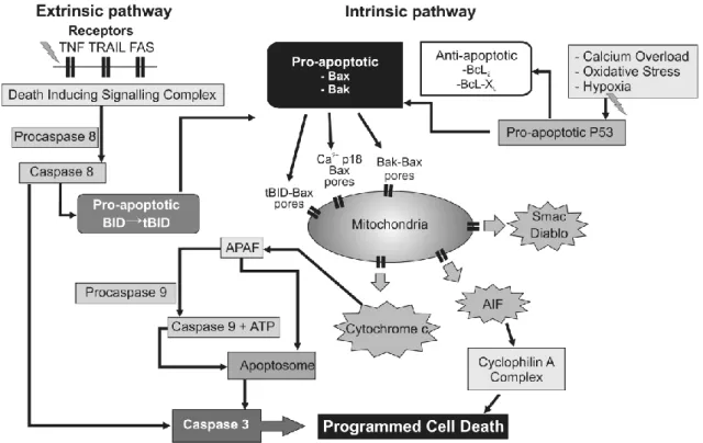
The precise pathomechanisms involved in this secondary energy failure are not fully understood yet, but they likely involve excessive calcium influx, inflammatory mediators, and activation of pro- and anti-apoptotic proteins as well.54 Mitochondrial failure appears to be a central feature of the SEF, upon which a number of these processes converge. Figure 2 summarizes some of these processes.54
Figure 2: Intracellular mechanisms associated with the permeabilization of the mitochondrial membrane leading to apoptosis, modified from Wassink et al.54 Upstream triggers such as inflammatory mediators and withdrawal of trophic support can activate the extrinsic pathway of apoptosis, while calcium accumulation and ROS can induce the intrinsic pathway. Some downstream members, such as Bid creates cross-activation between these two main pathways.
20
AIF, apoptosis inducing factor; Apaf-1, apoptotic protease-activating factor−1; ATP, adenosine triphosphate; BAK, Bcl2-antagonist/killer1; BAX, Bcl2-associated × protein; Bcl2, B-cell lymphoma 2 protein family; Bcl-XL, B-cell lymphoma-extra-large; BID, BH3 interacting- domain death agonist; Diablo, direct inhibitor of apoptosis binding protein with low Pi; P53, p53 tumor suppressor protein; Smac, Second mitochondria-derived activator of caspase; tBID, truncated BH3 interacting-domain death agonist; TNF, tumor necrosis factor receptor; TRAIL, TNF-related apoptosis-inducing ligand receptor
A central feature of secondary energy failure appears to be a cellular accumulation of calcium due to excessive glutamatergic activation. This calcium build- up can be buffered by the mitochondria up to a certain level.78 Above this, however, it can inhibit the function of the electron transport chain, uncouple oxidative phosphorylation and lead to a permeabilization of the mithochondrial membrane, which results in the activation of the intrinsic apoptotic pathway.72 This process occurs via the translocation of the cytosolic pro-apoptotic Bax to the mitochondrial membrane, where it forms transition pores together with Bak and Bid, all members of the apoptotic Bcl-2 family.79 This allows the leakage of pro-apoptotic proteins, such as the second mitochondria-derived activator of caspase (Smac), the direct inhibitor of apoptosis binding protein with low Pi (Diablo) and apoptosis inducing factor (AIF), as well as cytochrome c to the cytosol.79 While the loss of cytochrome c oxidase itself could impair energy metabolism, these processes also trigger further downstream effector mechanisms. These include the activation of the initiator caspase 9 and the subsequent formation of the apoptosome, which in turn will activate effector caspase 3 and ultimately lead to DNA fragmentation, membrane blobbing and apoptotic cell death.80 There is a good amount of evidence showing that apoptotic cell death is indeed an important contributor to HIE neuropathology.81 Additionally, a number of other pathological processes during this secondary energy failure stage can result in, or contribute to apoptotic neuronal death, including the loss of trophic support by astrocytic growth factors,82 inflammatory mediators,83 and the induction of the extrinsic apoptotic pathway via the activation of death receptors Fas and TRAIL.84 Recent consensus suggests, however, that the classification of injury as apoptotic vs. necrotic might be inadequate and instead there is probably a continuum of phenotypes between these two categories present after neonatal HIE.85
21
There is now accumulating evidence to show that following the SEF, a tertiary phase of active pathological processes takes place, which continues for weeks, months, even years.86 This is consistent with the observation that high levels of cerebral lactate and brain alkalosis can persist for over a year in babies with severe neurodevelopmental impairment.75 Pathomechanisms during this phase are yet poorly understood, but likely involve the proliferation of astrocytes and the formation of glial scar, persistent inflammatory activation and epigenetic changes.
3.4 Experimental neuroprotective interventions
Owing to the growing understanding of the pathomechanisms involved in neonatal HIE, there has been a number of experimental therapies designed to prevent or ameliorate its neurodevelopmental sequelae. Hypothermia, as the only therapeutic intervention to date with clinically proven safety and efficacy, will be described comprehensively in the following section. Before that, a brief overview is provided regarding the experimental therapies investigated in recent years.
One of the most intensively researched interventions has been the application of inhaled xenon.69 Xenon is a non-competitive NMDA-antagonist with a wide range of effects on downstream intracellular pathways, ultimately providing a strong anti- apoptotic effect.69 Several groups have demonstrated its neuroprotective effects in preclinical models.87-89 A recent clinical trial, however, failed to show the additional benefit of 30% xenon administration in addition to hypothermia.90 There are two other ongoing trials investigating the benefits of different xenon administration protocols (clinicaltrials.gov, NCT01545271 and NCT02071394).
As described before, the inhibition of xanthine-oxidase during hypoxia has been suggested as a potential way to decrease oxidative damage during HIE. Preclinical experiments demonstrated the benefits of preventive maternal allopurinol administration, an inhibitor of xanthine oxidase.69 Recent clinical trials, however, could not provide sufficient support for these claims.91
Another suggested experimental intervention has been the administration of melatonin.60 In addition to its well-known role as a regulator of circadian rhythms,
22
melatonin influences a wide range of physiological functions, including growth and development, reproduction and the immune response.60 Its neuroprotective effects have been demonstrated in a number of preclinical models92,93 and the mechanisms likely involve anti-oxidant, anti-apoptotic and anti-inflammatory effects.60 Clinical trials are now underway to provide information about optimal dosing regimens and potential neuroprotective effects (clinicaltrials.gov, NCT02621944).
Finally, erythropoietin (EPO) has also been suggested as a potential neuroprotective agent after neonatal HIE.60 In addition to its role as a hematopoietic growth factor, EPO is known to be highly important for normal brain development in utero and appears to be an important mediator of the brain’s endogenous protective mechanisms via anti-apoptotic, anti-inflammatory and anti-oxidative effects.60 A number of clinical trials involving various protocols of EPO administration are currently underway.94
Until today, however, the only intervention proven to be safe and effective was therapeutic hypothermia.
3.5 Therapeutic hypothermia
It is probably no overstatement that therapeutic hypothermia has been the most important recent development in the care of infants with HIE. The concept itself, however, is probably almost as old as medicine itself. The first description of intentional cooling to reduce neurological injury comes from Hippocrates, from approx. 400 BC, who suggested packing wounded soldiers in snow and ice.95 The first record of therapeutic hypothermia in newborns dates back to the 17th century, when Sir John Floyer described the immersion of a still-born infant into ice water in order to stimulate the onset of respiration:96
“Sarah Parks . . . gave still-birth to a baby boy . . . A young doctor assisting the Parks’
regular physician begged for an opportunity to experiment with an idea he had to rouse the lifeless infant. A tub of ice was ordered and the young doctor plunged the baby into it. Out came the screaming little Parks and he was named Gordon after the doctor who
23 prodded him to life.”
Sir John Floyer, 1697
The first scientific attempt to use hypothermia in the resuscitation of asphyxiated newborns was made by James Miller from New Orleans and Björn Westin from Stockholm in the 1950s. After conducting a series of animal experiments to demonstrate the beneficial effect of cooling on survival from asphyxia,97 they organized the first pilot study of therapeutic hypothermia. Ten severely depressed newborns who did not respond to resuscitation were immersed in a cold water bath until spontaneous respiration commenced or their rectal temperature decreased to 27 ˚C.98,99 Four of these infants also received oxygenated blood transfusions. After resuscitation, the infants were dried and allowed to rewarm spontaneously. One of the 10 infants died due to respiratory distress syndrome, but the other nine survived and did not show neurological impairment in a 10-year follow-up study, despite the fact that their periods of apnea ranged from 8 to 79 minutes.100 After their initial success Miller and Westin proceeded to conduct a larger study of cooling on 65 infants unresponsive to standard resuscitation, 52 of whom survived, which was an outstanding result at the time.97 Small scale clinical trials were also conducted elsewhere in the 1960s, from California to Switzerland and Finland.101-104
Despite these hugely promising results and the championing of cooling for severely depressed newborns by Westin and Miller, which they continued in the 1970s, the general international medical community did not adopt the concept of therapeutic hypothermia. One of the primary reasons for this was likely the publication of another scientific paper almost simultaneously with Miller and Westin’s first study. In 1958 Silverman and colleagues reported a study of 182 premature infants, who were kept in normothermic (31.9 ˚C) or hypothermic (28.9 ˚C) incubators for the first 5 days of life.105 The overall survival was 68% in the hypothermic group versus 83% in the normothermic group. Despite the fact that later re-analysis showed that most of the mortality in the hypothermic group occurred in infants <1000g and that there was a sampling bias between the two groups, which together could explain at least some of the results, this study propelled a large amount of research into neonatal thermoregulation and motivated clinicians to adopt strict monitoring and maintenance of rectal
24
temperatures of newborns.96 Even though the Silverman study was conducted on preterm babies, the extrapolation about the dangers of hypothermia to term newborns was generally accepted by clinicians.
It was only in the 1990s that neonatologists started to question the dogma concerning the dangers of hypothermia in the neonatal period. The causal relationship between postnatal hypothermia and mortality in preterm infants, suggested by the Silverman paper was called into question by a more detailed study by Hazan and colleagues in 1991.106 By the late 1990s a sufficient amount of data from animal studies have emerged which all supported the potentially beneficial effects of mild to moderate hypothermia after resuscitation from hypoxia-ischemia (HI).18
The first pilot safety study of selective head cooling in asphyxiated newborns was published in 1998 by Gunn and colleagues.107 Whole-body cooling was also successfully tested in a clinical pilot safety study two years later.108 The first randomized multicenter study evaluating the efficiency of selective head cooling for neonatal HIE was the CoolCap trial, the results of which were published in 2005.109 Even though that study only found significant improvement in neurological outcome in infants with moderate HIE, two other studies using whole-body hypothermia were published the same year, both of which found significant improvement in the risk of death or disability in infants with both moderate and severe HIE.110,111 The largest trial of whole-body hypothermia published interim results in 2008 7 and the final outcomes in 2009.11 After enrolling 325 infants, the TOBY study concluded that cooling to 33.5 C for 72 hours, if started within the first 6 hours of life, leads to improved neurological outcome in survivors, even though it could not identify significant reduction in the risk for death or severe disability which was the study’s primary outcome. A subsequent meta-analysis of hypothermia trials, encompassing 1320 infants was necessary to demonstrate with adequate statistical power that moderate hypothermia consistently improves both survival and neurological outcome at 18 months of age in infants with moderate HIE, but this effect was still not significant for infants with severe HIE.10 Only in 2012 and 2013 could two revised meta-analyses conclude that infants with severe HIE also benefit from therapeutic hypothermia and unequivocally put hypothermia in the standard protocol of care for infants with HIE.112,113 Still, concerns about the long-term benefits of hypothermia persist. The school-age follow ups of all
25
three major clinical trials has already been published and while all suggest benefits of hypothermia in this age group as well, the studies were not powered to draw such conclusions with statistical significance.12,114,115 Undoubtedly, an ongoing data collection from many centers employing similar standard protocol for cooling is necessary to further understand the benefits and limitations of therapeutic hypothermia.
3.6 Mechanisms of action in therapeutic hypothermia
The mechanisms of hypothermic neuroprotection are several-fold. Hypothermia reduces cerebral metabolism by approximately 5% for every 1 ˚C decrease in body temperature.116 Hypothermia during HI delays the onset of anoxic depolarization, reduces the amount of EEAs and potentially suppresses ROS and RNS formation.54 If hypothermia is initiated upon reperfusion/resuscitation, it appears to accelerate the normalization of extracellular EEAs, ROS sequestration and recovery of high-energy phosphates.117
Clinically, however, hypothermia treatment can be started realistically only a few hours after birth.118 The effect of hypothermia on secondary (and tertiary) injury is thus highly important. Microarray studies suggest that hypothermia can suppress a large number of gene responses to ischemia, many of which are involved in calcium homeostasis, cellular and synaptic integrity, inflammation and apoptosis.119 As described before, pro-apoptotic processes are incredibly complex and involve a large number of redundancies, therefore it is difficult to conduct and interpret studies including the manipulation of individual mediators. However, the caspase-dependent activation of apoptosis does generally converge on caspase-3 as a final executioner, which makes it a reasonably good indicator of upstream processes. Accordingly, delayed hypothermia was found to decrease caspase-3 activation after HI in the near- term fetal sheep.120 This finding was confirmed in both rodent121 and in piglet models.122 There is also evidence from adult animal studies that post-reperfusion hypothermia can suppress the mitochondrial permeability transition and prevent the release of cytochrome-c and other pro-apoptotic mediators.123
26
The inflammatory cascade induced by HI can cause injury via the activation of apoptotic cell death, or via direct damage from inflammatory cells, such as microglia or leukocytes.124 This inflammatory signaling appears to be interrupted by hypothermia at a number of sites, as cooling was found to decrease the levels of various pro- inflammatory cytokines as well as to directly suppress microglial activation.125
The fact that therapeutic hypothermia needs to be maintained for at least 48 hours in order to be effective suggests that a continued suppression of programmed cell death and inflammation is necessary until normal homeostasis can return.126 Additionally, hypothermia results in suppressed cerebral hyperperfusion, edema formation and seizure activity that could all indicate or contribute to neuroprotection.77 As these effects on a wide range of deleterious processes are combined, it is not hard to appreciate the level of protection offered by hypothermia. Hence it is reasonable to ask what may be the optimal setting for administering hypothermia in order to achieve maximal protection.
3.7 Application of therapeutic hypothermia
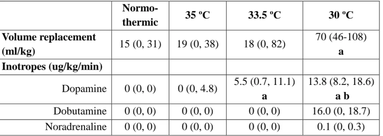
One important aspect of therapeutic hypothermia is the method of administration. One of the large clinical trials, the CoolCap study utilized selective head cooling,109 which was previously successfully used in large animal models as well.59 This method can produce selective head cooling with only mild systemic hypothermia. However, as discussed before, the CoolCap trial could only identify potential benefit of hypothermia in a selected subgroup of infants with HIE. Insight into the potential reasons for this was provided by piglet studies showing that different CNS structures have different optimum temperatures for protection.127 In these experiments optimal protection for cortical neurons was achieved at 35 ˚C, while deep gray matter structures required 33.5 ˚C for maximal neuronal sparing. These results, together with previous studies showing that it is challenging to achieve adequate cooling of deep brain structures by surface cooling alone,128 suggest that systemic hypothermia may offer a more reliable and effective protection.129
27
Another important aspect of the administration of therapeutic hypothermia is the initiation and the length of cooling. Inclusion criteria of the large multi-center trials involved the initiation of cooling within 6 hours after birth.7 This time was chosen as a practical compromise between the clinical reality of birth asphyxia (many such children are born out-of-hospital and require transport to a tertiary center) and the results of animal studies, which showed that delayed hypothermia can be neuroprotective, but only if started before the occurrence of seizures, i.e. during the latent phase.130 The length of cooling was set to 72 hours in order to achieve maximum protection, as preclinical studies indicated an inverse relationship between the depth and length of cooling necessary to achieve neuroprotection.131 72 hours of cooling was found to be safe and effective in translational large animal models of HIE.59 The potential benefits of stretching both ends of the cooling period are now being evaluated. Earlier start of cooling, for example during neonatal transport has been studied at a number of centers.
It appears to be feasible and safe using servo-controlled cooling devices132 and while definitive results regarding neurological outcomes are still lacking, preliminary data suggest improved neuroprotection with earlier initiation of cooling.133 On the other hand, the potential prolongation of cooling was evaluated in two recent clinical trials.
One of these trials was halted interim due to the lack of observed benefit in survival from either 120 hours vs 72 hours of cooling, 32 ˚C vs 33.5 ˚C target temperature, or both.134 The other trial examining the effect of late, prolonged cooling initiated between 6-24 hours after birth and maintained for 96 hours has not published its results yet.135
Although these recent results show a lack of benefit with cooling to 32 ˚C instead of 33.5 ˚C, animal studies have indicated that deeper cooling might offer better protection.136 However, it appears that there is be a clear trade-off between the depth of cooling and the systemic-side effects of hypothermia. For example, deep hypothermia (15 ˚C) after cardiac arrest in the adult dog was found to be detrimental,137 while moderate hypothermia (28-32 ˚C) offered no protection138 and only mild hypothermia (34-36 ˚C) resulted in consistently improved neurological outcomes.139 The question of whether deeper hypothermia might offer better protection is especially important now that a large number of physicians start to adopt therapeutic hypothermia as standard of care for infants with HIE, while currently no other neuroprotective interventions are
28
available. Therefore, it is essential to determine the optimal cooling temperature for infants with HIE.
3.8 Hypothermia as an endogenous protective mechanism
Even though recent successes with ‘artificial’ therapeutic hypothermia has raised a large amount of interest in the past decade, the first observations about ‘endogenous’
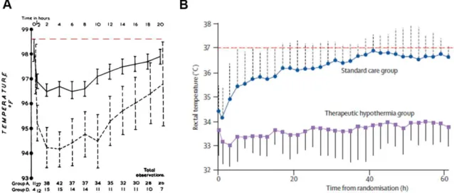
hypothermia as a response to hypoxia were made more than 50 years ago. In 1955 Cross, Tizard and Trythall observed that infants breathing 15% O2 had reduced metabolic rate and heat production.140 While their experiments could not show decreases in rectal temperature during the brief period of 15% hypoxia, another report showed that maintaining this O2 level for a week resulted in a significant decrease in temperature as well.141 Instead of this approach of induced hypoxia, Burnard and Cross decided to study infants after birth asphyxia, as natural model of hypoxia and found that asphyxic babies had significantly reduced body temperatures (approx. 35 ˚C) for several hours after birth (Figure 3A).142 This finding has also been confirmed more recently in a trial of therapeutic hypothermia under low-resource settings in a developing country, where this ‘natural cooling’ was present in the normothermic asphyxia group for an average of 15 hours, despite attempts of active rewarming (Figure 3B).143
Figure 3: Endogenous hypothermia in neonates after birth asphyxia. (A) shows the seminal work of Burnard and Cross from 1958, where they demonstrated that asphyxic infants (dashed
29
line) had significantly lower rectal temperatures than healthy newborns (solid line) for several hours after birth.142 Note the lack of quick rewarming even in healthy newborns. Data is displayed as mean and 95% confidence intervals. (B) is adopted from a recent clinical trial of hypothermia in low-resource settings, which shows that the normothermic control asphyxia group (blue line) remained at sub-normal temperatures for several hours after resuscitation, despite efforts of rewarming.143 Data is displayed as mean ± SEM. The dashed red line shows 37
˚C rectal temperature on both graphs. Adopted from Burnard and Cross, 1958 142 and Robertson et al, 2008 143.
Following the initial human observations in the 1950s, hypoxic hypometabolism was also studied in newborn lambs, puppies, kittens and rats.144,145 It appeared that since many of these species are born at a lower degree of maturity, endogenous protection against accidental and transient hypoxia might be evolutionarily beneficial. In the pursuing decades experimental research about the mechanism and role of hypoxic hypometabolism have expanded greatly and this body of knowledge have been the subject of a number of excellent reviews.146-150
Neonatal responses to hypoxia, including hypometabolism and a biphasic ventilatory response have often been considered as peculiarities by physicians, comparing them to adult physiology. It is important to note, however, that these processes have very similar analogues in lower species, which possess a greater tolerance to hypoxia than adult mammals.147 Studies confirmed that the decrease in metabolism in response to hypoxia is not due to the reduced oxygen availability, since lactate production was not increased, therefore anaerobic metabolism did not have to compensate for the reduced O2-supply.146 Another potential explanation of hypoxic hypometabolism might be the effect of decreasing temperature itself, which is known to slow down metabolism and result in a decreased metabolic rate. However, the drop in metabolic rate actually precedes the decrease in temperature, so hypothermia itself cannot cause the hypometabolism.148 Additionally, it was found that the magnitude of hypometabolic response to hypoxia is primarily determined by the level of baseline metabolic rate, which in turn depends on several factors, including body size, age and ambient temperature.151 Since newborns have limited thermal insulation, they require a higher basal metabolic rate and heat production to maintain a stable body temperature.