SYSTEMIC EFFECT OF PROLONGED MODERATE SYSTEMIC HYPOTHERMIA IN NEONATAL HYPOXIC
ISCHEMIC ENCEPHALOPATHY PhD thesis
Anikó Róka
Clinical Medicine Doctoral School Semmelweis University
Supervisor: Dr. Miklós Szabó MD, Ph.D
Official reviewers:
Dr. Ferenc Domoki MD, Ph.D Dr. Anna Beke MD, Ph.D
Head of the Final Examination Commitee:
Prof. Ferenc Paulin MD, D.Sc
Members of the Final Examination Commitee:
Dr. András Nobilis MD, Ph.D Dr. András Fogarasi MD, Ph.D
Budapest
2011
2 Contents
Abbreviations 4
1. Induction
1.1. Induction 6
1.2. Pathopysiology 7
1.3. Clinical evidence supporting therapeutic hypothermia in newborns 11 1.4 Organ dysfunction and cell necrosis in asphyxiated neonates 17
1.5. Markers for brain injury: S100 and NSE 17
1.6. Role of inflammation 19
1.7. Analgesia and drug metabolism during hypothermia 20
2. Aims 21
3. Methods
3.1. Patients and study protocol 23
3.2. Laboratory parameters indicating cell necrosis and organ failure 26
3.3. S100B and NSE measurements 26
3.4. Serum cytokine measurements 27
3.5. Serum morphine measurements 28
4. Results
4.1. Antropometric and clinical parameters in study group 29 4.2. Effect of hypothermia on cell necrosis and multiorgan failure 31 4.3 Effect of hypothermia on S100B and NSE levels, and their relationship
with the neurological outcome 35
4.4. Effect of hypothermia on serum cytokine levels 40 4.5. Effect of hypothermia on serum morphine concentrations 44
4.6. Follow up and outcome in our study group 48
3 5. Discussion
5.1. Hypothermia decreases the acute cell necrosis and may attenuate 50 organ dysfunction in neonatal asphyxia
5.2 Effect of hypothermia on S100B and NSE levels, and their relationship
with the neurological outcome 52
5.3. Hypothermia may suppresses the early increase of serum IL-6 levels 54 5.4. Elevated morphine concentrations in asphyxiated neonates treated with 58
prolonged moderate systemic hypothermia
6. Conclusion 62
7. Summary 63
8. Literature 65
9. List of publications 78
10. Acknowledgement 79
4 Abbreviations
HIE Hypoxic-ischemic encephalopathy
PCr Phosphocreatinine
Pi Inorganic phosphate
OFC Head circumference
NO Nitrogen monoxide
NOS NO synthase
IL1β Interleukin-1 beta
TNFα Tumor necrosis factor alpha CPR Cardiopulmonary resuscitation
EEG Electroencephalography
Ic Ca Intracellular Calcium
MRS Magnetic resonance spectroscopy
Glu Glutamate
aEEG amplitude integrated electroencephalograpy
RR Risk ration
CI Confidence interval
NICHD National Institute of Child Health and Human Development, USA
TOBY Total body hypothermia
NNT Number need to treat
MODS Multiorgan dysfunction syndrome ASAT aspartate aminotransferase
LDH lactate dehydrogenase
HT Hypothermia
NT Normothermia
MRI Magnetic resonance imaging NSE Neuron specific enolase
IL Interleukin
INFγ Interferon gamma
MCP-1 Monocyte chemotaxic protein 1
5 EGF Endothelial growth factor
VEGF Vascular endothelial growth factor
AUC Area under curve
CTG Cardiotocography
BE Base excess
CrUSS Cranial ultrasound scan
ECMO Extracorporal membrane oxigenisation
CPB Cardiopulmonary bypass
6 1. Induction
1.1. Induction
Neonatal hypoxic-ischemic encephalopathy (HIE) is an important cause of death and neurodevelopmental delay worldwide. Treatment for infants with hypoxic-ischemic encephalopathy was limited to supportive care for a long time, but efforts have been made to develop effective therapies. However, through several experimental therapies for hypoxic ischemic encephalopathy seemed promising none proved consistently successful in clinical studies [1],[2],[3]. Despite the improvement in neonatal care in the last decades, the incident of neurological disabilities related to perinatal brain injury remained unchanged affecting 2-3 in 1000 newborns. However accurate Hungarian national statistical data are not available, the estimated number of infants born with HIE is 150-200 in a year.
Over the past two decades, experimental and clinical evidence has accumulated that a 3-4oC reduction of body temperature maintained for at least 72 hours in newborns with hypoxic- ischemic encephalopathy may reduce cerebral injury and improve neurological outcomes [see later chapters]. Recent clinical trials have demonstrated that prolonged cooling of either the head or the whole body of neonates with HIE is safe and associated with reduced short-term mortality and morbidity at 18 months of age [4].
Neuronal damage and death after asphyxia occurs in different phases and pathways.
Initially primary energy failure and oxidative stress are responsible for tissue injury followed by secondary activated long term processes like excitotoxicity, inflammation, and apoptosis. Hypothermia, which decreases the cerebral metabolism and possibly affects on many other ways is the first therapeutic intervention proven being able to improve neurological outcome.
In this work we summarize the pathophysiology of hypoxic ischemic encephalopathy, the evidence behind hypothermia treatment and our observational studies during prolonged systemic hypothermia including markers of multiorgan failure and brain injury, cytokine and cortisol measurements, and changes in morphine metabolism.
7 1.2. Pathophysiology
The precise mechanism of neural rescue by moderate hypothermia is uncertain but may be related to the critical relationship between temperature and metabolic rate: for every 1oC lowering of the core temperature cerebral metabolism is reduced by approximately 7%, with consequently a lower glucose and oxygen demand [5] [6]. Both necrotic and apoptotic mechanisms are implicated in neuronal injury following neonatal hypoxia-ischemia and reperfusion.
The deprivation of oxygen and glucose caused by the reduction of cerebral blood flow is leading to severe decrease in high energy phosphate deserves including adenosine triphosphate. Due to the energy failure the sodium-potassium-adenosin pump is unable to maintain the polarity of the membrane in neurons and glia cells. The depolarisation results in excessive glutamate release from the axon terminals. The glutamate accumulates within the synaptic cleft, because of increased release and impaired reuptake mechanism. The excessive glutamate (phenomenon called glutamate excitotoxicity) acting on the receptor sites leads to a significant influx of sodium and calcium to the cells. The increased calcium level activates several enzymes including phospholipase, proteases and endonucleases or nitric oxide synthase. The combined effects of cellular energy failure, lactate acidosis, glutamate release, calcium accumulation and oxidative damage disrupt essential components of the cell leading to death.
During reperfusion phase the cerebral energy state usually recover, and the concentration of phosphate metabolites and the intracellular pH normalises. However 6 to 48 hours after the injury the secondary energy failure returns characterised by decrease in the ration of phosphocreatinine/ inorganic phosphate [7, Figure 1.].
8
In humans, the severity of the secondary energy failure correlates with adverse neurological outcome [8, Figure 2.]. Reperfusion causes excessive production of free radicals leading to oxidation of lipids, proteins, DNA, leading to failure in the mitochondria which can be one of the basic mechanisms of secondary energy failure. Two important sources of the oxygen radicals are the by-products of xanthine and prostaglandin synthesis.
Figure 1. Secondary disturbance of oxidative phosphorylation [7]. Changes in PCr/Pi ratio measured by MR spectroscopy after hypoxic insult and in control group.
Figure 2. Relation between changes in PCr/Pi and head growth (OFC) in the first year of life [8].
9
Oxygen free radicals contribute in tissue injury by membrane fragmentation with oxidation of the polyunsaturated fatty acid components. NO is a weak free radical produced by NO synthase (NOS) which is highly activated after hypoxia and reperfusion. NO combined with superoxide generates peroxynitrite radicals which activates lipid peroxidation and also increases glutamate release. During hypoxia free ferric iron is also released from complex proteins and reacts with peroxides producing hydroxyl radicals also responsible for tissue injury. Mitochondrial injury caused by membrane fragmentation results in energy failure, loss of cell membrane integrity and cytotoxic edema.
Inflammatory mediators play also crucial role in brain injury after hypoxia.
Expression of IL1β and TNFα mRNA was observed within 1 to 4 hours after initial insult along with the induction of a and b chemokines followed by neutrophil invasion [9], [10].
Brain microglia and macrophages can be activated rapidly after the initial insult by hypoxia, excess glutamate release or inflammation stimuli. Once activated, microglia/microphages can release various range of toxic mediators responsible for later neuronal damage, and axonal loss characteristic for white matter injury.
The delayed phase of neuronal cell death by apoptosis is lasting for several weeks of the initial insult. Apoptosis is considered to be a major cause of progressive neuronal injury following neonatal hypoxia ischemia however details are still not completely understood.
As mentioned previously, the neuroprotective effect of hypothermia is still not completely understood [11]. A reduction of 3-4oC core temperature clearly decreases the metabolic need of the cells, and maintaining cerebral high energy phosphate levels [12], [7, Figure 3.].
Figure 3. Effect of hypothermia on secondary energy failure [7]. Changes in nucleotide triphosphate and exchangeable phosphate pool ratio on MR spectroscopy after hypoxic insult in HT and NT piglets and control group.
10
Hypothermia is also associated with a reduction in free radicals and glutamate levels [7], [13] protecting mitochondrial function and also reduces inflammatory responses.
Animal studies showed that moderate hypothermia decreased apoptosis possibly via inhibiting caspase-3 activation [14] and increasing bcl-2 protein expression [15]. Figure 4. is summarizing the underlying processes in pathophysiology and possible target points for treatment.
PATHOPYSIOLOGY MARKERS POSSIBLE THERAPY
Hy poth erm ia
Figure 4. Pathophysiology of HIE, and possible target points for neuroprotective therapies
11
1.3. Clinical evidence supporting therapeutic hypothermia in newborns
In newborns, therapeutic hypothermia was first described as a method of reanimation by immersion in cold water [16], [17]. Later, experimental studies in adult models of hypoxic ischemic injury suggested that brief periods of post insult hypothermia offered neuroprotection. Moderate systemic or selective cooling of the brain has been shown to reduce brain injury in experimental human adult studies after events like stroke, trauma or cardiac arrest. These results led to a series of studies in piglets, neonatal rats and fetal sheep which showed repeatedly that moderate hypothermia significantly reduced cerebral injury following hypoxic ischemia. Investigators went on to perform a series of clinical studies, first to confirm the safety of prolonged moderate hypothermia in asphyxiated newborns and then to determine therapeutic effect by carrying out randomized controlled trials. However several questions had to be considered before initiating clinical study on neonates with hypoxic-ischemic encephalopathy about the length of the treatment, degree of the hypothermia, starting time of treatment and method of cooling (selective head or systemic). Selective head cooling was thought to be safer without adverse effects, but it is associated with temperature gradient in the brain with possible ineffective cooling of the deeper structures [18]. Major concerns about hypothermia were related to possible adverse effects like reduction of myocardial contractility, increased blood viscosity, coagulopathy, acid-base and electrolyte imbalance and increased risk of infection. To prove the safety of hypothermia pilot studies were performed using different cooling regimes in term neonates [19], [20], [21], [22], [23], [24], [25]. The studies used either selective head cooling (with mild systemic hypothermia) or whole body cooling methods using purposely designed cooling equipment or simple physical cooling with cold bag.
The pilot studies found no evidence of harm from prolonged moderate hypothermia particularly when body temperature was closely controlled; when temperature control was less accurate cardiovascular complications such as hypotension or severe bradycardia occurred [25], [26].
Subsequently several randomized clinical trials with neurological outcomes assessed over at least 18 months followed [27], [22], [28], [29], [30]. Most of these trials were carried out in highly developed countries with a low incidence of hypoxic ischemic
12
encephalopathy, and so required a large number of participating centers and study enrolment over several years. Each study aimed to determine whether therapeutic hypothermia improved survival but without increasing disability in survivors following neonatal hypoxic ischemic encephalopathy. The studies hypothesized that therapeutic hypothermia would result in a 30% reduction in the combined rate of death and disability in moderately or severely encephalopathic newborns. Uniform infant selection criteria were used: clinical evidence of asphyxia such as prolonged need for resuscitation at birth or severe metabolic acidosis, together with clinically assessed moderate or severe encephalopathy and additionally in most of the studies, abnormal cerebral activity confirmed by amplitude integrated electroencephalography (EEG). Enrollment of infants was complete or exceeded target in most studies but was terminated early in the two most recently reported studies because participating clinicians increasingly lost therapeutic equipoise as neurological outcome data from the completed studies accumulated. A remarkable achievement amongst the trials of therapeutic hypothermia in newborns was the almost complete (>95%) follow-up to 18 months of age of the study participants, which increases confidence in the study findings.
The Cool Cap study [27] applied selective head cooling with mild systemic hypothermia and enrolled 234 infants with signs of encephalopathy and abnormal aEEG.
116 neonates were randomised for head cooling for 72 hours started within 6 hours of life with aim rectal temperature 34-35 C and 118 neonates for conventional care. Primary outcome was a combined end point of death or moderate to several disabilities at 18 months of age. Death or moderate to severe disability occurred in 59 of 108 infants (55%) in the hypothermia group and 73 of 110 infants (66%) in the control group (relative risk 0.61 [95 percent confidence interval 0.34-1.09] p=0.1). However subgroup analysis showed that head cooling had no significant effect on infants with severe encephalopathy based on aEEG (n=46 relative risk 1.8 [95 percent confidence interval 0.49-6.4] p=0.51), but improved outcome of infants with less severe aEEG changes (n=172 relative risk 0.42 [95 percent confidence interval 0.22-0.80] p=0.009).
The NICHD trial by Shankaran et al [30] utilized systemic cooling of term neonates showing clinical evidence and signs of encephalopathy without aEEG monitoring. 208
13
infants were enrolled and randomised (102 in the hypothermia group and 106 in the control group). Cooling was commenced within 6 hours with blanket connected to a cooling system and was maintained for 72 hours. Aim oesophageal temperature was 33.5 C. Primary outcome was similar, combined death or several disability at 18 to 22 months of age.
Infants received hypothermia treatment had significantly less death or severe disability compared to control group (44% vs 62% relative risk 0.72 [95% CI 0.54-0.95] p=0.01).
Only this trial showed a significant reduction in the composite primary outcome.
The first two clinical trials showed no significant difference in physiological parameters or adverse events related to hypothermia, but were unable to provide clinically significant evidence for hypothermia treatment to be recommended as standard treatment.
To clarify the role of hypothermia, the TOBY trial [28], [31] was carried out which was the largest randomised multicentre trial, utilizing systemic hypothermia for 72 hours commenced at 6 hours of age enrolling 325 term neonates with clinical signs of hypoxic- ischemic encephalopathy confirmed by aEEG. Our unit was the second largest centre in the trial enrolling 24 infants over 2 years period between 2005 and 2007. Primary outcome was compound end point of death or severe disability at 18 months of age. Of 325 infants, 163 were allocated to cooling and 162 received standard care on normothermia. The rate of survival without neurological abnormality was significantly increased in the cooled group 71/163 (44%), compared to control group 45/162 (28%) (Relative risk 1.57 [95% CI 1.16- 2.12] p=0.003). Among survivals cooling resulted in reduced risk of cerebral palsy (RR 0.67 [95% CI 0.47-0.96] p=0.03) and improvement in the Bayley Mental Developmental Index (p=0.03), Bayley Psychomotor Index (p=0.03) and Gross Motor Function Classification Scale (p=0.01). The TOBY trial showed no significant difference in the combined rate of death and severe disability. In the hypothermia group 42 infants died and 32 survived with severe neurodevelopmental disability, whereas 44 normothermic infants died and 42 had severe disability (relative risk 0.86 [95% CI 0.68-1.07], p=0.17). Adverse events were minor and not associated with cooling. Although there was no significant difference in the primary outcome in the TOBY trial, hypothermia resulted in unequivocal improvement of neurological outcomes in survivors.
14
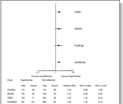
Recently, as individual trials were insufficient to provide conclusive evidence of efficiency, metaanalysis was carried out with studies identified from the Cochrane Central Register of Controlled Trials, the Oxford Database of Perinatal Trials, PubMed, previous reviews and abstract [4]. The synthesis and metaanalysis of the randomised trials using therapeutic hypothermia is showing striking consistency. The point estimates are remarkably similar and metaanalysis of three trials comprising 767 infants followed to 18 months shows highly significant improvements in neurological outcomes. In the three major trials comparing 767 term infants with asphyxia, cooling reduced the combined rate of death or severe disability (the primary outcome of all the studies) (risk ratio 0.81 [95%
CI 0.71-0.93] risk difference -0.11 [95% CI -0.18-0.04] with a number needed to treat of just 9 [95% CI 5-25] p=0.002) [4, Figure 5.].
Figure 5. Forrest plot of effect of hypothermia on main neurological outcomes at 18 months [4].
15
Hypothermia increased normal survival (RR 1.53 [95% CI 1.22-1.93] RD 0.12 [95% CI 0.06-0.18] NNT 8 [95% CI 5-17] p<0.0001) and in survivals reduced the rates of severe disability, cerebral palsy and both mental and psychomotor developmental index <70. In 10 trials comprising 1315 infants mortality was significantly reduced (RR 0.80 [95% CI 0.68- 0.94] RD -0.07 [95% CI -0.11, -0.02] with a number needed to treat of 14 [95% CI 8-47]
p=0.008) [4, Figure 6.]. However, there is some heterogeneity amongst the trials probably because of differences in practice about withholding or continuing intensive care in severely encephalopathic infants.
Reassuringly none of the large randomized trials reported clinically significant complications attributed to hypothermia: Bradycardia to 110-120 beats per minute is normal during therapeutic hypothermia but serious arrhythmia was rarely observed and occurred also in the non cooled groups. Inotropic support was used more commonly in cooled infants but this may have been due to physician bias. Extra cerebral hemorrhage was observed frequently in both cooled and non cooled infants and was mostly mild; however thrombocytopenia was significantly more common with cooling therapy. Cerebral sinus
Figure 6. Forrest plot of effect of therapeutic hypothermia on rate of intact survival at 18 months of age. Table gives total cases assessed and rates of events in each trial and on metaanalysis [4].
16
thrombosis occurred rarely and was not seemingly related to hypothermia. The only possible complication related to the hypothermia was reported in few cases as subcutaneous fat necrosis. Nonetheless none of the trials were powered to detect uncommon complications and although no clear evidence of harm attributed to therapeutic hypothermia has so far emerged from the registries of therapeutic hypothermia established since 2006.
However further experience needs to accumulate before clinicians can be reassured about the safety of therapeutic hypothermia especially when it is applied to infants with systemic complications associated with perinatal asphyxia such as pulmonary hypertension or myocardial ischemia.
The final synthesis of the collected data provides creditable evidence that hypothermia treatment is effective and improves neurological outcome in term neonates with hypoxic-ischemic encephalopathy without any known complication.
At this point, the long term follow up of previous clinical trials needs to be completed, but hypothermia has to be confirmed as standard care for neonates with hypoxic-ischemic brain injury in neonatology.
Despite these remarkable results there remain some uncertainties: First no study has reported outcomes yet beyond 18 months of age following therapeutic hypothermia but assessment of cognitive and behavioural functions requires assessment in later childhood.
The follow-up rates reported at 18 months may not be achievable later so the effect of therapeutic hypothermia on outcome in later childhood may be less clear cut. Secondly, although the therapeutic effect is remarkable, still over 40% of infants in the trials died or developed disabilities despite therapeutic hypothermia. This partly may be due to selection bias: Since this was a novel intervention, clinicians may have referred the more severely affected infants with prolonged or irreversible asphyxial injury. Thirdly, there is presently very little data on the application of therapeutic hypothermia in resource poor countries, where the incidence of hypoxic ischemic encephalopathy is greatly increased. In a pilot study in Uganda there was an increased mortality in the treatment group, but this may have been due chance allocation in a small study of more infants with severe encephalopathy to the treatment group [32]. Field trials are needed before the current data are extrapolated to different environments.
17
1.4. Organ dysfunction and cell necrosis in asphyxiated neonates
The most significant consequence of the hypoxic insult is irreversible cerebral injury. However, other organs are also severely distressed due to hypoxic-ischemic injury.
Cardiovascular instability, pulmonary dysfunction, hepatic impairment, gastrointestinal disorders and acute renal failure may evolve as characteristic components of multiorgan dysfunction (MODS) and failure [33], [34], [35]. MODS contributes significantly to the risk of death in the critically ill neonates including those subjected to asphyxia. Multiorgan failure is mainly due to acute cell necrosis in different organs. Cellular necrosis after hypoxia and reperfusion is the result of cell swelling, disruption of cytoplasmic organelles, loss of membrane integrity and unspecific activation of inflammatory cascade as it is discussed previously.
The diagnosis of MODS is based on clinical signs and laboratory parameters of tissue dysfunction and cell necrosis [33]. As mentioned previously, prolonged moderate systemic hypothermia (HT) is the first therapy proved to be effective in improving outcome of infants with HIE [4], [28]. The neuroprotective effect of HT has been excessively investigated in the last 10 years. However systemic effects have been not completely under focused research.
1.5. Markers for brain injury: S100 and NSE
Assessment of the severity of HIE to provide initial information for indication of hypothermia treatment and preliminary prognosis is usually very important in clinical situations. Hypothermia needs to be initiated as soon as possible within 6 hours of birth, at the moment indication is based on the severity of encephalopathy or abnormality of the amplitude integrated EEG (aEEG). Informing parents and clinical decisions like redirection of care can be particularly challenging. Up to date, brain MRI is considered to be the best tool to assess brain injury; however it is not widely available. Clinical indicators of the severity of asphyxia such as the degree of neurological depression or metabolic acidosis after birth are relatively imprecise predictors of subsequent neurological outcome.
Assessment of encephalopathy with aEEG is more and more frequently used, but interpretation can be difficult, especially if sedative or paralytic agents have been used
18
earlier, so accurate additional sentinel markers are highly awaited. S100B and neuron specific enolase (NSE) may be promising biomarkers of the severity of brain injury and prognosis following asphyxia.
S100B protein is cytosolic calcium binding protein consisting of two monomers, α and β in different combinations. S100B (β β) and S100A (α β) are mainly present in glial cells and specific neuron populations in the central nervous system, but expressed in some extra CNS tissues. Due to its molecular weight (21 kDa), S100B may be only detected in peripheral blood if the integrity of the blood-brain barrier is disrupted. The biological half- life is about 30 minutes. S100B protein can be measured in the blood and urine and is considered a reliable marker of brain damage in adults and newborns. Elevated serum S100B levels are reported after perinatal asphyxia [36], [37], [38], [39], [40], [41], stroke [42], [43], traumatic brain injury [44], cardiopulmonary arrest [45], cardiopulmonary surgery [46].
Neuron specific enolase (NSE) is the neuronal form of intracytoplasmic glycolytic enzyme enolase. NSE is specific for neurons and neuroectodermal cells. The dimeric enzyme consists of 2 subunits with a molecular weight of 78 kDa and biologic half-life about 24 hours. Like S100B, following neuronal injury and impairment of blood-brain barrier integrity NSE is detectable in peripheral blood. Elevated NSE levels are reported after stroke [43], brain injury [44], cardiac surgery [46] cardiopulmonary arrest [47], and perinatal asphyxia [48].
Previous studies mentioned above have shown that serum S100B and NSE may be useful markers for brain injury in infants. Measurements of NSE and S100B proteins are rapid, non-expensive, simple to perform and widely available. These markers may be useful for early assessment of cerebral injury following asphyxia and possibly for monitoring the effect of new therapeutic agents. The influence of moderate hypothermia on these markers in infants is unknown.
19 1.6. Role of inflammation
As mentioned previously experimental studies indicate that inflammation is involved in the pathogenesis of hypoxic-ischemic brain injury in the neonate [49], [50]. Both cerebral and peripheral immune responses occur following asphyxia. Microglia and astrocytes become activated and release proinflammatory cytokines and chemokines.
Disruption of the blood brain barrier allows infiltration of peripheral monocytes and macrophages into the brain that further enhances the inflammatory response. Immune activation is characterized by increased synthesis of chemokines and cytokines such as interleukin (IL)-1, IL-2, IL-6, tumor necrosis factor (TNF)-α and interferon (IFN)-γ [51].
Levels of inflammatory cytokines are dramatically increased in serum following perinatal asphyxia [52], [53], [54]. The progressing inflammatory response leads to neuronal injury and apoptosis and, therefore, may be a target for neuroprotective therapy in the nearest future.
As demonstrated previously, prolonged moderate hypothermia improves neurological outcome of term infants with HIE. However data about the exact effect of hypothermia on inflammation like cytokine levels in asphyxiated neonates undergoing hypothermia treatment are not reported yet, although one mechanism by which this treatment exerts a neuroprotective effect may be by reducing systemic inflammation.
Simultaneously with inflammatory response, marked endocrine changes may also occur with a possible immune modulator effect. There are increasing data about the significance of low cortisol levels and relative adrenal insufficiency in critically ill preterm and term neonates [55], [56], [57]. These data suggest that hypocortisolaemia can be associated with increased mortality and morbidity. Persistent hypocortisolaemia can also have an adverse effect on systemic inflammatory processes. Data about cortisol levels in asphyxiated neonates are not reported yet, however these infants are often requiring high level of intensive care because of multiorgan failure and are at the risk of relative adrenal insufficiency.
20
1.7. Analgesia and drug metabolism during hypothermia
The induction and maintenance of hypothermia may be stressful for the patient, which may counteract the benefits of hypothermia. Thoresen et al demonstrated in piglets that the neuroprotective effect of hypothermia is abolished in the absence of adequate analgesia [58]. Therefore, it is considered highly important to maintain adequate sedation and analgesia during hypothermia. The protocol of the Total Body Hypothermia (TOBY) Study recommends routine treatment with morphine for all infants in the study who required ventilation or showed signs of distress and this is also a common practice in our unit. Assessment of the stress response can be difficult in encephalopathic neonates during hypothermia. Signs of distress include tachycardia, irritability, facial grimacing and shivering. A heart rate consistently above 110-120 beats per minute during hypothermia suggests that analgesia or sedation is required.
Hypothermia influences cellular functions, especially the rate of enzymatic processes. In mammals, cerebral metabolism is reduced by 7% when the body temperature is lowered by 1°C [5], [6]. Data obtained in adults indicate that even short-term hypothermia may have an effect on the metabolism of major analgesics and other drugs [59],[60],[61]. No data are available for neonates concerning the impact of hypothermia on the pharmacokinetics of morphine. However, Thoresen et al reported as unpublished data that the half-life of phenobarbitone in neonates with HIE treated with hypothermia is double that of normothermic infants. The abnormal liver and renal function that often occurs after asphyxia is also likely to alter drug metabolism and excretion.
21 2. Aims
Our aim was to perform several observational studies on our study group while participating in the international, randomized TOBY trial. We felt that this trial was one of the last opportunities to do observations comparing a randomized hypothermic and normothermic study group.
As mentioned in the induction, there are several questions not fully investigated in hypothermia. Our plan was to collect more information during prolonged systemic hypothermia about markers of multiorgan failure and brain injury, cytokine responses, and changes in morphine metabolism before hypothermia introduced as standard care. For this purpose additionally to the TOBY protocol we collected serial blood samples at fixed time points in term infants treated with HIE.
Our aim was to find answers for four hypotheses:
1. We hypothesized that as the main pathogenic processes of brain injury and the dysfunction of other organs are partly similar after asphyxia, hypothermia believed to attenuate hypoxic cerebral injury could also protect organs other than brain exposed to hypoxia. There are just few data concerning this issue. Therefore we investigated the effect of hypothermia on some laboratory parameters reflecting cellular necrosis and organ dysfunction characteristic for the failure of internal organs in asphyxiated neonates.
2. Secondly we wanted to evaluate the effect of systemic moderate hypothermia on levels of serum S100B and NSE, their time course, and association with the aEEG and neurodevelopmental outcome as no data was available before.
22
3. One of the mechanisms via hypothermia believed to act is the reduction of the inflammation. The aim of our next substudy was to explore the influence of therapeutic hypothermia on serum cytokine and cortisol concentrations.
4. During our pilot study before the TOBY trial we observed severe late hypotension in some infants treated with hypothermia without clear cardiovascular cause.
Discussing this we found that we use morphine treatment more often and in higher dose than other study groups, and the late cardiovascular instability can be related to morphine toxicity. Our hypothesis was that morphine pharmacokinetics are altered during prolonged moderate systemic hypothermia in asphyxiated neonates, resulting in excessively high morphine concentrations compared with infants kept at normothermia.
23 3. Methods
3.1. Patients and study protocol
Between January 2005 and December 2007, 64 term infants were admitted to the regional level 3 neonatal care unit at the First Department of Pediatrics, Semmelweis University with the diagnosis of hypoxic-
ischemic encephalopathy. These infants were screened for evidence of encephalopathy according to a 3-step eligibility system based on clinical and neurologic criteria as used in the TOBY Study [28], [31]. Infants were enrolled within 6 hours of birth if each of the following criteria was fulfilled. (1) infants were ≥36 weeks’ gestation with ≥1 of the following: (a) Apgar score of ≤5 at 10 minutes after birth; (b) continued need for resuscitation, including endotracheal or mask ventilation, at 10 minutes after birth; (c) acidosis defined as pH ≤7.0 and/or base deficit ≥16 mmol/L in umbilical cord blood sample or any blood sample within 60 minutes of birth (arterial or venous blood); (2) moderate to- severe encephalopathy consisted of altered state of consciousness (irritability, lethargy, stupor, or coma) and ≥1 of the following: (a) hypotonia, (b) abnormal reflexes including oculomotor or pupillary abnormalities, (c) an absent or weak suck, or (d) clinical seizures;
and (3) ≥30 minutes duration of amplitude-integrated electroencephalogram recording showed moderately abnormal or suppressed background amplitude-integrated electroencephalogram activity or seizures. Exclusion criteria from the TOBY were prematurity, congenital malformations, suspected metabolic disorders, absence of parental consent and age of more than six postnatal hours.
From the 64 admitted infants 24 with HIE were enrolled into the multinational, randomized and prospective TOBY Study (Total Body Hypothermia for the Treatment of Perinatal Asphyxial Encephalopathy, ISRCTN 89547571). The study was approved by the national Ethical Committee for Medical Research (591/KO/2004). 40 infants were not enrolled to the study because of the following reasons: no evidence or mild encephalopathy 22/40, unstable infants with severe HIE 9/40, admission after 5 hours of life 2/40, congenital abnormality 1/40 (diaphragmal hernia), parental consent not given 5/40, lack of human resources 1/40.
24
Before treatment, each patient’s parents provided informed consent to participate in this study. Patient allocation was by central telephone randomization provided by the National Perinatal Epidemiology Unit (Oxford, United Kingdom). Infants allocated to treatment with standard intensive care and hypothermia were cooled to an aim rectal temperature of 33.5°C for 72 hours, called the hypothermia group ((HT) n =13 ).
Hypothermia was maintained by using a cooling mattress (Tecotherm, Munich, Germany) (Figure 7.). Infants allocated to the control group (normothermia (NT); n = 11) were treated with standard intensive care on normothermia (37°C). In both groups, rectal temperatures were monitored continuously and recorded each hour for the 72 hours intervention period.
Both groups were treated with the
same regimen, except for the hypothermia. All of the infants received morphine- hydrochloride (Biogal-Teva, Budapest, Hungary), as a single loading dose of 50 to 150 μg/kg of body weight before 6 hours of age, followed by continuous infusion at 5 to 30 μg/kg per h. The maintenance dose was adjusted according to physical signs and symptoms of discomfort, such as excessive movement, irritability, or tachycardia assessed by the attending neonatologist. Continuous morphine infusions were stopped after 72 hours or earlier if the infant was extubated and was not distressed.
After randomization, aEEG monitoring was continued and the background activity was assessed in the time points of blood sampling according the following evaluation system and scores were given. Flat = 1, burst suppression = 2, moderately depressed = 3, normal ± sleep-awake cycling = 4. The total score (minimum 5, maximum 20) was calculated in all infants as a marker of recovery on aEEG.
Seizures were controlled with phenobarbital in both groups (starting dose: 20 mg/kg; maintenance dose: 5–10 mg/kg per d). If the infant remained agitated or seizures
Figure 7. Cooling equipment and setting used in the TOBY trial.
25
persisted, a single dose of midazolam (0.1– 0.2 mg/kg) was administered. The daily cumulative doses of morphine and other drugs were recorded.
Cardiovascular instability was defined as mean arterial blood pressure ≤40 mmHg and was treated with a single or repeated dose of saline (10–20 mL/kg). If hypotension persisted, dobutamine (5–20 μg/kg per min), dopamine (2–10 μg/kg per min), or norepinephrine (0.1– 0.3 μg/kg per min) was administered. When this was insufficient, hydrocortisone (1-2 mg.kg-1) was administered, but infants requiring hydrocortisone supplementation were not included for cytokine measurements.
Blood and outer ear swab cultures were obtained at admission from all infants and bacterial infection was excluded. All infants received a regular antibiotic regimen i.e.
ampicillin and amikacin during the study period.
Venous blood samples were taken at 6, 24, 48 and 72 h after birth for laboratory measurements of full blood count, electrolytes, liver function (ASAT, ALAT), lactate dehydrogenase, creatine kinase, creatinine, uric acid, coagulation and for further investigations (S100, NSE, cytokine and morphine measurements). Samples were collected via venous umbilical catheter. Remaining blood was centrifuged; sera were separated and stored at -80°C until further measurements.
Urine output was monitored in each patient. Enteral feeding was not started during investigation period. Dysfunction of individual organs was characterized according to the following parameters during the first 72 h: catecholamine requirement to maintain blood pressure in normal range (cardiovascular instability); need for oxygen supplementation during the first day (pulmonary dysfunction); elevation of the transaminases (liver involvement); rate of diuresis and serum creatinine levels (renal function) and abdominal distension, gastrointestinal bleeding (gastrointestinal damage). The diagnosis of MODS was established if the investigated parameters suggested the significant impairment of two or more organs additionally to that of the brain.
The severity of encephalopathy during the first 24 hours of age was assessed by Sarnat score and again at 4 days of age by using the TOBY protocol modified encephalopathy score.
26
Cranial ultrasound scan was performed daily during the investigation period and reported by radiologist consultants. Brain MRI was performed between day 5 and 14 on a Philips 3T scanner obtaining T1 weighted, T2 weighted, and diffusion weighted images.
Images were reported by radiologist consultant. Some patients also had MR spectroscopy.
Neurodevelopmental assessment (Bayley Scales of Infant and Toddler Development TM III) was performed between 18-22 months according to TOBY study protocol. Infants were classified into two groups depending on the results of the follow up examination:
Survival without severe disability; and severe developmental delay (MDI and PDI <70) or death.
The analysis of the data usually was performed by the statistical software Statistica 8.0 (StatSoft Inc, Tulsa, USA). Anthropometric and clinical parameters were compared with Mann-Whitney and Fischer tests. The level of significance was set at p<0.05.
3.2. Laboratory parameters indicating cell necrosis and organ failure
Laboratory measurements of 21 infants were analysed in this substudy by the commercially available tests on a Roche Hitachi 912 system Hitachi 714 automated system.
The 3 patients of the 24 enrolled infants who died during the 72 hours investigation period were excluded from this substudy. The analysis of the data was performed by the statistical software Statistica 8.0 (StatSoft Inc, Tulsa, USA). For statistical analysis comparing the markers and area under curve (AUC) we used Kruskal–Wallis test with Dunn’s post hoc test and Mann–Whitney U test.
3.3. Markers for brain injury: S100 and NSE
S100B and NSE were measured by enzyme-linked immunosorbent assay (Roche) from frozen sample (100 µl sera) according to the manufactures instructions. The assay’s detection limit was 0.02 µg/l for S100B and 1 µg/l for NSE. The coefficient of variation of the control tests was <10% for both measurements. Samples were available in 24 patients (n=13 in HT group and n=11 in NT group). The analysis of the data was performed by the
27
statistical software Statistica 8.0 (StatSoft Inc, Tulsa, USA). Friedman test with Bonferroni correction was used to assess time-related differences. The analyses were performed both for treatment temperature and outcome data. Spearman correlation was used to assess the relationship between clinical prognostic indicators and S100B and NSE levels.
3.4. Serum cytokine and cortisol measurements
12 cytokines and vasoactive agents (interleukin (IL)-1-α, IL-1-β, IL-2, IL-4, IL-6, IL-8, IL-10, MCP-1, EGF, VEGF, IFN-γ, TNF-α), were measured from frozen sample (100 µl sera) with the Randox Cytokine and Growth factors array (Randox Lifesciences, Crumlin, United Kingdom). The protein chip utilizes a biochip as a reaction platform. This biochip is a 9 mm2 solid substrate with multiple specific ligands attached at pre-defined sites on the surface and utilizes competitive, sandwich and antibody capture immunoassay formats. The competitive assay uses an enzyme labeled analyte for signal production whereas the sandwich assay is an enzyme labeled antibody. The chemiluminescence signals are simultaneously detected for the full array of tests on each chip. Intra-assay coefficients of variations for each analytes are below 10%. Cortisol levels were measured by a two-step competitive immunoassay with streptavidine microparticles and electro-chemiluminescence detection (Hoffmann-La Roche Ltd, Basel). Intra-assay coefficient of variation was 6.2%.
In this substudy, 6 infants of the 24 enrolled were excluded because of the following reasons: early death: 2, insufficient samples: 1, corticosteroid supplementation because of pressor resistant systemic hypotension: 3 infants.
The analysis of the data was performed by the statistical software Statistica 8.0 (StatSoft Inc, Tulsa, USA). Finally, due to large number of missing data at 48 and 72 hours, these time points were not included into the analysis. Logarithmic transformation was used to normalize cytokine and cortisol data. Cytokine and cortisol levels were analyzed by One Way Repeated Measure ANOVA, where treatment (hypothermia and normothermia) effect was compared over time-points (6, 12 and 24 hours), respectively. Newman-Keuls post hoc tests were also performed. Spearman correlation was used to assess relationship between duration (hours elapsed from the time of introduction of hypothermia to the time of the sampling) of hypothermia treatment and cytokine levels.
28 3.5. Serum morphine measurements
Serum morphine concentrations were determined with an enzyme-linked immunosorbent assay (Opiates Reagent Pack, Abbott Diagnostics, Abbott Park, IL) from the frozen samples. The assay used a stored 6-point calibration curve, and it was linear within the concentration range of 50 and 1000 ng/mL. Control tests were conducted before the analysis of morphine concentrations. The coefficient of variation of the control tests was <10%. SPSS 12, Apache Software Foundation (SPSS Inc, Chicago, IL), was used for statistical analysis.
Morphine measurements were carried out before the end of the TOBY trial from samples available from the first 16 patients (n=10 HT, n=6 NT group). We calculated the area under the curve (AUC) of the serum morphine concentrations and calculated clearance by dividing the total morphine administered by the AUC. Serum morphine concentrations were considered to reach a steady state when the difference between measurements at successive time points was <15%. If morphine concentrations reached a steady state, we also estimated clearance by dividing the infusion rate by the steady-state serum morphine concentration.
A 2-tailed t test was used to compare means of normally distributed continuous data, and we used the Mann-Whitney U and Kruskal-Wallis tests for nonparametric data.
Categorical data were compared with Fisher’s exact test. Correlation of the serum morphine concentrations with the morphine infusion rate and treatment with hypothermia was computed by multiple regression analysis.
29 4. Results
4.1. Antropometric and clinical parameters in study group
The clinical characteristics of the infants in the HT and NT groups were similar (Table 1). The presumed cause of asphyxia was prolonged second stage labour (17/24), shoulder distocia (1/24), placental abruption (3/24), and prolonged fetal bradycardia on CTG (3/24).
Table 1. Anthropometric and initial clinical parameters of 24 asphyxiated newborns treated with systemic hypothermia or on normothermia enrolled to the TOBY trial.
There were no significant differences in the parameters except core temperature at the 6th hour of life. Values are median [range], and number of patients. * p≤ 0.05
HYPOTHERMIA N = 13
NORMOTHERMIA
N = 11 p
GA (wk) 38 [36-41] 39[36-41] ns
Birthweight (g) 3500[2300-4390] 3450[2540-4040] ns
Vaginal delivery 12/13 7/11 ns
Emergency cesarian section 1/13 4/11 ns
Return of spont. breathing (min) 15[5-60] 32[7-120] ns
Apgar score 5 min ≤5 8/13 8/11 ns
Apgar score 5 min 5[0-8] 3[0-7] ns
Apgar score 10 min 6[1-7] 5[0-8] ns
First measured pH 7.19[6.9-7.34] 7.05[6.8-7.29] ns
First measured BE (mmol/l) -12.6[-21- -3.9] -18.1[-28- -7.0] ns First measured lactate (mmol/l) 9.7[0.9-15] 8.3[1.8-15] ns Time of randomisation (hrs) 3.3[2.5-5.3] 3.1[2.5-5.5] ns
Sarnat 1-2 (n/N) 11/13 7/11 ns
Sarnat 3 (n/N) 2/13 4/11 ns
Temperature at 6th hrs (0C) 33.5 [33.2-33.7]* 36.7 [35.7-36.9]* * 0.0001
30
19 of the 24 deliveries were vaginal, including two infants delivered with ventouse extraction, while five infants were by emergency cesarean section (placental abruption in three and fetal bradycardia in two infants). 20 of the 24 infants were boys. 12 of 13 infants survived in the hypothermia group while eight of 11 survived in the normothermia group.
The time of admission was (median [range]) 1.8 [0.8-4.4] hours of age in the HT and 1.3 [1.0-4.5] hours in NT groups. The time of randomization was 3.3 (2.5-5.3) hours of age in the HT and 3.1 (2.2-5.5) hours in NT groups. Clinical parameters used to assess the severity of HIE (Apgar scores, pH base excess and lactate within the first hour of life, Sarnat scores) showed no significant difference between the two groups (Table 1.).
The rectal temperature of the two groups was significantly different at the 6th hour of life and throughout the observation period (p=0.0001).
31 4.2. Effect of hypothermia on multiorgan failure
Laboratory parameters during the first postnatal 72 hours are summarized in Table 2. None of the investigated parameters differed significantly at 6 hours of life except uric acid. Shortly, during the investigated period significant differences were obtained in the AUC values (Figure 8.) of ASAT median [range] (4314 [1983–22 580] vs. 12 140 [3930–
37 440] p = 0.0014), LDH (81 430 [45 780–143 100] vs. 194 300 [80 050–540 700] p = 0.007), creatinine (4520 [3321–6393] vs. 6786 [4317–14 560] p = 0.005) and uric acid (17 720 [9084–24 230] vs. 28 020 [9897–49 670] p = 0.024). ALAT values were significantly different at 72 h of life (19 [11–159] vs. 106.5 [39–214] IU/L p = 0.004). In addition to AUC, postnatal kinetics also differed. LDH values reached their maximum in HT and NT neonates at 24 and 48 h, respectively; peak uric acid levels were measured in HT and NT neonates at 6- and 24 h, respectively.
6 24 48 72
0 500 1000 1500
(h) Hypothermia
ASAT IU/l
6 24 48 72
0 2500 5000 7500 10000
Hypothermia
LDH IU/l
(h) 6 24 48 72
0 100 200 300
Hypothermia
creatininemol/l
(h) 0 6 24 48 72
250 500 750 1000
Hypothermia
uric acid mmol/l
(h)
6 24 48 72
0 500 1000 1500
Normothermia
ASAT IU/l
(h) 0 6 24 48 72
2500 5000 7500
10000
Normothermia
LDH IU/l
(h) 6 24 48 72
0 100 200 300
Normothermia
creatininemol/l
(h) 6 24 48 72
0 250 500 750 1000
Normothermia
uric acid mmol/l
(h)
a b c d
Figure 8. Changes of serum ASAT (a), LDH (b), creatinine (c) and uric acid (d) during the first 72 h of life in asphyxiated neonates treated with hypothermia or on normothermia.
Values are given as median [range]. *significant difference between hypothermic and normothermic group, p < 0.05.
32
Table 2. Median values for investigated laboratory parameters in hypothermic newborns compared to newborns treated on normothermia with standard intensive care in the MODS substudy. Values are median [range]. * p≤ 0.05
Time of blood sampling
HYPOTHERMIA N = 12
NORMOTHERMIA
N = 9 p
ASAT (U/l)
6 80.0 [30-350] 199.1 [53-1144] ns
24 61.5 [34-424] 198.0 [54-530] * 0.020
48 47.0 [29-398] 148.5 [54-878] * 0.012
72 32.0 [20-209] 185.5 [53-265] * 0.006
AUC 4314 [1983-22580] 12140 [3930-37440] * 0.014 ALAT (U/l)
6 21.0 [10-269] 57.0 [16-717] ns
24 24.0 [9-248] 55.0 [11-270] ns
48 19.0 [8-187] 68.5 [8-365] ns
72 19.0 [11-159] 106.5 [39-214] * 0.004
AUC 1374 [576-12820] 4086 [711-20650] 0.056
LDH (U/l)
6 1427 [803-3470] 2345 [1159-7714] ns
24 1454 [787-2242] 2675 [1312-9040] * 0.014
48 1109 [693-1883] 3111 [1084-8443] * 0.002
72 1074 [471-4374] 2487 [1048-7000] * 0.010
AUC 81430 [45780-143100] 194300 [80050-540700] * 0.007 CK (U/l)
6 1399 [308-3962] 2320 [595-4989] ns
24 705 [104-2127] 2394 [336-5505] ns
48 445 [59-1454] 870 [236-2602] ns
72 202 [43-1541] 438 [204-2967] ns
AUC 56000 [4428-122100] 124000 [25050-255200] 0.08
33 Uric acid (mmol/l)
6 346 [36-509] 482 [40-619] * 0.046
24 246 [161-442] 533 [254-731] * 0.011
48 233 [117-416] 421 [195-800] * 0.040
72 241 [125-394] 327 [159-733] ns
AUC 17720 [9084-24230] 28020 [9897-49670] * 0.024
Creatinine (μmol/l)
6 94 [68-120] 99 [79-133] ns
24 73 [45-112] 99 [69-219] * 0.011
48 58 [24-92] 101 [59-241] * 0.006
72 49 [42-66] 71 [42-215] ns
AUC 4520 [3321-6393] 6786 [4317-14560] * 0.005
Diuresis (ml/kg/h)
0-23 1.9 [0.5-2.8] 1.7 [0.0-2.3] ns
24-47 3.2 [1.2-5.6] 2.7 [1.1-4.0] ns
48-72 3.1[2.3-6.1] 3.7 [0.5-5.3] ns
AUC 168 [74-238] 142 [74-178] 0.337
The prevalence of organ dysfunction is presented in Table 3. It is worth mentioning that acute renal failure and liver impairment affected less HT than NT neonates (p = 0.03, and p
= 0.08, respectively). Four of the 12 HT and 6 of the 9 NT neonates developed MODS (p = 0.20).
Table 3. Organ involvements in the hypothermic and normothermic newborns in the MODS substudy. Values are number of patients. *p≤ 0.05. Brain: signs of encephalopathy.
Cardiovascular: hypotension treated with catecholamine to maintain blood pressure above 40 mmHg for at least 24 hours. Pulmonary: FiO2 > 0.4 for at least 24 hours without primer pulmonary disease. Liver: ASAT> 200 U/l at any time during the first 72 hours of life.
Renal: diuresis < 1 ml/kg h-1 for a least 24 hours or creatinine > 100 μmol/l at any time during the first 72 hours of life. Gastrointestinal: abdominal distension, gastrointestinal
34
bleeding, clinical and radiographic signs of necrotizing enterocolitis. MODS: involvement of two or more organs additionally to the brain.
Organ HT NT p
Brain 12/12 9/9 ns
Cardiovascular 9/12 5/9 ns
Pulmonary 3/12 1/9 ns
Liver 3/12 6/9 0.08
Renal 3/12 7/9 *0.03
Gastrointestinal 0/12 0/9 ns
MODS 4/12 6/9 ns
35
4.3. Effect of hypothermia on S100B and NSE levels, and their relationship with the neurological outcome
Serum S100B levels were greatly increased above the range reported in healthy infants [62], [63] during the 72 hours but were lower in infants treated with hypothermia compared to normothermic infants, although this reached statistical significance only at 48 hours of age (p=0.047) (Table 4., Figure 9.). Compared to values measured at 6 hours of age, S100B values decreased over time in both groups (NT: p=0.002, HT: p=0.04).
Table 4. Serum S100B and NSE levels in asphyxiated infants treated with systemic hypothermia or normothermia. Values are given as median [range].
Postnatal age at blood sampling (hour)
HYPOTHERMIA N =13
NORMOTHERMIA
N =11 P
S100B µg/l 6 1.03 [0.52 - 52.90] 4.58 [0.39 – 64.74] Ns
12 0.77 [0.0 - 20.06] 1.04 [0.31 – 31.82] Ns
24 0.46 [0.29 – 4.66] 0.78 [0.37 – 20.3] ns
48 0.36 [0.23 – 1.20] 0.61 [0.42 – 1.91] *0.047
72 0.43 [0.29 – 1.19] 0.70 [0.52 – 0.77] ns
NSE ng/ml 6 43.86 [23.36 – 132.50] 46.96 [23.92 – 99.20] ns 12 43.12 [32.14 – 166.1] 38.58 [26.98 – 141.80] ns 24 36.13 [13.94 – 175.1] 38.76 [17.14 – 129.20] ns 48 29.44 [21.50 – 110.30] 38.38 [13.82 – 94.42] ns 72 23.72 [13.96 – 119.3] 45.16 [22.20 – 62.18] ns
36
At all-time points S100B values were very significantly lower in infants with normal outcome compared to levels obtained from infants with severely abnormal neurological outcome or death (6 h p<0.001, 12 h p=0.003, 24 h p=0.002, 48 h p=0.007, 72 h p=0.04) (Table 5., Figure 10.). The association of S100B levels with outcome was greatest at 6 hours of age.
Table 5. Serum S100B and NSE levels in asphyxiated infants with normal neurodevelopmental outcome or severe brain injury/ death. Values are given as median [range].
6 12 24 48 72
0 5 10 15 20 25 30 35
Hypothermia
ug/ L
6 12 24 48 72
0 5 10 15 20 25 30 35
Normothermia
ug/ L
Figure 9. Serum S100B levels in hypothermia and normothermia treatment groups. The median value (indicated by the horizontal lines) and individual values were shown. (At 6 hours of life one outliner value >50 ug/ L in each group is not shown, but these values were included in the analysis).
37
6 12 24 48 72
0 10 20 30 40 50 60 70
No severe disability
ug/ L
6 12 24 48 72
0 10 20 30 40 50 60 70
Severe disability or death
ug/ L
Figure 10. Serum S100B levels in the groups with or without adverse outcome. Median (indicated by the horizontal line) and individual values were shown.
Postnatal age at blood sampling (hour)
NO SEVERE DISABILITY
N =18
SEVERE DISABILITY OR DEATH
N =6 p
S100B µg/l 6 1.01 [0.39 – 14.30] 18.22 [6.16 – 64.74] *<0.001 12 0.70 [0.0 – 7.68] 20.06 [3.0 – 31.82] *0.003 24 0.48 [0.29 – 3.86] 4.12 [1.82 – 20.32] *0.002 48 0.45 [0.23 – 1.24] 1.20 [1.04 – 1.91] *0.007 72 0.52 [0.29 – 0.77] 0.77 [0.67 – 1.19] *0.039 NSE ng/ml 6 38.5 [23.36 – 114.70] 71.14 [32.86 – 132.50] ns
12 39.0 [27.34 – 166.1] 42.88 [26.98 – 141.80] ns 24 33.82 [13.94 – 175.10] 88.85 [43.78 – 129.20] *0.036 48 30.20 [13.82 – 110.30] 68.98 [35.52 – 94.42] ns 72 28.56 [13.96 – 119.3] 45.16 [25.24 – 45.58] ns
38
6 12 24 48 72
0 25 50 75 100 125 150 175 200
Hypothermia
ng/ L
6 12 24 48 72
0 25 50 75 100 125 150 175 200
Normothermia
ng/ L
Figure 11. NSE levels in hypothermia and normothermia treatment group. Median (indicated by the horizontal line) and individual values were shown.
Serum NSE levels also greatly increased above the range measured in healthy infants in both HT and NT groups. NSE values in infants treated with hypothermia appeared to be lower compared to levels in normothermic infants but the difference was not significant statistically at any time point (Table 4., Figure 11.).
NSE values were significantly lower in infants with good outcome at 24 hours of age (p=0.036) but not at other time points (Table 5., Figure 12.).
In the combined cooled and non cooled groups, a significant correlation was found between pH within 1st hour of life and S100B values at 6, 24, 72 hours (6 h: R=0.69 p<0.001, 24 h: R=0.61 p=0.005, 72 h: R=0.60 p=0.013), and also with BE within 1st hour of life and S100B values at similar time points (6 h: R=0.69 p<0.001, 12 h: R=0.62 p=0.003, 24 h: R=0.69 p=0.001, 72 h: R=0.67 p=0.004).
Similar significant correlation was found between pH and BE within 1st hour of life and NSE levels at 24 and 72 hours of age (pH 24h: R=0.47 p=0.044, 72h: R=0.69 p=0.004, BE 24h: R=0.60 p=0.006, 72h: R=0.53 p=0.04)
![Figure 1. Secondary disturbance of oxidative phosphorylation [ 7 ]. Changes in PCr/Pi ratio measured by MR spectroscopy after hypoxic insult and in control group](https://thumb-eu.123doks.com/thumbv2/9dokorg/1380900.113899/8.892.248.617.157.417/figure-secondary-disturbance-oxidative-phosphorylation-changes-measured-spectroscopy.webp)
![Figure 3. Effect of hypothermia on secondary energy failure [ 7 ]. Changes in nucleotide triphosphate and exchangeable phosphate pool ratio on MR spectroscopy after hypoxic insult in HT and NT piglets and control group](https://thumb-eu.123doks.com/thumbv2/9dokorg/1380900.113899/9.892.173.739.818.1044/hypothermia-secondary-changes-nucleotide-triphosphate-exchangeable-phosphate-spectroscopy.webp)

![Figure 5. Forrest plot of effect of hypothermia on main neurological outcomes at 18 months [ 4 ]](https://thumb-eu.123doks.com/thumbv2/9dokorg/1380900.113899/14.892.268.596.538.917/figure-forrest-plot-effect-hypothermia-neurological-outcomes-months.webp)