Rosacea Is Characterized by a Profoundly Diminished Skin Barrier
Barbara Medgyesi1,2,3,7, Zsolt Dajnoki1,2,7, Gabriella Be´ke1,2, Krisztia´n Ga´spa´r1,2, Imre L}orinc Szabo´1,2, Eszter Anna Janka2, Szila´rd Po´liska4, Zolta´n Hendrik5, Ga´bor Me´hes5, Da´niel To¨r}ocsik2, Tama´s Bı´ro´6, Aniko´ Kapita´ny1,2,8and Andrea Szegedi1,2,8
Rosacea is a common chronic inflammation of sebaceous glanderich facial skin characterized by severe skin dryness, elevated pH, transepidermal water loss, and decreased hydration levels. Until now, there has been no thorough molecular analysis of permeability barrier alterations in the skin of patients with rosacea. Thus, we aimed to investigate the barrier alterations in papulopustular rosacea samples compared with healthy seba- ceous glanderich skin, using RNA sequencing analysis (n ¼ 8). Pathway analyses by Cytoscape ClueGO revealed 15 significantly enriched pathways related to skin barrier formation. RT-PCR and immunohisto- chemistry were used to validate the pathway analyses. The results showed significant alterations in barrier components in papulopustular rosacea samples compared with sebaceous glanderich skin, including the cornified envelope and intercellular lipid lamellae formation, desmosome and tight junction organizations, barrier alarmins, and antimicrobial peptides. Moreover, the barrier damage in papulopustular rosacea was unexpectedly similar to atopic dermatitis; this similarity was confirmed by immunofluorescent staining. In summary, besides the well-known dysregulation of immunological, vascular, and neurological functions, we demonstrated prominent permeability barrier alterations in papulopustular rosacea at the molecular level, which highlight the importance of barrier repair therapies for rosacea.
Journal of Investigative Dermatology(2020)-,-e-;doi:10.1016/j.jid.2020.02.025
INTRODUCTION
Rosacea is a common chronic immune-mediated inflamma- tory skin disease of unknown cause (Buhl et al., 2015; Gallo et al., 2018a; Steinhoff et al., 2011). Rosacea mainly affects sebaceous glanderich (SGR) skin regions, particularly the central face, nose, chin, and forehead of light-skinned peo- ple, aged 30e50 years (Buechner, 2005). The prevalence of rosacea in Europe is 2e10% (Gallo et al., 2018b). Clinical features of rosacea include flushing (transient erythema),
persistent erythema, telangiectasia, papules, pustules, pla- ques, edema, and phyma (Gallo et al., 2018b). The four major clinical subtypes of rosacea are erythemato- telangiectatic, phymatous, ocular, and papulopustular rosa- cea (PPR) (Crawford et al., 2004; Gallo et al., 2018b).
Although rosacea occurs in SGR skin regions, affected skin of patients is frequently observed as severely dry and sensitive by dermatologists. Moreover, several functional studies indicate that the skin permeability barrier is dysfunctional in rosacea. The lesional skin of patients is characterized by significantly increased pH and trans- epidermal water loss, whereas skin hydration levels are significantly decreased (Darlenski et al., 2013; Powell and Ni Raghallaigh, 2011). Although recent studies demon- strate that a dysfunctional barrier is capable of either initi- ating or augmenting inflammatory skin diseases, barrier damage in the skin of patients with rosacea has not been thoroughly investigated.
Therefore, in this study, in the lesional skin of patients suffering from PPR, we characterized the major groups of molecules involved in permeability barrier formation including cornified envelope and intercellular lipid lamellae formation, desmosome and tight junction organizations, barrier alarmins, and antimicrobial peptides (AMPs). We performed whole transcriptomic analysis using RNA sequencing (RNASeq) and confirmed our results with quan- titative real-time reverse transcriptaseePCR and immuno- histochemistry (IHC). In addition, we compared PPR barrier damage to that of atopic dermatitis (AD), another inflam- matory skin disease with a well-described dysfunctional skin barrier. Because different healthy skin regions have unique immune and barrier characteristics, exclusively SGR skin was
1Division of Dermatological Allergology, Department of Dermatology, Faculty of Medicine, University of Debrecen, Debrecen, Hungary;
2Department of Dermatology, Faculty of Medicine, University of Debrecen, Debrecen, Hungary;3Doctoral School of Clinical Immunology and Allergology, University of Debrecen, Debrecen, Hungary;4Genomic Medicine and Bioinformatics Core Facility, Department of Biochemistry and Molecular Biology, Faculty of Medicine, University of Debrecen, Debrecen, Hungary;5Department of Pathology, Faculty of Medicine, University of Debrecen, Debrecen, Hungary; and6Department of Immunology, Faculty of Medicine, University of Debrecen, Debrecen, Hungary
7These authors contributed equally to this work.
8These authors contributed equally to this work.
Correspondence: Andrea Szegedi, Division of Dermatological Allergology, Department of Dermatology, Faculty of Medicine, University of Debrecen, Hungary, 4032, Nagyerdei street 98, Debrecen, Hungary. E-mail:aszegedi@
med.unideb.hu
Abbreviations: AD, atopic dermatitis; AMP, antimicrobial peptide; CDSN, corneodesmosin; DEG, differentially expressed gene; DSG1, desmoglein 1;
KLK, kallikrein; IHC, immunohistochemistry; PPR, papulopustular rosacea;
RNASeq, RNA sequencing; sfTSLP, short form thymic stromal lymphopoietin;
SGP, sebaceous glandepoor; SGR, sebaceous glanderich; Th, T helper type Received 9 December 2019; revised 17 January 2020; accepted 7 February 2020; accepted manuscript published online XXX; corrected proof published online XXX
ORIGINAL ARTICLE
ª2020 The Authors. Published by Elsevier, Inc. on behalf of the Society for Investigative Dermatology. This is an open access
article under the CC BY-NC-ND license (http://creativecommons.org/licenses/by-nc-nd/4.0/). www.jidonline.org 1
used as a healthy control for comparison with PPR specimens (Be´ke et al., 2018; Dajnoki et al., 2017; Jenei et al., 2019).
According to our results, all major components of the skin permeability barrier in patients with PPR were severely affected and were unexpectedly similar to that of affected AD skin. To confirm the similarities between the two inflamma- tory skin diseases, we performed immunofluorescent staining on selected molecules important in the formation of the permeability skin barrier. Our findings also highlight the importance of skin barrier restoring therapies for the man- agement of rosacea. Based on our results, we recommend the incorporation of skin barrier targeted therapies into clinical guidelines for rosacea, similar to the recommendations for AD.
RESULTS
RNASeq reveals prominent barrier differences between PPR and SGR skin samples
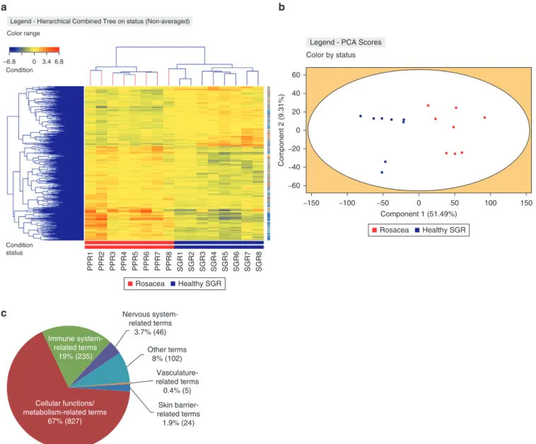
Heatmap, principal component analysis. To identify the in- depth differences in gene expression patterns between SGR and PPR skin samples, RNASeq analysis was performed on lysates of eight healthy SGR skin samples and eight PPR samples. The PPR and SGR sample groups were clearly separated in the heatmap as well as in the principal component analysis of the RNASeq data (Figure 1a and b).
The following statistical cut-off level was applied: a minimum of 1.5-fold alteration (fold change; higher or lower levels in PPR over SGR) in the average expression of the given mo- lecular transcript in all donors atP<0.05 statistical signifi- cance. A total of 5,136 genes were differentially expressed in PPR compared with SGR skin. Of the differentially expressed genes (DEGs), 3,133 genes showed higher expression, whereas 2,003 genes were expressed at lower levels in PPR as compared with SGR (Supplementary Table S1).
Pathway analysis 1. To identify the function of the DEGs, multiple bioinformatics analyses were performed using the Cytoscape ClueGO bioinformatics tool (Bindea et al., 2009).
First, to identify the general biological function of DEGs, we performed a pathway enrichment analysis on all DEGs with fold change1.5. To identify the significantly enriched (P 0.05) terms and/or pathways with global functions, the following criterion was applied: all terms contain at least 50 genes from our input gene set (the detailed parameters of the analysis can be found in the Materials and Methods section).
By using the above approach, 1,239 significantly enriched terms were found by ClueGO. Unsurprisingly, the identified terms were mostly involved in cellular/metabolic functions (e.g., ion transport, lipid biosynthetic process, and transferase activity) and innate (e.g., response to external stimulus, response to stress, cytokine secretion, defense response to bacterium, NOD-like receptor signaling pathway, and com- plement activation) and adaptive (e.g., T-cell activation, leukocyte migration, TNF production, IFN-gamma produc- tion, and chemokine signaling pathway) immune mecha- nisms. In addition, genes taking part in vascularization (e.g., regulation of vasculature development and angiogenesis) and the nervous system (e.g., nervous system development) were differentially expressed (Figure 1c,Supplementary Table S2).
Most importantly, 24 terms related to skin barrier were also
differentially expressed (e.g., epithelium development, morphogenesis of an epithelium, epithelial cell differentia- tion, and epithelial cell proliferation) (Figure 1c, Supplementary Table S2).
Pathway analysis 2. Next, we performed a second pathway enrichment analysis to determine the specific functions of DEGs; thus, a stricter analytical approach was applied and the up- and downregulated DEGs were analyzed as two different clusters by ClueGO (the detailed parameters of the analysis can be found in the Materials and Methods section).
This analytical approach revealed 426 significantly enriched terms and/or pathways (Supplementary Table S3) and most of them (291) belonged to innate and adaptive immune mech- anisms (e.g., T helper type [Th] 17 cell differentiation, toll- like receptor cascades, T-cell selection, and neutrophil migration). In addition, several terms were involved in cellular and metabolic functions (63 terms; e.g., transport along microtubule, exocytosis of specific granule membrane proteins, and response to cAMP) and took part in vasculari- zation (2 terms; positive regulation of vasculature develop- ment and positive regulation of angiogenesis) and the nervous system (21 terms; e.g., axonogenesis, glial cell dif- ferentiation, and axon guidance) were differentially expressed (Supplementary Table S3). Notably, 15 terms were found to be related to skin barrier function (e.g., keratiniza- tion, cornification, and tight junction) (Figure 1d and e, Supplementary Table S3). Hereafter, we focused on the skin barrier-related pathways (Figure 1d).
RT-qPCR and IHC validation confirm the significant alterations in the major skin barrier components in PPR To gain further insight into the permeability barrier differ- ences in PPR versus healthy SGR skin, we examined the expression of genes belonging to the major groups of skin barrier molecules by RT-qPCR. These major groups included (i) cornified envelope formation (FLG, KRT1, KRT10, LCE1D, LCE1F, LOR, SPRR1A, SPRR2A, TGM1, TGM3, and TGM5), (ii) intercellular lipid lamellae formation (ABCA12), (iii) desmosome organization (CDH1, corneodesmosin [CDSN], desmoglein 1 [DSG1], DSC1, and PKP1), (iv) corneocyte desquamation (kallikrein [KLK]5, KLK7, and KLK14), (v) tight junction formation (CLDN1, CLDN16, CLDN23, and OCLN), (vi) barrier alarmins (KRT6, KRT16, and KRT17), and (vii) AMPs (S100A7, S100A8, S100A9, DEFB4B, LCN2, and cathelicidin). Furthermore, we also assessed the expression of selected molecules at the protein level by using IHC.
Cornified envelope formation. First, we focused on inves- tigating the mRNA levels of molecules composing the cor- nified envelope in healthy SGR and PPR skin samples. Using RT-qPCR, we verified the RNASeq results. Most structural molecules (KRT1, KRT10, FLG, LOR, LCE1D, and LCE1F) were downregulated in PPR samples, whereas SPRR1Aand SPRR2AmRNA levels were higher in the diseased samples compared with the healthy controls (Table 1,Supplementary Figure S1). The differences were statistically significant in all cases, except for LCE1D, LCE1F, and SPRR1A. Enzymes crucial for peptide cross-linking (TGM1 and TGM3) were expressed at similar levels in PPR and SGR samples, except for TGM5. TGM5 was significantly downregulated in PPR B Medgyesiet al.
Skin Barrier Alterations of PPR
Journal of Investigative Dermatology (2020), Volume-
2
samples compared with SGR samples (Table 1, Supplementary Figure S1).
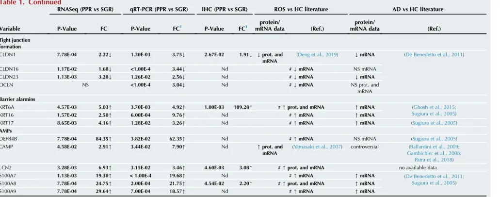
Next, we analyzed cornified envelope components at the protein level using IHC. LOR and KRT1 protein levels were significantly lower in PPR than SGR skin, whereas FLG levels were similar in the sample groups (Table 1,Figure 2). Among the previously mentioned enzymes, no significant differences in TGM5 were detected between healthy SGR and PPR samples (Table 1,Figure 2).
Intercellular lipid lamellae formation. Among molecules with a pivotal role in composing intercellular lipid lamellae, the gene expression level of ABCA12 was assessed by RT- qPCR in PPR and SGR samples. According to our results,
ABCA12 was significantly downregulated in the diseased specimens (Table 1,Supplementary Figure S1).
Desmosome organization. The gene expression levels of desmosome components (DSG1, DSC1, CDSN, PKP1, and CDH1) were examined by RT-qPCR. We found that all investigated molecules were highly and significantly down- regulated in PPR specimens, except forCDSN.CDSNmRNA levels were not significantly different between the two groups (Table 1,Supplementary Figure S1). To validate our results at the protein level, we also examined two junction compo- nents, DSG1 and CDSN, by IHC. DSG1 protein levels were significantly lower in PPR compared with SGR samples, whereas CDSN protein was expressed at a similar level in the two sample groups (Table 1,Figure 2).
b a
c
Rosacea Healthy SGR
Rosacea Healthy SGR
Component 2 (9.31%)
60 40 20
–20 0
–40 –60
Component 1 (51.49%) –150
Legend - PCA Scores Color by status
–100 –50 0 50 100 150
Condition status Condition
−6.8 0 3.4 6.8 Color range
Legend - Hierarchical Combined Tree on status (Non-averaged)
SGR8
SGR6 SGR7
SGR5
SGR4
SGR3
SGR2
SGR1PPR8
PPR7
PPR6
PPR5
PPR4
PPR3
PPR2
PPR1
Skin barrier- related terms 1.9% (24) Vasculature- related terms 0.4% (5) Nervous system-
related terms 3.7% (46)
Other terms 8% (102)
Cellular functions/
metabolism-related terms 67% (827) Immune system-
related terms 19% (235)
Figure 1. RNASeq analyses revealed significant skin barriererelated differences between PPR and healthy SGR skin samples.(a) Heat map was created by analyzing genes showing significantly different expression (P<0.05) between SGR (n¼8) and PPR (n¼8) skin. (b) Principal component analysis generated by StrandNGS software could distinguish the two sample groups unambiguously. (c) The distribution of significantly enriched terms based on their functions on the basis of the first-round enrichment analyses of significantly differentially expressed genes with FC>1.5 between SGR and PPR by Cytoscape and ClueGO (www.cytoscape.org). (d) Barrier-related significantly enriched terms revealed by Cytoscape and ClueGO between SGR and PPR in the second pathway analysis of significantly differentially expressed genes with FC>1.5. (e) Representative barrier-related terms visualized by ClueGO. CASP, caspase; CDSN, corneodesmosin; FC, fold change; Nr., number; PPR, papulopustular rosacea; RNASeq, RNA sequencing; SGR, sebaceous glanderich.
B Medgyesiet al.
Skin Barrier Alterations of PPR
www.jidonline.org 3
Corneocyte desquamation. We measured mRNA levels for skin barriererelated enzymes having a crucial role in desquamation (KLK5, KLK7, and KLK14). The expression levels forKLK5,KLK7, andKLK14were similar in PPR versus SGR samples (Table 1, Supplementary Figure S2). We immunostained for KLK5 in healthy SGR and PPR samples, and no significant differences between PPR and SGR were detected (Table 1,Figure 2).
Tight junction formation. The mRNA levels of tight junc- tion components (CLDN1, CLDN16, CLDN23, and OCLN) were measured. The tight junction molecules were signifi- cantly downregulated in PPR samples compared with healthy SGR skin (Table 1,Supplementary Figure S2). To confirm our results at the protein level, we measured CLDN1 by IHC and detected significantly lower levels in PPR than SGR samples (Table 1,Figure 2).
Barrier alarmins. KRT6, KRT16, and KRT17 are highly induced in response to an epidermal barrier breach.
Therefore, we investigated the gene expression levels of these barrier alarmins. According to our results, the mRNA levels of KRT6,KRT16, and KRT17were significantly upregulated in PPR compared with SGR (Table 1,Supplementary Figure S2).
Immunostaining of KRT6 confirmed our mRNA data. KRT6 was almost absent from SGR skin but highly expressed in PPR (Table 1,Figure 2).
AMPs. We also examined the gene expression levels for key components of the immunological barrier, including the following AMPs: S100A7, S100A8, S100A9, DEFB4B, LCN2, and cathelicidin. All of the investigated AMPs were signifi- cantly upregulated in PPR samples compared with healthy SGR skin samples (Table 1, Supplementary Figure S2). To confirm that the increased mRNA levels resulted in increased protein, we measured the protein levels of S100A8 and LCN2. S100A8 and LCN2 protein levels were significantly higher in PPR samples compared with controls (Table 1, Figure 2).
d
e
43,81%
43,40%
42,99%
41,38%
40,70%
40,70%
40,31%
35,25%
29,67%
24,77%
24,04%
23,81%
23,81%
23,21%
20,47%
Plasma membrane resorption Late envelope proteins bind cornified envelope: CDSN CASP14 cleaves filaggrin CDSN binds the cornified envelope Reinforcement of the Cornified Envelope Lamellar bodies bind the early cornified envelope Formation of the cornified envelope Cornification Cell junction organization Keratinization Keratinocyte differentiation Epidermal cell differentiation Skin development Morphogenesis of a branching epithelium Tight junction
46 Nr. Associated Genes
% Associated Genes
46 46 36 35 35 52 43 27 53 88 105 120 55 35 0% 5% 10% 15% 20% 25% 30% 35% 40% 45%
Downregulated genes Upregulated genes Figure 1.Continued
B Medgyesiet al.
Skin Barrier Alterations of PPR
Journal of Investigative Dermatology (2020), Volume-
4
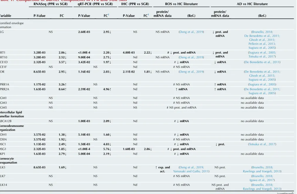
Table 1. Comparison of Barrier Components in PPR and SGR Skin
Variable
RNASeq (PPR vs SGR) qRT-PCR (PPR vs SGR) IHC (PPR vs SGR) ROS vs HC literature AD vs HC literature
P-Value FC P-Value FC1 P-Value FC1
protein/
mRNA data (Ref.)
protein/
mRNA data (Ref.)
Cornified envelope formation
FLG NS 2.60E-03 2.95Y NS NS mRNA (Deng et al., 2019) Yprot. and
mRNA
(Brunello, 2018;
De Benedetto et al., 2011;
Ghosh et al., 2015;
Pellerin et al., 2013;
Sugiura et al., 2005)
KRT1 3.28E-03 2.86Y <1.00E-4 2.20Y 4.80E-03 2.22Y #Yprot. and mRNA Yprot. and
mRNA
(Sugiura et al., 2005;
Totsuka et al., 2017)
KRT10 3.28E-03 2.52Y 9.00E-04 2.75Y Nd NS mRNA (Deng et al., 2019)
LCE1D 2.32E-03 3.57Y 3.42E-02 1.97Y Nd #YmRNA YmRNA (De Benedetto et al., 2011)
LCE1F NS NS Nd # NS mRNA
LOR 8.65E-03 2.95Y 1.16E-02 2.03Y 2.11E-02 1.81Y NS mRNA (Deng et al., 2019) YmRNA (De Benedetto et al., 2011;
Ghosh et al., 2015;
Sugiura et al., 2005)
SPRR1A 1.17E-02 3.26[ NS Nd # NS mRNA [mRNA (Sugiura et al., 2005)
SPRR2A 1.63E-03 8.64[ 2.59E-02 4.96[ Nd [mRNA [mRNA (De Benedetto et al., 2011;
Sugiura et al., 2005)
TGM1 NS NS Nd # NS mRNA no available data
TGM3 NS NS Nd # NS mRNA no available data
TGM5 NS NS NS # NS prot. and mRNA no available data
Intercellular lipid lamellae formation
ABCA12B NS 1.00E-03 2.09Y Nd #YmRNA no available data
Corneodesmosome organization
CDH1 3.57E-02 1.38Y 5.10E-03 1.60Y Nd #YmRNA no available data
CDSN 3.57E-02 1.92Y NS NS # NS mRNA no available data
DSC1 1.13E-03 2.49Y 1.50E-03 4.03Y Nd #YmRNA Yprot. (Totsuka et al., 2017)
DSG1 2.32E-03 1.85Y <1.00E-4 5.76Y 1.60E-03 2.86Y #Yprot. and mRNA
PKP1 1.63E-03 2.79Y 5.00E-04 2.19Y Nd #YmRNA no available data
Corneocyte desquamation
KLK5 8.65E-03 1.69Y NS Nd [exp. and
act.
(Deng et al., 2019;
Yamasaki and Gallo, 2011)
NS prot. (Brunello, 2018;
Rawlings and Voegeli, 2013)
KLK7 NS NS Nd # NS mRNA NS prot. (Brunello, 2018;
Igawa et al., 2017)
KLK14 NS NS Nd # NS mRNA NS prot. and
mRNA
(Brunello, 2018;
Rawlings and Voegeli, 2013) (continued)
BMedgyesietal.SkinBarrierAlterationsofPPR
www.jidonline.org5
Table 1. Continued
Variable
RNASeq (PPR vs SGR) qRT-PCR (PPR vs SGR) IHC (PPR vs SGR) ROS vs HC literature AD vs HC literature
P-Value FC P-Value FC1 P-Value FC1
protein/
mRNA data (Ref.)
protein/
mRNA data (Ref.)
Tight junction formation
CLDN1 7.78E-04 2.22Y 1.30E-03 3.75Y 2.67E-02 1.91Y Yprot. and
mRNA
(Deng et al., 2019) YmRNA (De Benedetto et al., 2011)
CLDN16 1.17E-02 1.68Y <1.00E-4 3.44Y Nd #YmRNA NS mRNA
CLDN23 1.13E-03 3.28Y 1.26E-02 2.56Y Nd #YmRNA YmRNA
OCLN NS <1.00E-4 3.04Y Nd #YmRNA NS prot. and
mRNA Barrier alarmins
KRT6A 4.57E-03 5.03[ 3.70E-03 4.92[ 1.00E-03 109.28[ #[prot. and mRNA [mRNA (Ghosh et al., 2015;
Sugiura et al., 2005)
KRT16 1.57E-02 2.50[ 6.00E-04 9.76[ Nd #[mRNA [mRNA
KRT17 8.65E-03 4.16[ 1.28E-02 3.26[ Nd #[mRNA [mRNA (Sugiura et al., 2005)
AMPs
DEFB4B 7.78E-04 84.35[ 3.82E-02 62.35[ Nd #[mRNA NS mRNA (Sugiura et al., 2005)
CAMP 4.58E-02 2.91[ 3.44E-02 7.90[ Nd [prot. and
mRNA
(Yamasaki et al., 2007) controversial (Ballardini et al., 2009;
Gambichler et al., 2008;
Patra et al., 2018)
LCN2 3.28E-03 6.93[ 3.15E-02 3.46[ 4.60E-03 3.08[ #[prot. and mRNA no available data
S100A7 1.13E-03 19.30[ <1.00E-4 19.68[ Nd #[mRNA [mRNA (De Benedetto et al., 2011;
Sugiura et al., 2005)
S100A8 7.78E-04 24.75[ 2.00E-04 21.75[ 4.54E-02 2.20[ #[prot. and mRNA [mRNA
S100A9 7.78E-04 29.64[ 7.00E-04 18.57[ Nd #[mRNA [mRNA
Abbreviations: act., activity; AD, atopic dermatitis; CAMP, cathelicidin; CDSN, corneodesmosin; DSG1, desmoglein 1; exp., expression; FC, fold change; HC, healthy control; IHC, immunohistochemistry; KLK, kallikrein; nd, not determined; NS, not significant; PPR, papulopustular rosacea; prot., protein; Ref., reference; RNASeq, RNA sequencing; ROS, rosacea; SGR, sebaceous glanderich.
Boxes represent the studied seven major groups of barrier composing molecules. The first column contains the molecules that have been investigated in our study. The second, third, and fourth columns summarize our findings according to the applied methods: RNASeq, qRT-PCR and IHC, respectively. Eight samples were examined in each group regarding all the investigated molecules.
Available literature data about papulopustular rosacea regarding all the investigated molecules, as well as our present findings from column 1e3 are compiled in Column 5. In this column,#represents that to our knowledge our study provides novel, previously unreported data, whereas the absence of # represents preexisting literature data. Available literature data about atopic dermatitis regarding all the investigated molecules are referenced in column 6. Comparison of column 5 and 6 highlights the similarities between the barrier alterations of these two skin disorders.
Statistical analyses between protein and mRNA levels were determined by two-samplet-test. Bold type indicates data with significant differences.
1Arrows indicate the direction of significant changes.
BMedgyesietal.SkinBarrierAlterationsofPPR
JournalofInvestigativeDermatology(2020),Volume-6
SGR
FLG
LOR
KRT1 Cornified
envelope formation
Desmosome organization
Tight junction formation
Barrier alarmin
Antimicrobial peptides
TGM5
CDSN
DSG1
CLDN1
KRT6
S100A8
LCN2
PPR
SGR skin (n=8)
FLG
PPR skin (n=8) LOR
*
SGR skin (n=8)
PPR skin (n=8) Epidermal KRT1
**
SGR skin (n=8)
PPR skin (n=8) Epidermal TGM5
SGR skin (n=8)
PPR skin (n=8) Epidermal CDSN
SGR skin (n=8)
PPR skin (n=8)
*
SGR skin (n=8)
PPR skin (n=8) Epidermal S100A8
***
SGR skin (n=8)
PPR skin (n=8) Epidermal KRT6
*
SGR skin (n=8)
PPR skin (n=8) Epidermal CLDN1
*
SGR skin (n=8)
PPR skin (n=8) Epidermal DSG1
*
SGR skin (n=8)
Epidermal LCN2
PPR skin (n=8)
Epidermal FLG levelsEpidermal LOR levelsEpidermal KRT1 levelsEpidermal TGM5 levelsEpidermal CDNS levelsEpidermal S100A8 levelsEpidermal KRT6 levelsEpidermal CLDN1 levelsEpidermal DSG1 levelsEpidermal LCN2 levels
5 4 3 2 1 0
8 6 4 2 0
30
20
10
0
15
10
5
0
15
10
5
0
50 40 30
10 0 20 30
20
10
0 20 15 10 5 0 20 15 10 5 0
20 15 10 5 0
Figure 2. Prominent differences in the protein expression levels of skin barriererelated molecules between PPR and healthy SGR skin samples.Representative images for immunostaining and quantification of epidermal levels of FLG, LOR, KRT1, TGM5, CDSN, DSG1, CLDN1, KRT6, S100A8, and LCN2 in SGR and PPR skin sections. The graphs show the mean95% confidence interval of measured protein levels (*P<0.05; **P<0.01; ***P<0.001, as determined by two-samplet-test). CDSN, corneodesmosin; DSG1, desmoglein 1; PPR, papulopustular rosacea; SGR, sebaceous glanderich. Bar¼50mm.
B Medgyesiet al.
Skin Barrier Alterations of PPR
www.jidonline.org 7
Disrupted skin barrier features of PPR are similar to that of AD
Literature search. As barrier alterations in PPR seemed to be similar to that of lesional AD skin, we reviewed the literature on AD barrier damage and compared barrier al- terations of the two disorders (Table 1). According to the literature search, the skin barrier alterations were similar in PPR and AD skin compared with the respective healthy control skin (SGR and sebaceous glandepoor [SGP]).
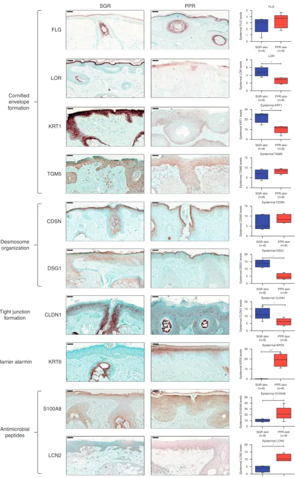
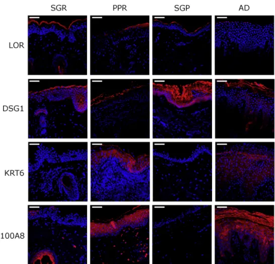
Immunofluorescent staining. To confirm similarities in skin barrier changes between PPR and AD, immunofluorescent staining was performed in PPR and AD samples and the respective SGR and SGP skin specimens. According to the immunostaining results, a cornified envelope structural molecule (LOR) and key components of desmosomes (DSG1) and tight junctions (CLDN1) were highly downregulated in PPR and AD samples compared with controls (Figure 3). In contrast, the barrier alarmin KRT6 and the AMP S100A8 were almost absent from healthy skin types and were expressed at high levels in PPR and AD skin specimens (Figure 3).
Notably, the expression patterns of these proteins were similar in PPR and AD skin (Figure 3).
DISCUSSION
Clinical features and functional studies on the affected facial skin of patients with rosacea indicate the presence of
permeability barrier alterations (Darlenski et al., 2013;
Powell and Ni Raghallaigh, 2011). However, no detailed analysis of skin barrier disruption in rosacea at the molecular level has been conducted so far. In the present study, we performed whole transcriptomic analysis (RNASeq) of PPR skin samples, and gene expression profiles of diseased skin were compared with that of healthy SGR skin. According to our findings, all major components of the skin barrier are severely altered in PPR.
An intact skin barrier is essential for maintaining homeo- stasis as it protects the body against external agents and mi- crobes and provides a waterproof cover. According to the literature, stratum corneum, the outermost layer of the epidermis, and tight junctions are considered the most important components of the skin permeability barrier (Egawa and Kabashima, 2016; Egawa and Kabashima, 2018).
Molecules forming the stratum corneum can be further divided into four groups: cornified envelope formation, intercellular lipid lamellae formation, corneodesmosome organization, and corneocyte desquamation (Egawa and Kabashima, 2016; Egawa and Kabashima, 2018).
Cornified envelope is built up from several types of intra- cellular structure proteins, including FLG, LOR, LCE, and SPRR proteins; envoplakin; periplakin; involucrin; and KRT filaments that are derived from keratohyalin granules (Carregaro et al., 2013; Egawa and Kabashima, 2016; Koch et al., 2000; Palmer et al., 2006). These proteins undergo
Figure 3. Highly similar barrier alteration occurs in PPR and AD.PPR and AD, as well as their respective control samples (SGR and SGP), were immunostained. Representative images for immunofluorescent staining of epidermal levels of LOR, DSG1, KRT6, and S100A8 in SGR, PPR, SGP, and AD skin sections. AD, atopic dermatitis; DSG1,
desmoglein1; PPR, papulopustular rosacea; SGP, sebaceous glandepoor;
SGR, sebaceous glanderich. Bar¼50 mm.
B Medgyesiet al.
Skin Barrier Alterations of PPR
Journal of Investigative Dermatology (2020), Volume-
8
cross-linking, which is catalyzed by TGM1, TGM3, and TGM5 (Eckert et al., 2005). According to our results, FLG, LOR, KRT1, KRT10, and LCE1D were significantly down- regulated, whereas SPRR2A was significantly upregulated in PPR skin compared with healthy SGR skin. The significant decrease in LOR and KRT1 levels was also confirmed at the protein level. Regarding the expression of TGMs, no signifi- cant difference was detected either at mRNA or protein levels.
Lipid lamellae formation is also a critical event in the development of intact stratum corneum, in which lip- oxygenases (ALOX12B and ALOXB3) and lipid transporters (ABCA12) are essential to lipid synthesis and transport (Iwai et al., 2012; Kelsell et al., 2005; Krieg and Fu¨rstenberger, 2014). In our study, ABCA12 was significantly down- regulated at the mRNA level in PPR compared with controls.
Another component of the stratum corneum is the desmosome apparatus, which is responsible for cell adhesion of corneocytes (Egawa and Kabashima, 2016; Egawa and Kabashima, 2018). Based on our findings, desmosomes are highly disrupted in PPR, as demonstrated by the significantly decreased gene expression levels of CDH1, CDSN, DSC1, DSG1, andPKP1. The significant decrease in DSG1 was also confirmed at the protein level by IHC.
In the stratum corneum, corneocytes are shed as part of an event called desquamation, which is primarily regulated by KLKs (KLK5, KLK7, and KLK14) via a pH-dependent proteo- lytic cascade (Brattsand et al., 2005; Rippke et al., 2004). In this study, we detected similar gene expression levels of KLK5, KLK7, and KLK14 in lesional PPR compared with healthy skin; however, the enzymes’ protein levels and ac- tivity were not examined.
In addition to the stratum corneum, tight junctions are also key components for the integrity of the skin barrier by sealing adjacent keratinocytes in the stratum granulosum and acting as a barrier for water and solutes. Tight junctions are composed of transmembrane proteins, particularly the CLDNs and OCLN (Kirschner et al., 2010; Yokouchi et al., 2015). In our study, mRNA levels for CLDN1, CLDN16, CLDN23, and OCLN were significantly decreased in PPR compared with controls. The significant decrease in CLDN1 was confirmed at the protein level by IHC.
In parallel with the stratum corneum and tight junction components, we focused on investigating two additional groups of molecules, key barrier alarmins and AMPs, because the expression levels of these molecules are tightly connected with the level of permeability barrier structural components (Borkowski and Gallo, 2011; Zhang et al., 2019). In our study, all investigated barrier alarmins (KRT6A, KRT16, and KRT17) and AMPs (cathelicidin, hBD-2, LCN2, S100A7, S100A8, and S100A9) were significantly upregulated in PPR skin compared with controls. The significantly higher pres- ence of LCN2, S100A8, and KRT6 in PPR was confirmed at the protein level.
To our knowledge, until now, only one study has investi- gated rosacea samples at a whole transcriptomic level. In this previous study, the authors focused on examining the immunological characteristics of different rosacea subtypes, and no data were published on barrier components (Buhl et al., 2015). Another study, performed by Deng et al.,
focused on mRNA expression levels of cornified envelope components, including KRT10, FLG, and LOR in different rosacea subtypes (Deng et al., 2019). They also studied the tight junction protein CLDN1 and could detect significantly lower gene expression and protein levels of CLDN1 in PPR samples compared with controls, in agreement with our re- sults. Regarding the mRNA levels of KRT10 and LOR, the authors of this study did not find significant differences when comparing PPR to controls, and protein levels were not assessed (Deng et al., 2019). The expression of KLK5 in ro- sacea has been studied previously by another workgroup, and its mRNA and protein levels were demonstrated to be significantly increased, in parallel with elevated enzyme activity; however, the authors did not specify which rosacea subtype had been assessed (Morizane et al., 2010; Yamasaki and Gallo, 2011). In our study, in PPR, we could not detect significantly elevatedKLK5mRNA levels, but protein levels and enzyme activity were not assessed. Among AMPs, only the expression of cathelicidin was assessed previously in PPR, whereas data regarding hBD-2 is available only in ocular rosacea (Go¨kc¸nar et al., 2019; Morizane et al., 2010;
Yamasaki and Gallo, 2011). In line with these previous re- sults, in our study, cathelicidin and hBD-2 were significantly upregulated in PPR as compared with SGP skin at the mRNA level.
As a whole, our in-depth molecular biological investiga- tion, using whole transcriptomic and bioinformatics analyses, qRT-PCR, and quantitative IHC, indicate that all major components of the permeability skin barrier are severely disrupted in PPR, which may significantly contribute to dis- ease pathophysiology. Barrier alterations have gained far- reaching importance in recent years. In AD, it has been proven that barrier disruption is able to induce skin inflam- mation, and, in this manner, can be considered as an initiator of disease pathogenesis. From this point of view, it is quite interesting that we could detect highly similar barrier alter- ations in PPR and AD. According to our literature search and results, these similarities included the altered expression of cornified envelope components, desmosome and tight junc- tion molecules, barrier alarmins, and AMPs. Moreover, we could confirm some of these similarities at the protein level by immunofluorescent staining of selected molecules. The demonstrated similarity between the barrier disruptions in PPR and AD is surprising because, up until now, mainly the differences in clinical features and immune composition of these diseases have been highlighted (Figure 4).
Although it is well known that barrier dysfunction is a potent initiator of inflammation in AD, on the basis of our results, we cannot determine the role of barrier alterations in rosacea, if it is the result or the cause of inflammation. Barrier damage may occur because of the Th1/Th17 type inflam- mation that is characteristic in rosacea, and IL-17A recently has been shown to downregulate FLG, LOR, and KRT10 mRNA levels in organotypic three-dimensional skin equiva- lents (Pfaff et al., 2017). In contrast, it is also possible that dysfunctional barrier is the initiating factor of rosacea development. Although barrier alteration is an adjuvant fac- tor for Th2 type inflammation in AD, the reason why similar barrier disruption may precipitate Th1/Th17 type inflamma- tion in rosacea could be that AD has an SGP region B Medgyesiet al.
Skin Barrier Alterations of PPR
www.jidonline.org 9
b
c Although in AD and PPR permeability barrier alteration is similar, the different types of immune inflammation result major distinction between the
two diseases Due to the unique immune milieu, permeability barrier alterations of distinct skin regions may initiate different types of immune responses
and immune-mediated diseases
Barrier dysfunction of SGP:
IfTSLP ↑, IL-33 ↑, IL-25 ↑
Immune response:
Th2/Th22, IL-4, IL-13, IL-5 ↑ AD
disease of SGP skin
a
Different homeostatic immune milieu of distinct
skin regions
PPR disease of SGR skin Barrier dysfunction of SGR:
sfTSLP ↓, AMPs ↑
Immune response:
Th1/Th17(23), IFN-γ, IL-17
papules, plaques, papulosus infiltration, scaling, excoriations
Atopic dermatitis Papulopustular rosacea
Major differences of atopic dermatitis and papulopustular rosacea
usually in childhood, less frequently in adolescence or
adulthood mainly on SGP skin Th2/Th22 cells, DCs, proliferation
of KCs, high eosinophil and low neutrofil count high inflammatory lfTSLP
expression in AD skin
Th2/Th22 cells: IL-4/IL-13/IL-5/
IL-22
Th1/Th17 T cells: IL-17/IFNγ /TNFα
Th1/Th17 cells, DCs, high macrophage, mast cell and
neutrofil counts loss of homeostatic sfTSLP
expression in PPR in adulthood flushing, persistent erythema,
teleangiectasia, papules and pustules
on SGR skin
Key players of pathomechanism
Histology Localisation Age at onset Clinical features
low TLR2 expression high TLR2 expression
TARC+ inflammatory DCs TARC-DCs
low AMP level high AMP level
SGP skin SGP skin (no sfTSLP, low DC, Th17(
(no sfTSLP, low DC, Th17(ββ)) and Treg count) and Treg count)
SGR skin SGR skin
(sfTSLP, higher noninflammatory (sfTSLP, higher noninflammatory
DC, Th17(
DC, Th17(ββ) and Treg counts,) and Treg counts, IL10/IL17 milieu) IL10/IL17 milieu) SGP skin
(no sfTSLP, low DC, Th17(β) and Treg count)
SGR skin
(sfTSLP, higher noninflammatory DC, Th17(β) and Treg counts,
IL10/IL17 milieu)
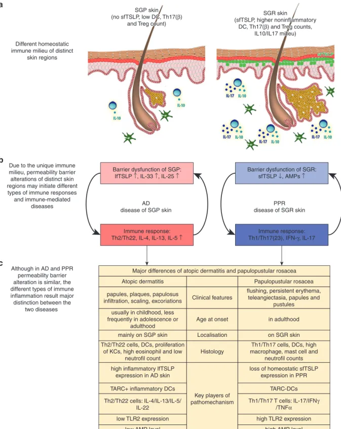
Figure 4. The possible role of permeability barrier alteration in the pathogenesis of PPR.Until now, when comparing AD and PPR, mainly the differences in their clinical and immunological characteristics were pronounced by dermatologists. However, based on our results, these two skin disorders can be considered from another point of view, because both are characterized by severe and notably similar skin barrier damage, although being developed on distinct skin areas.
We propose that the prominently different steady-state immune and barrier features of (a) the skin regions (SGR and SGP skin) where these diseases prefer to localize can influence (b) the barrier damageeconnected inflammation differently, leading to (c) region-specific immune-mediated skin diseases and explaining the prominent distinctions between AD and PPR. AD, atopic dermatitis; AMP, antimicrobial peptide; DC, dendritic cell; KC, keratinocyte; lfTSLP, long form thymic stromal lymphopoietin; sfTSLP, short form thymic stromal lymphopoietin; PPR, papulopustular rosacea; SGP, sebaceous glandepoor; SGR, sebaceous glanderich; Th, T helper; Th17(b), noninflammatory Th17 cell; Th17(23), inflammatory Th17 cell; TLR2, toll-like receptor 2; Treg, regulatory T cell.
B Medgyesiet al.
Skin Barrier Alterations of PPR
Journal of Investigative Dermatology (2020), Volume-
10
preference, whereas rosacea localizes exclusively on SGR skin. SGR and SGP skin areas have prominently different homeostatic immune and barrier characteristics (Be´ke et al., 2018; Dajnoki et al., 2017). Whereas SGR skin regions dispose significantly higher AMP levels, noninflammatory Th17(b) and regulatory T-cell counts, and constitutive expression of homeostatic short form thymic stromal lym- phopoietin (sfTSLP), SGP skin is characterized by low AMP levels and T-cell counts, without TSLP presence under steady- state (Be´ke et al., 2018; Dajnoki et al., 2017). These features of the skin seem to be very similar to that of another barrier, namely the gut, where the regional immune-related differ- ences are well known. Th17(b) cells are enriched and sfTSLP is expressed in the small intestine, in contrast to the colon, where Th17(b) cells are absent and sfTSLP is present only in the proximal part (Iliev et al., 2009; Rimoldi et al., 2005). It has been raised that these unique immune and barrier char- acteristics of different gut sections can lead to distinct types of immune-mediated diseases. In Crohn’s disease, the loss of sfTSLP from the small intestine can promote the activation of inflammatory Th1/Th17 cells (Iliev et al., 2009; Rimoldi et al., 2005), whereas in ulcerative colitis, significantly increased inflammatory long form TSLP levels initiate Th2 type inflammation in the colon (Fornasa et al., 2015). On the basis of these findings, we hypothesize that similar barrier alter- ations of SGR and SGP skin regions, because of their different homeostatic immune and barrier milieu, initiate distinct immune-mediated skin diseases with unique clinical features driven by different Th subsets (AD on SGP and PPR on SGR areas) (Figure 4). Notably, De Benedetto et al. already raised the possibility that barrier defects can lead to the production of keratinocyte-derived mediators including pro-Th2 and pro- Th17 types that affect the characteristics of Th response (De Benedetto et al., 2012).
To determine if PPR barrier damage is the initiator of the dis- ease or a consequence of manifested inflammation, the exact time of barrier disruption should be studied and a detailed analysis of the perilesional and/or nonlesional skin of patients with PPR should be performed in the future. Moreover, analo- gous experiments are needed in other subtypes of rosacea.
In summary, our results unambiguously prove the presence of severe barrier alterations in the facial skin of patients with PPR; thus, we suggest that skin barrier restoring therapies should be incorporated into clinical guidelines for rosacea management, similar to that of AD.
MATERIALS AND METHODS Skin biopsies
Skin punch biopsies (0.5e1 cm2) were taken from normal SGR skin sites of eight healthy individuals (mean ageSD: 56.25 13.11 years) undergoing plastic surgery and from lesional skin of eight patients with PPR (mean age SD: 59.38 13.59 years) (Supplementary Table S4). Written informed consent according to the Declaration of Helsinki principles was obtained by all in- dividuals before participating in the study. The study was approved by the local ethics committee of the University of Debrecen.
RNA isolation and reverse transcription
Samples were homogenized in Tri reagent (Sigma-Aldrich, St. Louis, MO) with Tissue Lyser (Qiagen, Hilden, Germany) using previously
autoclaved metal beads (Qiagen). RNA concentration and purity were measured and cDNA was synthesized using the High Capacity cDNA Archive Kit (Invitrogen, Thermo Fisher Scientific, Waltham, MA).
RT-qPCR
RT-PCR was carried out in triplicate using pre-designed MGB assays (Thermo Fisher Scientific). The assessed TaqMan Gene Expression assays are listed inSupplementary Materials and Methods. All re- actions were performed with an ABI PRISM 7000 Sequence Detec- tion System. Relative mRNA levels were calculated using the 2-DDCt method normalized to the expression of PPIA mRNA.
RNASeq analysis
Complementary DNA library for RNASeq was generated from 1mg total RNA using TruSeq RNA Sample Preparation Kit (Illumina, San Diego, CA) according to the manufacturer’s protocol. A single read 50 base pair sequencing run was performed on Illumina HiScan SQ instrument (Illumina), and 16e18 million reads per sample were obtained. Sequenced reads were aligned to Human Genome v19 using TopHat and Cufflinks algorithms and bam files were generated.
StrandNGS software was used for further statistical analysis. To identify statistically significant gene expression patterns, nonpara- metric WilcoxoneManneWhitney test was used. Library prepara- tions, sequencing, and data analysis were performed at the Genomic Medicine and Bioinformatics Core Facility of University of Debre- cen. RNA sequencing data of our samples were deposited to the Sequence Read Archive database (https://www.ncbi.nlm.nih.gov/
sra), under accession numbers PRJNA421246 and PRJNA592080.
Pathway analyses
Multiple bioinformatics analyses were performed by Cytoscape ClueGO bioinformatics tool. Pathway enrichment analyses were performed on all DEGs with fold change1.5. The details of our analytical approach can be found in theSupplementary Materials and Methods.
Immunohistochemistry
For IHC analyses, paraffin-embedded sections from patients and healthy controls were deparaffinized. Heat-induced antigen retrieval was performed. Sections were stained with primary antibodies listed inSupplementary Materials and Methods. Subsequently, anti-mouse/
rabbit horseradish peroxidaseeconjugated secondary antibody (Biogenex, Fremont, CA) was employed. Staining was detected with the Vector NovaRed Kit (Vector Laboratories, Burlingame, CA).
Sections were counterstained with methylene green and digitized by Whole Slide Imaging, and Pannoramic Viewer software was used for the evaluation of the degree of staining of the slides.
Statistical analysis
Data distribution was analyzed by Kolmogorov-Smirnov test.
Because our data showed normal distribution, two groups of sam- ples were compared statistically by two-samplet-test. Differences between the groups were demonstrated using mean95% confi- dence interval. P-values <0.05 were considered statistically sig- nificant (*P<0.05; **P<0.01; ***P<0.001). Statistical data was analyzed using GraphPad Prism v6 (GraphPad Software Inc., La Jolla, CA) and SPSS 25 (SPSS package for Windows, Chicago, IL).
Additional details are provided in theSupplementary Information online.
Data availability statement
The data that support the findings of this study are available from the corresponding author upon reasonable request.
B Medgyesiet al.
Skin Barrier Alterations of PPR
www.jidonline.org 11
ORCIDs
Barbara Medgyesi:https://orcid.org/0000-0001-8800-7693 Zsolt Dajnoki:https://orcid.org/0000-0002-7787-1002 Gabriella Be´ke:https://orcid.org/0000-0003-3113-2287 Krisztia´n Ga´spa´r:https://orcid.org/0000-0001-6530-7052 Imre L}orinc Szabo´:https://orcid.org/0000-0002-9628-4372 Eszter Anna Janka:https://orcid.org/0000-0003-0724-5281 Szila´rd Po´liska:https://orcid.org/0000-0002-9722-251X Zolta´n Hendrik:https://orcid.org/0000-0001-8546-2148 Ga´bor Me´hes:https://orcid.org/0000-0003-2503-7451 Da´niel To¨r}ocsik:https://orcid.org/0000-0002-6094-6266 Tama´s Bı´ro´:https://orcid.org/0000-0002-3770-6221 Aniko´ Kapita´ny:https://orcid.org/0000-0003-4540-5258 Andrea Szegedi:https://orcid.org/0000-0003-2109-9014
CONFLICT OF INTEREST
The authors state no conflict of interest.
ACKNOWLEDGMENTS
The publication is supported by Hungarian Research Grant (NKFIH K-128250 and NKFIH PD-131689) and the GINOP-2.3.2-15-2016-00050 and EFOP- 3.6.1-16-2016-00022 projects. The project is cofinanced by the European Union and the European Regional Development Fund and the European Social Fund. This project was supported by the Ja´nos Bolyai Research Scholarship of the Hungarian Academy of Sciences (AK and DZ) and is also supported by the U´ NKP-18-4 New National Excellence Program of the Ministry of Human Capacities (AK) and the U´ NKP-19-4 New National Excellence Program of the Ministry for Innovation and Technology (AK and DZ).
SUPPLEMENTARY MATERIAL
Supplementary material is linked to the online version of the paper atwww.
jidonline.org, and athttps://doi.org/10.1016/j.jid.2020.02.025.
REFERENCES
Ballardini N, Johansson C, Lilja G, Lindh M, Linde Y, Scheynius A, et al.
Enhanced expression of the antimicrobial peptide LL-37 in lesional skin of adults with atopic eczema. Br J Dermatol 2009;161:40e7.
Be´ke G, Dajnoki Z, Kapita´ny A, Ga´spa´r K, Medgyesi B, Po´liska S, et al.
Immunotopographical differences of human skin. Front Immunol 2018;9:
424.
Bindea G, Mlecnik B, Hackl H, Charoentong P, Tosolini M, Kirilovsky A, et al.
ClueGO: a Cytoscape plug-in to decipher functionally grouped gene ontology and pathway annotation networks. Bioinformatics 2009;25:
1091e3.
Borkowski AW, Gallo RL. The coordinated response of the physical and antimicrobial peptide barriers of the skin. J Invest Dermatol 2011;131:
285e7.
Brattsand M, Stefansson K, Lundh C, Haasum Y, Egelrud T. A proteolytic cascade of kallikreins in the stratum corneum. J Invest Dermatol 2005;124:
198e203.
Brunello L. Atopic dermatitis. Nat Rev Dis Primers 2018;4:2.
Buechner SA. Rosacea: an update. Dermatology 2005;210:100e8.
Buhl T, Sulk M, Nowak P, Buddenkotte J, McDonald I, Aubert J, et al. Mo- lecular and morphological characterization of inflammatory infiltrate in rosacea reveals activation of Th1/Th17 pathways. J Invest Dermatol 2015;135:2198e208.
Carregaro F, Stefanini AC, Henrique T, Tajara EH. Study of small proline-rich proteins (SPRRs) in health and disease: a review of the literature. Arch Dermatol Res 2013;305:857e66.
Crawford GH, Pelle MT, James WD. Rosacea: I. Etiology, pathogenesis, and subtype classification. J Am Acad Dermatol 2004;51:327e41:quiz 42e4.
Dajnoki Z, Be´ke G, Kapita´ny A, Mo´csai G, Ga´spa´r K, Ru¨hl R, et al. Sebaceous gland-rich skin is characterized by TSLP expression and distinct immune surveillance which is disturbed in rosacea. J Invest Dermatol 2017;137:
1114e25.
Darlenski R, Kazandjieva J, Tsankov N, Fluhr JW. Acute irritant threshold correlates with barrier function, skin hydration and contact hypersensitivity in atopic dermatitis and rosacea. Exp Dermatol 2013;22:752e3.
De Benedetto A, Kubo A, Beck LA. Skin barrier disruption: a requirement for allergen sensitization? J Invest Dermatol 2012;132:949e63.
De Benedetto A, Rafaels NM, McGirt LY, Ivanov AI, Georas SN, Cheadle C, et al. Tight junction defects in patients with atopic dermatitis. J Allergy Clin Immunol 2011;127:773e86.e1e7.
Deng Z, Chen M, Xie H, Jian D, Xu S, Peng Q, et al. Claudin reduction may relate to an impaired skin barrier in rosacea. J Dermatol 2019;46:
314e21.
Eckert RL, Sturniolo MT, Broome AM, Ruse M, Rorke EA. Transglutaminase function in epidermis. J Invest Dermatol 2005;124:481e92.
Egawa G, Kabashima K. Multifactorial skin barrier deficiency and atopic dermatitis: essential topics to prevent the atopic march. J Allergy Clin Immunol 2016;138:350e8.e1.
Egawa G, Kabashima K. Barrier dysfunction in the skin allergy. Allergol Int 2018;67:3e11.
Fornasa G, Tsilingiri K, Caprioli F, Botti F, Mapelli M, Meller S, et al. Di- chotomy of short and long thymic stromal lymphopoietin isoforms in in- flammatory disorders of the bowel and skin. J Allergy Clin Immunol 2015;136:413e22.
Gallo RL, Granstein RD, Kang S, Mannis M, Steinhoff M, Tan J, et al.
Rosacea comorbidities and future research: the 2017 update by the National Rosacea Society Expert Committee. J Am Acad Dermatol 2018a;78:167e70.
Gallo RL, Granstein RD, Kang S, Mannis M, Steinhoff M, Tan J, et al. Standard classification and pathophysiology of rosacea: the 2017 update by the National Rosacea Society Expert Committee. J Am Acad Dermatol 2018b;78:148e55.
Gambichler T, Skrygan M, Tomi NS, Othlinghaus N, Brockmeyer NH, Altmeyer P, et al. Differential mRNA expression of antimicrobial peptides and proteins in atopic dermatitis as compared to psoriasis vulgaris and healthy skin. Int Arch Allergy Immunol 2008;147:17e24.
Ghosh D, Ding L, Sivaprasad U, Geh E, Biagini Myers J, Bernstein JA, et al. Multiple transcriptome data analysis reveals biologically relevant atopic dermatitis signature genes and pathways. PLOS ONE 2015;10:
e0144316.
Go¨kc¸nar NB, Karabulut AA, Onaran Z, Yumus‚ak E, Budak Yldran FA. Elevated tear human neutrophil peptides 1e3, human beta defensin-2 levels and conjunctival cathelicidin LL-37 gene expression in ocular rosacea. Ocul Immunol Inflamm 2019;27:1174e83.
Igawa S, Kishibe M, Minami-Hori M, Honma M, Tsujimura H, Ishikawa J, et al. Incomplete KLK7 secretion and upregulated LEKTI expression un- derlie hyperkeratotic stratum corneum in atopic dermatitis. J Invest Der- matol 2017;137:449e56.
Iliev ID, Spadoni I, Mileti E, Matteoli G, Sonzogni A, Sampietro GM, et al.
Human intestinal epithelial cells promote the differentiation of tolerogenic dendritic cells. Gut 2009;58:1481e9.
Iwai I, Han H, den Hollander L, Svensson S, Ofverstedt LG, Anwar J, et al. The human skin barrier is organized as stacked bilayers of fully extended ceramides with cholesterol molecules associated with the ceramide sphingoid moiety. J Invest Dermatol 2012;132:
2215e25.
Jenei A, Dajnoki Z, Medgyesi B, Ga´spa´r K, Be´ke G, Kinyo´ A´, et al. Apocrine gland-rich skin has a non-inflammatory IL-17-related immune milieu, that turns to inflammatory IL-17-mediated disease in hidradenitis suppurativa.
J Invest Dermatol 2019;139:964e8.
Kelsell DP, Norgett EE, Unsworth H, Teh MT, Cullup T, Mein CA, et al. Mu- tations in ABCA12 underlie the severe congenital skin disease harlequin ichthyosis. Am J Hum Genet 2005;76:794e803.
Kirschner N, Houdek P, Fromm M, Moll I, Brandner JM. Tight junctions form a barrier in human epidermis. Eur J Cell Biol 2010;89:839e42.
Koch PJ, de Viragh PA, Scharer E, Bundman D, Longley MA, Bickenbach J, et al. Lessons from loricrin-deficient mice: compensatory mechanisms maintaining skin barrier function in the absence of a major cornified en- velope protein. J Cell Biol 2000;151:389e400.
Krieg P, Fu¨rstenberger G. The role of lipoxygenases in epidermis. Biochim Biophys Acta 2014;1841:390e400.
Morizane S, Yamasaki K, Kabigting FD, Gallo RL. Kallikrein expression and cathelicidin processing are independently controlled in keratinocytes by calcium, vitamin D(3), and retinoic acid. J Invest Dermatol 2010;130:
1297e306.
Palmer CN, Irvine AD, Terron-Kwiatkowski A, Zhao Y, Liao H, Lee SP, et al.
Common loss-of-function variants of the epidermal barrier protein filaggrin
B Medgyesiet al.
Skin Barrier Alterations of PPR
Journal of Investigative Dermatology (2020), Volume-
12