1Department of Rheumatology and Immunology, University of Szeged, Faculty of Medicine;
2Department of Pulmonology, University of Szeged, Faculty of Medicine;
3Department of Internal Medicine, Cardiology Centre and Department of Family Medicine, University of Szeged, Faculty of Medicine, Szeged, Hungary.
Rita Adrienn Hemelein, Assist. Prof.
Imre Lajkó, Head Physician Kristóf Baráth, Resident János Varga*, Head Physician Gergely Ágoston, Assoc. Prof.
Daniella Hulló, Assist. Prof.
Márta Bocskai, Assist. Prof.
Diána Dobi, Clin. Fellow Zoltán Rózsavölgyi&, Assoc. Prof.
Ágnes Milassin+, Clin. Fellow Albert Varga, Prof.
Attila Somfay, Prof.
László Kovács, Prof.
*Current affiliation:
National Korányi Institute of Pulmonology, Budapest, Hungary.
&Current affiliation:
Markusovszky Hospital of Vas County, Szombathely, Hungary.
+Current affiliation:
University of Szeged, Faculty of Medicine, 1st Department of Internal Medicine, Szeged, Hungary.
Please address correspondence to:
László Kovács,
Department of Rheumatology and Immunology,
University of Szeged, Faculty of Medicine, Kálvária sgt. 57, 6725 Szeged, Hungary.
E-mail: kovacs.laszlo@med.u-szeged.hu Received on July 27, 2020; accepted in revised form on January 4, 2021.
Clin Exp Rheumatol 2021; 39 (Suppl. 131):
S94-S102.
© Copyright CliniCaland
ExpErimEntal rhEumatology 2021.
Key words: systemic sclerosis, cardiopulmonary exercise test, disease progression, desaturation Competing interests: none declared.
ABSTRACT
Objective. Cardiopulmonary exercise test (CPET) is a widely used examina- tion to predict the prognosis of many chronic pulmonary diseases, and it has also been tested in systemic sclerosis (SSc) with a focus on the development of pulmonary hypertension. CPET is a highly informative non-invasive tool that provides a more complex informa- tion than conventional lung function tests to predict the course of cardiopul- monary diseases, as it provides a gener- al overview of the aerobic metabolism, influenced by pulmonary, cardiovascu- lar and peripheral muscle function. The purpose of this investigation was to as- sess if the progression and the develop- ment of poor overall disease outcome in SSc can be predicted by this method.
Methods. Twenty-nine SSc patients were investigated prospectively with standard follow-up plus CPET for a mean of 3.7 years to match the results of conventional evaluation modalities and CPET. A composite end-point of several serious outcomes reflecting SSc-related vascular and cardiopulmonary dam- age was set up, and the predictive value of and correlations between the CPET parameters and resting lung function and echocardiography variables were assessed.
Results. None of the clinical param- eters, resting lung function or echocar- diographic test results proved to be pre- dictive of the development of the end- point of poor prognosis in this cohort.
In contrast, several CPET parameters were found to discriminate between SSc patients with or without adverse outcome. The detection of desaturation (at any CPET test) was associated with a higher risk of poor prognosis (OR:
5.265). VO2 and VE/VCO2 at baseline correlated with the annual decrease in FVC, anaerobic threshold with the development of digital ulcers, and VE/
VO2 with the increase in pulmonary arterial pressure.
Conclusion. Several CPET parameters obtained at the beginning of follow-up are informative of the appearance of various adverse end-points. CPET is a feasible examination in the care of SSc patients and provides excess infor- mation to current standard follow-up examinations.
Introduction
Systemic sclerosis (SSc), also termed scleroderma, is a systemic autoimmune disease with multi-organ involvement and frequently with a destructive, pro- gressive disease course. Key pathways in the pathogenesis are vascular, fi- brotic and immunological dysfunction, leading to progressive organ damage and increased mortality (1, 2). Leading causes of morbidity and mortality are interstitial lung disease (ILD), pulmo- nary arterial hypertension (PAH) and obliterative vasculopathy, in particular digital ulcers, gangraene and macro- vascular complications. Predictors of such poor outcome are hard to defini- tively identify by the physician at the first encounter, but it is obviously of crucial importance in the determination of follow-up strategy and of the inten- sity of immunosuppressive therapy.
Current guidelines prompt the treating physicians to regularly (every 6 to 12 months) perform resting lung function tests, high-resolution computed tomo- graphy (HRCT) of the lung and resting echocardiography to estimate disease progression (3-5).
Cardiopulmonary exercise test (CPET) is a widely used examination to predict the prognosis of chronic obstructive pulmonary disease (6), post-transplant lung dysfunction (7, 8), or PAH (9, 10), and it has also been examined in pa- tients suffering from SSc in the context of circulatory dysfunction, PAH, and
prediction of disease progression in systemic sclerosis
R.A. Hemelein
1, I. Lajkó
2, K. Baráth
2, J. Varga
2, G. Ágoston
3, D. Hulló
1, M. Bocskai
1,
D. Dobi
1, Z. Rózsavölgyi
2, Á. Milassin
1, A. Varga
3, A. Somfay
2, L. Kovács
1exercise-triggered left ventricular di- astolic disorder (11-22). As CPET pro- vides a general overview of the aerobic metabolism, influenced by pulmonary, cardiac and vascular function (23), the purpose of this investigation was to as- sess if the development of poor over- all disease outcome could be predicted by this non-invasive examination, and whether CPET may provide additional prognostic information to standard rest- ing follow-up examinations. As the complexity of the disease is based upon the various individual organ involve- ments, we hypothesised that this syn- thetical examination is useful to survey the actual state of aerobic metabolism and predict the development of poten- tial organ failures. As the predictive value of CPET has not been studied in SSc, neither to the progression of the individual organ damage nor to mor- tality, we have initiated a prospective study with a focus on patients of a rela- tively short disease duration and with- out advanced-stage organ damage. Our aim was to assess correlations between CPET and traditional measurements, and to compare the predictive ability of CPET to multiple adverse disease out- comes.
Patients and methods Patients
Twenty-nine patients were consecu- tively enrolled at the Department of Rheumatology and Immunology, Uni- versity of Szeged, Hungary into this prospective study from 2010 to 2017.
The diagnosis of SSc had been set up by ACR/EULAR diagnostic criteria (24). The standard exclusion criteria of CPET (NYHA stage III-IV heart fail- ure, neurological comorbidities and unstable blood pressure) were applied, as well as severe musculoskeletal in- volvement, which had led to inability to participate at the examination. Each patient had at least two CPET exami- nations with at least one year differ- ence. A composite end point was set up to define poor disease outcome.
Our composite end point comprised:
death, digital ulceration necessitating endothelin receptor antagonist therapy, pulmonary hypertension (estimated systolic pulmonary arterial pressure /
PAP/ >40mmHg), change of carbon- monoxide diffusion capacity (deltaDL- CO) >2%/year, or change in PAP (del- taPAP) >3mmHg/year.
Procedures
All patients attended spirometry to measure dynamic lung volumes (forced vital capacity, /FVC/ and forced ex- piratory volume in 1 s, /FEV1/), body pletysmography to measure total lung capacity /TLC/, and the measurement of diffusing capacity of the lung meas- ured by carbon monoxide (DLCO), which informs about alveolo-capillary gas exchange. Regular, yearly non- invasive examinations were performed in parallel with the CPETs. The pres- ence, extent and activity of interstitial lung disease were assessed by HRCT, to explore (subclinical) alveolitis and progression of pulmonary fibrosis.
Chest HRCT was performed at the first visit, and repeated if necessitated by worsening lung function tests or other relevant clinical findings. HRCT inter- pretation was based on visual analysis.
The examinations, evaluated by expe- rienced senior radiologists, determined the presence and distribution of the following CT signs suggestive of SSc involvement: Alveolitis was verified if areas of isolated ground-glass opaci- ties were observed on high-resolution chest CT (HRCT) scans. Fibrosis was diagnosed when fine or coarser re- ticular pattern of pathology was noted within areas of ground-glass opacities, with or without honeycombing, traction bronchiectasis and/or bronchiolectasis.
Estimated pulmonary pressure was fol- lowed by echocardiography once year- ly, or at six-month intervals if neces- sitated by the clinical course. Systolic PAP values >40 mmHg were regarded as pulmonary hypertension (PH). We avoided the use of pulmonary arterial hypertension (PAH) in this study, since the primary focus of this study was not the specific examination in changes in PAP, but rather of a composite end- point as mentioned above, and there- fore we regarded it ethical to perform right heart catheterisation only in the event of a significant elevation of PAP (>50 mmHg), as in routine care, follow- ing national guidelines. We therefore
used the term pulmonary hypertension (PH). The same cardiologist performed all the echocardiographic examinations in order to eliminate inter-observer var- iability. In addition to the above-listed examinations, patients were under reg- ular gastroenterological surveillance to upper gastrointestinal involvement, in- cluding gastrointestinal reflux disease (GERD), Barrett’s metaplasia or gastric antral vascular ectasia, following inter- national guidelines (25, 26).
Resting lung function and CPET meas- urements were performed at the De- partment of Pulmonology, University of Szeged. Spirometry, body pletys- mography and DLCO measurements were carried out on a Vmax229 Auto- box 6200 (Sensormedics, Yorba Linda, California) equipment. Normal values were defined by NHANES (National Health and Nutrition Examination Sur- vey) (27). Systolic PAP was estimated from the degree of tricuspid valve re- gurgitation by echocardiography. Other parameters of left ventricular systolic (ejection fraction), diastolic (E/A ratio) and right ventricular systolic (tricus- pid annular plane systolic excursion – TAPSE) function were also recorded.
CPET was implemented on a bicycle ergometer with an electric brake (Er- goline 900) by continuously increasing (RAMP) load. A three-minute resting phase was followed by a three-minute consistent intensity warm-up phase on 20 W, next the performance was raised by 10 W/min consistently. Tid- al volume (VT), expired volume (VE), oxygen uptake (VO2), carbon dioxide production (VCO2) were measured breath by breath by a mass flow me- ter on a metabolic load system (VMAX
29C Respiratory Analyzer, Sensormed- ics). Before every test, the system had been calibrated. Anaerobic threshold (AT) was defined by V-slope method.
Heart rate, twelve-lead ECG (Cardi- osoft, Sensormedics) and oxygen satu- ration (SatTrak, Sensormedics) were monitored during the tests. No medi- cation was interrupted in the period of the test. Breathing reserve (BR%) was determined and if the value was 15%
or below, it indicated that the test was ventilation-limited. Heart rate reserve (HRR%) was also defined, and in cases
in which the value was 10% or below, circulation-limited exercise capacity was considered. In the present study, the following parameters were analysed:
peak workrate (Wmax [Watt]), heart rate (HR), aerobic capacity as measured by oxygen uptake (VO2 [L/min]), specific aerobic capacity (VO2/kg [mL/min/
kg]), carbon dioxide output (VCO2 [L/
min]), minute ventilation (VE [ref%]), anaerobic threshold (AT [L/min]), oxy- gen saturation at rest, oxygen saturation at maximum load (SpO2), and the dif- ference of these two values (desatura- tion), respiratory equivalent for carbon dioxide output (VE/VCO2) determined at anaerobic threshold, which inform about ventilator efficiency (ventilation- perfusion mismatch). Normal values were the following: VO2max> 84 ref%, AT (VO2 at AT / VO2pred,max) >40%, HR-
max: > 90% of the expected maximum (calculated by 220-age/years/), VE/
VCO2<30. Following the American Medical Association (AMA) standard- ised values, if VO2max is between 15 and 20 mL/kg/min or DLCO is between 41 and 59%, pulmonary functional dam- age is between 26 and 50%. If VO2max is
<15 mL/kg/min or DLCO is <40%, the degree of pulmonary damage is more than 50% (27). In healthy individuals, SpO2 and the partial pressure of oxy- gen in arterial blood remain stable dur- ing exercise, and saturation does not decrease more than 2%. Desaturation means SpO2 decreases ≥4% or becomes less than 88% (i.e. it is a categorical variable). The maximum difference (change) in SpO2 during exercise (i.e.
the degree of desaturation) is represent- ed as DeltaSatmax (i.e. it is a continuous variable).
Statistical methods
Continuous variables were compared between groups with t-test or analysis of variance, or, in the event of non- normal distribution of the values, with Mann-Whitney U-test. Categorical variables, including frequencies, were compared with chi2 test. The predictive value of baseline categorical variables was assessed with Fisher exact test, where the odds ratio of the develop- ment of the endpoint was determined.
The predictive value of continuous
variables was estimated with receiver- operated curve (ROC) analysis. Corre- lations between certain CPET param- eters with resting lung function tests or clinical variables were examined with Pearson’s correlation test. For the anal- ysis of the association between certain continuous variables and the develop- ment of the end-point, logistic regres- sion was applied. A p-value of <0.05 was regarded as statistically significant throughout the study.
Results
Patient characteristics
Demographic data, organ manifesta- tions, autoantibody-positivities and some specific immunosuppressive or vasoactive medications applied in the patients are presented in Table I. Dif- fuse and limited cutaneous SSc sub- sets were represented in comparable numbers, and the disease had a rela- tively short duration from the onset of the first non-Raynaud symptom, with some exceptions. Immunosuppressive therapy was applied in the majority of the patients, especially in those with dcSSc, as per international guidelines, with rapid progression of skin involve- ment, ILD or some other inflammatory manifestations of the disease being the most frequent indications. ILD was detected with HRCT in more than half
of the patients, whereas the prevalence of PH was relatively low. Scleroderma renal crisis did not occur ever in the en- rolled patients.
In addition to anti-Scl-70 and anti- centromere antibodies, other autoanti- body positivities have been revealed in a total of 6 patients: anti-nucleosome antibody (n=4), anti-SSA/Ro (n=3), anti-mutated citrullinated vimentin (n=2), and anti-double stranded DNA (n=2). We note that we did not examine further SSc-specific autoantibodies in this cohort. Four patients fulfilled the criteria of another autoimmune disease too: two patients had an overlapping rheumatoid arthritis (RA), one had sys- temic lupus erythematosus (SLE), and one had myositis. The overlapping RA or SLE were in remission at study en- try, and occasional relapses manifest- ed as polyarthritis responded well to transient low dose corticosteroid, and to immunosuppressive therapies indi- cated for SSc. The overlapping myosi- tis was also controlled throughout the follow-up and did not lead to signifi- cant muscle atrophy. We did not clas- sify any of the patients with Sjögren’s syndrome overlap.
Immunosuppressive therapy was ap- plied in the majority of the patients, especially in those with dcSSc, as per international guidelines, with rapid pro- Table I. Clinical characteristics of the SSc cohort.
Age (yr) 49 (16-68)
Female : male (n) 24:5
DcSSc : LcSSc (n) 15:14
anti-Scl-70: anti-centromere-B antibody positivity (n) 14:5
Time since Raynaud’s phenomenon (yr) 8.2 (1-28)
Time since first non-Raynaud symptom (yr) 4.8 (0-28)
Alveolitis (n) 15
Lung fibrosis(n) 16
Pulmonary hypertension (n) 10
Digital ulcers (n) 12
Calcinosis (n) 2
Macroangiopathy (n) 7
GERD (n) 26
Barrett oesophagus (n) 21
GAVE (n) 5
Motility disorder (n) 11
Hypercholesterolaemia (n) 6
Hypertension (systemic) (n) 7
Immunsupressive therapy (n) 18
Endothelin receptor antagonist therapy 5
Malignancy (n) 2
Overlap syndrome (n) 4
Numbers indicate mean (range) or absolute numbers.
GERD: gastro-oesophageal reflux disease; GAVE: gastric antral vascular ectasia.
gression of skin involvement, ILD or some other inflammatory manifestations of the disease being the most frequent indications. A detailed presentation of the medications of the patients, includ- ing immunosuppressants, cardiovas- cular agents (indicated for Raynaud’s phenomenon or systemic hypertension), treatment of obstructive airway symp- toms and other are presented in Sup- plementary Table S1. Changes in these treatments were made for immunosup- pressants as required by the clinical course of SSc, and in a few cases for the cardiovascular medication, but we note that none of them were specifically in- dicated for pulmonary hypertension or congestive heart failure.
The patients were followed-up for a median of 94 (range: 33-110) months.
An average of 3.93 (range: 2-8) lung function and CPET examinations were performed per patient. During the study period, one patient died (sudden cardiac death, advanced microvascular damage, lung and myocardium fibro- sis), five received endothelin-antago- nist treatment necessitated by recur- rent, severe digital ulcerations, in three of them did the estimated PAP increase to values over 40 mmHg, in 4 of them was the annual increase of PAP higher than 3 mmHg, and in 12 patients was the annual decline of DLCO higher than 2%. Overall, 16 patients met the pre-defined endpoint of a severe dis- ease outcome. Comparisons between the groups who did and who did not meet the endpoint revealed no statisti- cally significant difference in terms of age, gender, dcSSc versus lcSSc sub- set, disease duration, the presence of ILD, PH, digital ulcer or calcinosis.
Resting lung function tests and PAP measurements
Resting lung function test and echocar- diography results at enrolment and dur- ing follow-up are summarised in Table II. Pletysmographic values were, on average, close to the normal range, but with significant variability, and mark- edly decreased lung volumes were also noted in a few patients. In contrast, mean DLCO value was in the mod- erately decreased range, again, with high variability. PAP values were in the
normal or moderately elevated range.
Annual changes in the DLCO and PAP values were, on average, modest, but changes in both directions occurred during follow-up. When the direction of worsening was examined separately, decreasing DLCO was observed in 19 of the 29 patients, whereas PAP in- creased in 10 of 14 patients in whom repeated PAP measurements could be performed. It is to be noted that PAP was estimated from the degree of tri- cuspid regurgitation, and, therefore, was not feasible to assess in the ab- sence of tricuspid retrograde blood flow. None of the resting parameters, nor their changes displayed a statisti- cally significant difference between the
subgroups with or without meeting the end-point.
Cardiopulmonary exercise test results Mean values and ranges of the CPET results at first and last visits of the study are presented in Table III.
It can be concluded that the values at inclusion reflect moderately decreased work-rate and aerobic capacity with slightly impaired ventilation-perfusion mismatch (higher VE/VCO2) compared to age-matched healthy individuals.
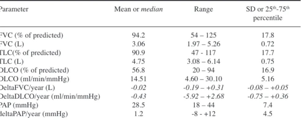
Baseline desaturation (i.e. a drop in SpO2 of at least 4% during the exercise test) occurred in 6 patients, and during follow-up, it was detected in further 8 patients. The degree of desaturation Table II. Summary of the most important resting lung function and pulmonary arterial pressure values at baseline, and the change of selected variables.
Parameter Mean or median Range SD or 25th-75th
percentile
FVC (% of predicted) 94.2 54 – 125 17.8
FVC (L) 3.06 1.97 – 5.26 0.72
TLC(% of predicted) 90.9 47 - 117 17.7
TLC (L) 4.75 3.08 – 6.14 0.75
DLCO (% of predicted) 56.8 20 – 94 16.9
DLCO (ml/min/mmHg) 14.51 4.60 – 30.10 5.16
DeltaFVC/year (L) -0.02 -0.19 – +0.31 -0.08 – +0.05
DeltaDLCO/year (ml/min/mmHg) -0.43 -5.92 – +2.68 -0.75 – +0.36
PAP (mmHg) 28.5 18 – 44 7.4
deltaPAP/year (mmHg) 1.2 -8 - +12 4.5
FVC: forced vital capacity; DLCO: diffusion capacity of carbon monoxide; TLC: total lung capac- ity; PAP: pulmonary arterial pressure; L: litre. Delta values indicate changes from the first to the last measurement.
Values are presented as mean, range and SD (normal distribution) or median range and 25th and 75th percentile (non-normal distribution – in italics).
Table III. Cardiopulmonary exercise test results.
Parameter First measurement Last measurement Annual
change
WR 101.4 46-187 98.52 62-189 -0.09
VO2 (L/min) 1.34 0.5-2.4 1.47 0.84-2.81 0.03
VCO2 (L/min ) 1.485 0.45-2.53 1.58 0.83-2.79 0.04
VO2/kg(ml/min/kg) 19.64 9-5-36.5 20.2 10.7-33.1 0.43
VE(L/min) 51.42 17.6-85.6 48.3 28.1-83.5 -0.74
VE/VCO2 33.24 26-42 29.43 21-44 -0.85*
AT (L/min) 1.00 0.58-1.91 0.92 0.46-1.67 -0.02
AT (% of VO2max,pred) 47.96 25-77 46.04 25-69 -0.39
DeltaSatmax (Decrease in SpO2) (%) 4.5 1-12 5 1-12 0.12
Desaturation (n) 6 14
WR: workrate; VO2: aerobic capacity; VO2/kg: specific aerobic capacity; VCO2: CO2 output; VE:
minute ventilation; VE/VCO2: respiratory equivalent for CO2output; AT: anaerobic threshold (L/min), AT% (% of predicted VO2max); SpO2: haemoglobin O2 saturation measured by pulse-oximetry. De- crease in SpO2 indicates the degree of desaturation, whereas Desaturation denotes the number of pa- tients in whom desaturation (decrease in SpO2 ≥ 4%) occurred ever during follow-up. Comparison of values at first and last visits. Numbers indicate mean (first and third column) and range (second and fourth column) or absolute number of involved patients.
*p<0.05 between first and last visit results.
(mean change in SatO2 during CPET) did not change significantly during follow-up. Anaerobic threshold values were close to normal both at baseline and follow-up. Average changes in the different variables were relatively small indicating relatively stable cardiopul- monary function during exercise at the group level. Significant decrease in the VE/VCO2 values during follow-up re- vealed improved ventilator efficiency.
In order to confirm the clinical validity of CPET in our SSc cohort, first we cor- related the values of its parameters at enrolment with the resting lung function test results measured at the same time.
Significant correlations (p<0.05) are presented in Table IV (second column).
Many of the baseline CPET param- eters correlated with baseline DLCO (in most instances with both absolute values expressed in ml/min/mmHg and
% of predicted values), a gold-standard parameter most commonly used during the follow-up of SSc patients to screen for and monitor pulmonary parenchy- mal or vascular progression. As it can be seen, WR, VO2, VO2/kg, VE, AT and AT% all correlated positively, whereas VE/VCO2 negatively with DLCO, indi- cating that CPET is a feasible and valid examination also in SSc patients, cor- relating well with the well-established measurement method of gas exchange.
WR, VO2, VO2/kg, VE and AT also pos- itively correlated with FVC.
Next, we assessed the correlation be- tween baseline CPET variables and several scleroderma-specific clinical outcome variables, as our aim was to examine whether CPET performed at the diagnosis of SSc may identify dis- ease parameters that require special focus during follow-up. Demographic data, clinical parameters included in the composite end-point, and the annual decrease in DLCO, FVC and desatura- tion were analysed, the latter three were selected because they are of special im- portance with regard to disease progres- sion (2). These data are also presented in Table IV (third column). Lower AT% and higher DeltaSatmax correlated with the development of digital ulcers.
Higher VE/VO2 values were in asso- ciation with a higher annual increase in PAP. A higher decline in FVC during
follow up correlated with higher VO2
and lower VE/VCO2 values at baseline.
Furthermore, the presence of desatura- tion at baseline also positively correlat- ed with the subsequent annual change (increase) in the degree of desaturation (r=+0.57, p<0.05, data not shown). Not only the baseline value of desatura- tion (DeltaSatmax), but also its annual change (i.e. increase) correlated with the annual change in DLCO (p<0.05, data not shown). Finally, age correlated negatively (data not shown), whereas disease duration positively with several CPET parameters.
As mentioned before, the complexity of CPET offers the opportunity to use it as a general predictor of poor prognosis is SSc. We therefore compared the two
patient groups separated as to whether the patients met the serious outcome endpoint (endpoint+, n=16) or did not meet it (endpoint-, n=13). Selected pa- rameters are presented in Table V. None of the clinical variables were different among the two groups, and although the dcSSc subset and patients with ILD were apparently represented in higher prevalence in the endpoint+ group, not even this comparison was statistically significant. PAP was also comparable in the two patient groups. Similarly, none of the resting lung function test vari- ables either as measured at enrolment, nor the change in any of the parameters, were found to differentiate patients with poor outcome from those with a better prognosis. In contrast, desatu- Table IV. Correlations of CPET parameters at enrolment with those of resting lung function tests and with standard follow-up parameters of SSc.
Results of the Pearson’s correlation tests with baseline CPET results.
CPET parameters Resting lung function Standard follow-up parameters Correlation at enrolment parameter at enrolment (including changes in resting (Pearson’s r)
lung function parameters) (p<0.05)
WR DLCO (% pred) +0.60
DLCO (ml/min/mmHg) +0.77
FVC (L) +0.76
VO2 DLCO (% pred) + 0.52
DLCO (ml/min/mmHg) +0.76
FVC (L) +0.72
DeltaFVC/year (%pred) - 0.43 DeltaFVC/year (L) -0.42
VO2/kg FVC (L) +0.33
DLCO (ml/min/mmHg) +0.46
VE DLCO (%pred) +0.45
DLCO (ml/min/mmHg) +0.58
FVC (L) +0.58
AT DLCO (%pred) +0.40
DLCO (ml/min/mmHg); +0.61
FVC +0.44
duration of Raynaud’s phenomenon +0.43
AT% DLCO (%pred) +0.42
duration of Raynaud’s phenomenon +0.41 digital ulcers -0.44
VE/VO2 TLC (L) -0.46
DeltaPAP/year +0.73
VE/VCO2 DLCO (%pred) -0.43
DLCO (ml/min/mmHg) -0.50
VE/VCO2 DeltaFVC/year (%pred) +0.58
DeltaFVC/year (L) +0.57
DeltaSatmax digital ulcers +0.53
For the explanation of the abbreviations, please refer to Materials and methods section, or the legends to Tables II and III. In the fourth column + Pearson’s r values mean positive correlation, whereas – means inverse correlation.
p<0.05 for all correlations presented in the Table.
ration occurred less frequently in the patients who did not meet the endpoint (endpoint- patients) (p<0.05 with chi2 test, odds ratio: 5.63 [CI: 1.08-29.38]).
The mean maximal desaturation at any CPET examination (i.e., the highest SpO2 reduction value during exercise) was actually higher in the endpoint- pa- tients than in the endpoint+ group, but this is counterbalanced by the fact that the number of patients who experienced desaturation was lower in the endpoint- group (4 of 13 vs. 10 of 14). Logistic regression of baseline CPET values did not identify any CPET parameter to in- dicate a significantly higher likelihood of the development of endpoint, but the annual change in desaturation dis- played a near-significant risk (p=0.07).
We also determined the specificity and sensitivity of the baseline CPET param- eters to differentiate patients in whom a subsequent endpoint will evolve with ROC analysis, but none of the calculat- ed cut-off values provided a statistically significant discriminative power.
Since the CPET values displayed a significant variation within the cohort, including a considerable number of patients with markedly increased and decreased values, resulting in near- normal mean values, we supposed that this may confound meaningful changes.
In order to eliminate this problem, we
have identified patients from the cohort with severely or moderately abnormal CPET results and compared them with the rest of the cohort. For this purpose, two groups were separated: 1.) patients with at least two measurement values below the 25th percentile of WR, VO2, VO2/kg, AT (L/min) or AT% at inclu- sion (severely abnormal – n=4), and 2.) patients with at least three results below the 50th percentile of the above- mentioned parameters (moderately ab- normal – n=10). Comparisons of these groups with the respective remaining parts of the cohort, however, revealed only one significant difference: the degree of desaturation/year proved to be on average negative (decreasing) in the patients with severely abnormal baseline CPET values (-1.0 [-1.2-0%]), whereas it increased in the rest of the cohort (1.15 [0.76-2.0]) (p=0.01). Age, disease duration, and poor prognostic values, including ILD, digital ulcer, need for immunosuppressive therapy and the composite endpoint were not different in any of the comparisons.
Finally, our data indicate that resting lung function parameters reflecting the lung parenchymal involvement (TLC, FVC) on average do not display a no- ticeable change during follow-up in our cohort, and therefore the moderate decrease of DLCO at baseline (mean:
57%) suggests that we should focus on pulmonary vascular involvement when assessing the value of CPET. We therefore separated the patients with either an increase of >3 mmHg in PAP or >2% decrease in DLCO annually (n=12), and compared their CPET re- sults with the remaining patients. This comparison also revealed one remark- able difference: VCO2 at baseline was markedly lower in the group of patients in whom these parameters subsequently worsened more rapidly (mean: 1.29 vs.
1.66, p=0.06), indicating that CO2 out- put might as well be a useful predictor of pulmonary vascular insufficiency.
Discussion
An unmet need in the management of SSc patients is the accurate early iden- tification of patients with rapid disease progression and with a high risk of ir- reversible organ damage and a conse- quent poor prognosis. The diagnostic delay is still rather long in scleroderma, and therefore early signs of lung, heart or vascular involvement are detectable in many patients at diagnosis. However, the identification of patients for exam- ple with progressive ILD – in contrast to those, in whom the initial interstitial lung changes remain relatively stable – typically requires repeated assessments of resting lung function, of the extent of involvement with HRCT, and of other clinical variables such as Rodnan skin score. Biomarkers (28-32) or 18 fluoro-desoxy-glucose-positron-emis- sion tomography (33) have also been tested for such purposes, but informa- tion is still limited (34, 35).
In the present study, we did not wish to focus on specific organ involvements of SSc, but rather we intended to ex- ploit the complexity of the evaluation of CPET, and hypothesised that it may thus provide prognostic information about multiple adverse disease out- comes. We have, therefore designed a composite end-point in which the indi- vidual parameters reflect the value of CPET to inform about the full spectrum of aerobic metabolism starting from oxygen uptake (deltaDLCO) through pulmonary and systemic circulation to tissue oxygenation (digital ulcers).
Our end-point is somewhat arbitrary, Table V. Comparison of selected variables between the patient subgroups who met and who
did not meet the composite end-point of unfavourable outcome.
Endpoint + (n=16) Endpoint – (n=13)
Age 48.56 (16-64) 50.69 (29-62)
dcSSc/lcSSc 10/6 5/8
ILD 11 (69%) 5 (39%)
Digital ulcer 7 (44%) 5 (39%)
Calcinosis 2 (6%) 1 (8%)
PAP 28.82 (18-44) 28.22 (24-35)
DeltaPAP/year 1.675 (-8 - 12) 0.6083 (-0.4-2)
DLCO 58 (31-94) 55 (20-79)
WR 107.6 (46-187) 93.17 (66-126)
VO2 (L/min) 1.45 (0.5-2.40) 1.21 (0.96-1.69)
VCO2 (L/min ) 1.54 (0.45-2.53) 1.43 (1.10-1.85) VO2/kg (ml/min/kg) 19.47 (9.5-28.2) 19.85 (14.2-36.5)
VE (L/min) 52.19 (17.6-85.6) 50.4 (35.8-81)
VE/VCO2 32.73 (28-41) 33.8 (26-42)
AT (L/min) 1.01 (0.58-1.56) 0.99 (0.65-1.91)
AT (% of VO2max,pred) 47.07 (27-66) 49.00 (37-76)
Decrease in SpO2 (%) 5.3 (3-12) 11.75 (7-20)*
Desaturation (n) 10 4*
DeltaDesat/year (SpO2%) 1.07 (-1 – 2.5) 0.80 (-1.4-6.6) Numbers indicate mean (range) or absolute number (percentage).
*p<0.05; p=0.07.
but composite end-points are not at all unique in SSc research. Several stud- ies have used combinations of death, event-free survival (the lack of major cardiovascular or pulmonary or renal complications), worsening in the Scle- roderma-HAQ functional index, time until major organ damage, or initia- tion of parenteral prostanoid therapy or long-term oxygen therapy due to wors- ening of PAH as components of com- posite end-points (36-39). In addition, our outcome measures have widely been used as single end-points in SSc studies too (40-44). We have found that the detection of desaturation at the first CPET examination, at the beginning of the follow-up indicates worse overall prognosis, i.e. the development of the composite end-point. Higher degree of desaturation at onset also predicted the development of digital ulcers, as well as a more rapid worsening of DLCO, and a faster subsequent increase in desatu- ration at exercise, rendering the meas- urement of oxygen saturation as a good indicator of the development of wors- ening exercise capacity and cardiovas- cular function. Patients with multiple and marked abnormalities in CPET (in particular, WR, VO2, VO2/kg and AT) at enrolment also have a higher likelihood to develop exercise-induced severe de- saturation during follow-up. VCO2, AT and VE/VO2 also appear as useful pre- dictors of selected outcome measures:
low VCO2 appeared as an indirect indi- cator of the development of pulmonary vascular insufficiency. Low AT at base- line correlated with the development of digital ulcers and high VE/VO2 with that of pulmonary hypertension.
CPET parameters also correlated well with many of the standard resting as- sessment tools, indicating that this test is feasible in SSc patients too. It is im- portant to note that CPET provides a general overview about the state of the aerobic metabolism, and is influenced by ventilation, alveolo-capillary gas- exchange, systemic and pulmonary cir- culation, cardiac output, microvascular capillary structure and flow and the state of intracellular oxidative processes. Al- though some CPET parameters can be more or less associated with specific organ dysfunction (23), it is considered
more useful to predict survival or gen- eral function, as it has been shown in lung-transplant patients (7) or chronic obstructive pulmonary disease (6). This is the reason why we have constructed a composite endpoint of frequent and widely-used cardiovascular or pulmo- nary complications or death.
Our study indicated that CPET can pro- vide additional information to identify SSc patients in whom the disease could run a progressive course and a poor prognosis may develop. If SSc patients display desaturation during exercise, they have a higher likelihood to experi- ence progressive ILD, PH or peripheral vascular lesions. Importantly, standard resting lung function and echocardio- graphic parameters were insufficient to predict the development of such unfa- vourable outcome. This makes CPET a promising non-invasive examination method to estimate the progression of SSc.
Several clinical parameters, includ- ing diffuse cutaneous subset, various autoantibody-positivities, the presence of ILD or increased PAP at presenta- tion have been proven to be helpful in identifying patients who need a closer follow-up and more aggressive therapy (1, 2, 4, 25-27). The value of decreased or decreasing DLCO or FVC and el- evated or progressively increasing PAP to select patients at risk of high morbid- ity or mortality has also been convinc- ingly confirmed and regular measure- ment of these parameters is strongly advocated during the follow-up of SSc patients (25, 26, 45). Our cohort in- cluded consecutive SSc patients with heterogeneous, but often relatively mild clinical presentation, and patients with advanced cardiopulmonary or other organ damage were excluded. Most of the patients had relatively short disease duration since the first non-Raynaud symptom. Although the follow-up time was acceptably long, these factors, to- gether with the fact that the cohort is not as high as those in whom the above- mentioned predictive factors were identified, may explain why we did not find the standard clinical and lung function tests or echocardiography to show a significant correlation with the occurrence of the serious outcome end-
point. With these limitations in mind, it is even more noteworthy that vari- ous CPET parameters, being strongly linked to oxygen utilisation, were able to discriminate between patients with or without endpoint (desaturation during exercise), or to correlate with adverse clinical outcomes, such as a more rapid decline in FVC or increase in PAP, or the development of digital ulcers (VO2, VE/VCO2, VE/VO2, desaturation and anaerobic threshold).
There is a number of studies that have previously already addressed the value of CPET in SSc (11-22). Unlike our investigation, these studies have ad- dressed almost exclusively two is- sues: First, they have searched whether CPET can help to discriminate among causes of reduced exercise capacity in SSc, and found certain CPET param- eters to be associated with pulmonary vasculopathy, left ventricular dysfunc- tion or respiratory limitation, respec- tively (12, 13, 15, 16), in some cases even in those patients in whom resting lung function tests and echocardiog- raphy failed to disclose such evident causes. The other area of research was to assess whether CPET may aid in the detection of PAP in SSc patients (11, 14, 18, 19). Dumitrescu et al. have pro- spectively analysed 173 SSc patients, in whom PAP was verified by right heart catheterisation, and found peakVO2 and VE/VCO2 to show the highest correla- tions with PAP, transpulmonary pres- sure gradient and pulmonary vascular resistance (11). Of several parameters with high sensitivity and specificity for PH detection, peakVO2 showed highest diagnostic accuracy (sensitivity 87.5%, specificity 74.8% at a threshold level of 13.8 mL/min/kg). These investiga- tions confirm that CPET is a feasible examination in SSc patients too, simi- larly to chronic lung disease patients.
However, the above-cited publications concluded that although CPET adds certain benefits during the work-up of dyspnea or fatigue in SSc, the standard assessment techniques, such as HRCT, resting echocardiography or right heart catheterisation for the verification of the particular cardiopulmonary organ manifestations are still not replaced by CPET. Since the highest value of CPET
is that it provides a comprehensive overview about oxygen utilisation from the alveolo-capillary gas exchange to cellular metabolism, we have decided to set up endpoints that are the most relevant to patient mortality and mor- bidity, rather than to analyse individual organ involvements. Indeed, data about the value of CPET to assess functional outcome or survival in SSc patients is rather limited. We have found two publications about the association of between composite functional indices and CPET: Cuomo et al. have found that VO2/kg was the only parameter that showed a significant correlation between the HAQ-DI functional index and various CPET parameters (20), whereas in another study, VE/VCO2
correlated positively with both the Dis- ease Activity Index and the Disease Se- verity Scale (21). One further study ex- amined the connection between CPET and patient survival. Similarly to our results, Swigris et al. also demonstrated that desaturation (a drop in SpO2max below 89% or >4 percentage points) increased the risk of death during a me- dian follow-up of 7.1 years, but it has to be noted that this study focused exclu- sively on SSc patients with interstitial lung disease, and it was a retrospective study (22). The recently published pro- spective follow-up study on the patients from the PHAROS registry, although it did not apply CPET, also confirmed that desaturation on exercise (6 minute walking test) is a predictor of mortality in SSc (46).
Our study has some weaknesses and strengths. The patient number is not as high as in many previous examina- tions especially in multicentre cohorts, but still it was sufficient for the iden- tification of some statistically signifi- cant findings. We did not include right heart catheterisation in the protocol to corroborate the measurement of pul- monary arterial pressure with this most accurate method because, as we men- tioned, our primary aim was not to fo- cus on PH, and because most of the pa- tients had relatively early disease with no significant risk factors for PH, and we decided that right heart catheteri- sation was not indicated and ethically established. However, we did perform
right heart catheterisation in patients in whom the estimated PAP was >50 mmHg, and there was only one such patient in this cohort. A strength of the investigation is that to our knowledge, this is the first study with prospective follow-up of SSc patients and repeat- ed CPET examinations, so we could provide data about the time-course of CPET parameters during several years of follow-up in SSc. Unlike most of the previous studies, we did not selectively analyse various organ manifestations in SSc, but concentrated on the most cru- cial outcomes of SSc, and we believe that such approach could help to cor- rectly define the place of CPET in the care of SSc patients: to predict the ap- pearance of complications that are re- lated to oxygen-dependent processes, from tissue necrosis and PAP to patient survival.
References
1. RUBIO-RIVAS M, ROYO C, SIMEÓN CP, COR- BELLA X, FONOLLOSA V: Mortality and survival in systemic sclerosis: systematic review and meta-analysis. Semin Arthritis Rheum 2014; 44: 208-19.
2. POKEERBUX MR, GIOVANNELLI J, DAU- CHET L et al.: Survival and prognosis factors in systemic sclerosis: data of a French mul- ticenter cohort, systematic review, and meta- analysis of the literature. Arthritis Res Ther 2019; 21: 86.
3. WELLS AU: High-resolution computed to- mography and scleroderma lung disease.
Rheumatology 2008; 47: v59-v61.
4. MATHAI SC, HUMMERS LK, CHAMPION HC et al.: Survival in pulmonary hypertension associated with the scleroderma spectrum of diseases: impact of interstitial lung disease.
Arthritis Rheum 2009; 60: 569-77.
5. COGHLAN JG, DENTON CP, GRÜNIG E et al.:
Evidence-based detection of pulmonary arte- rial hypertension in systemic sclerosis: the DETECT study. Ann Rheum Dis 2014; 73:
1340-9.
6. STRINGER W, MARCINIUK D: The role of cardiopulmonary exercise testing (CPET) in pulmonary rehabilitation (PR) of chronic obstructive pulmonary disease (COPD) pa- tients. COPD 2018; 15: 621-31.
7. LAYTON AM, ARMSTRONG HF, KIM HP, MEZA KS, D’OVIDIO F, ARCASOY SM: Cardi- opulmonary exercise factors predict survival in patients with advanced interstitial lung disease referred for lung transplantation.
Respir Med 2017; 126: 59-67.
8. DUDLEY KA, EL-CHEMALY S: Cardiopulmo- nary exercise testing in lung transplantation:
A review 2012. Pulm Med 2012: 237852 9. PTASZYŃSKA-KOPCZYŃSKA K, KREN-
TOWSKA A, SAWICKA E et al.: The strengths and weaknesses of non-invasive parameters obtained by echocardiography and cardio-
pulmonary exercise testing in comparison with the hemodynamic assessment by the right heart catheterization in patients with pulmonary hypertension. Adv Med Sci 2017;
62: 39-44.
10. ZHAO QH, WANG L, PUDASAINI B et al.:
Cardiopulmonary exercise testing improves diagnostic specificity in patients with echo- cardiography-suspected pulmonary hyper- tension. Clin Cardiol 2017; 40: 95-101.
11. DUMITRESCU D, NAGEL C, KOVACS G et al.:
Cardiopulmonary exercise testing for detect- ing pulmonary arterial hypertension in sys- temic sclerosis. Heart 2017; 103: 774-82.
12. BOUTOU AK, PITSIOU GG, SIAKKA Pet al.:
Phenotyping exercise limitation in systemic sclerosis: the use of cardiopulmonary exer- cise testing. Respiration 2016; 91: 115-23.
13. MARTIS N, QUEYREL-MORANNE V, LAU- NAY Det al.: Limited Exercise Capacity in Patients with Systemic Sclerosis: Identifying Contributing Factors with Cardiopulmonary Exercise Testing. J Rheumatol 2018; 45: 95- 14. 102.NINABER M, HAMERSMA W, SCHUERWEGH
A, KOVACS G, OLSCHEWSKI H, STOLK J: Oxygen pulse slope analysis during exercise testing identifies patients with systemic scle- rosis at a possible risk for developing pulmo- nary vasculopathy. Eur Respir J 2013; 42:
P3969.
15. TZILAS V, BOUROS D: Cardiopulmonary ex- ercise testing in systemic sclerosis: ‘Ars lon- ga, vita brevis’. Respiration 2016; 91: 202-3.
16. WALKEY AJ, IEONG M, ALIKHAN M, FARBER HW: Cardiopulmonary exercise testing with right-heart catheterization in patients with systemic sclerosis. J Rheumatol 2010; 37:
1871-7.
17. SCHWAIBLMAIR M, BEHR J, FRUHMANN G: Cardiorespiratory responses to incremental exercise in patients with systemic sclerosis.
Chest 1996; 110: 1520-5.
18. DUMITRESCU D, OUDIZ RJ, KARPOUZAS G et al.: Developing pulmonary vasculopathy in systemic sclerosis, detected with non- invasive cardiopulmonary exercise testing.
PLoS One 2010; 5: e14293.
19. KOVACS G, MAIER R, ABERER E et al.:
Assessment of pulmonary arterial pressure during exercise in collagen vascular disease:
echocardiography vs right-sided heart cath- eterization. Chest 2010; 138: 270-8.
20. CUOMO G, SANTORIELLO C, POLVERINO F, RUOCCO L, VALENTINI G, POLVERINO M: Impaired exercise performance in systemic sclerosis and its clinical correlations. Scand J Rheumatol 2010; 39: 330-5.
21. ROSATO E, ROMANIELLO A, MAGRÌ D et al.: Exercise tolerance in systemic sclero- sis patients without pulmonary impairment:
correlation with clinical variables. Clin Exp Rheumatol 2014; 32 (Suppl. 86): S103-8.
22. SWIGRIS JJ, ZHOU X, WAMBOLDT FS et al.: Exercise peripheral oxygen saturation (SpO2) accurately reflects arterial oxygen saturation (SaO2) and predicts mortality in systemic sclerosis. Thorax 2009; 64: 626-30.
23. ALBOUAINI K, EGRED M, ALAHMAR A, WRIGHT DJ: Cardiopulmonary exercise testing and its application. Postgrad Med J 2007; 83: 675-82.
24. vanden HOOGEN F, KHANNA D, FRANSEN J et al.: 2013 classification criteria for system- ic sclerosis: an American College of Rheu- matology/European League against Rheuma- tism collaborative initiative. Ann Rheum Dis 2013; 72: 1747-55.
25. SMITH V, SCIRÈ CA, TALARICO R et al.:
Systemic sclerosis: state of the art on clini- cal practice guidelines. RMD Open 2019; 4:
e000782.
26. KOWAL-BIELECKA O, FRANSEN J, AVOUAC J et al.: Update of EULAR recommendations for the treatment of systemic sclerosis. Ann Rheum Dis 2017; 76: 1327-39.
27. HANKINSON JL, ODENCRANTZ JR, FEDAN KB: Spirometric reference values from a sample of the general U.S. population. Am J Respir Crit Care Med 1999; 159: 179-87.
28. DE LAURETIS A, SESTINI P, PANTELIDIS P et al.: Serum interleukin 6 is predictive of early functional decline and mortality in interstitial lung disease associated with systemic sclero- sis. J Rheumatol 2013; 40: 435-46.
29. CARULLI MT, HANDLER C, COGHLAN JG, BLACK CM, DENTON CP: Can CCL2 serum levels be used in risk stratification or to mon- itor treatment response in systemic sclerosis?
Ann Rheum Dis 2008; 67: 105-9.
30. HOFFMANN-VOLD AM, TENNØE AH, GAREN T et al.: High level of chemokine CCL18 is associated with pulmonary function deterio- ration, lung fibrosis progression, and reduced survival in systemic sclerosis. Chest 2016;
150: 299-306.
31. KUMÁNOVICS G, GÖRBE E, MINIER T, SIMON D, BERKI T, CZIRJÁK L: Follow-up of serum KL-6 lung fibrosis biomarker levels in 173 patients with systemic sclerosis. Clin Exp Rheumatol 2014; 32 (Suppl. 86): S138-44.
32. VOLKMANN ER, TASHKIN DP, ROTH MD et al.: Changes in plasma CXCL4 levels are as-
sociated with improvements in lung function in patients receiving immunosuppressive therapy for systemic sclerosis-related inter- stitial lung disease. Arthritis Res Ther 2016;
18: 305.
33. BELLANDO-RANDONE S, TARTARELLI L, CAVIGLI E et al.: 18F-fluorodeoxyglucose positron-emission tomography/CT and lung involvement in systemic sclerosis. Ann Rheum Dis 2019; 78: 577-8.
34. ORLANDI M, LEPRI G, DAMIANI A et al.: One year in review 2020: systemic sclerosis. Clin Exp Rheumatol 2020; 38 (Suppl. 125): S3- 35. 17.MERKEL PA, CLEMENTS PJ, REVEILLE JD,
SUAREZ-ALMAZOR ME, VALENTINI G, FURST D: Current status of outcome meas- ure development for clinical trials in sys- temic sclerosis. Report from OMERACT 6.
J Rheumatol 2003; 30: 1630-47.
36. SULLIVAN KM, GOLDMUNTZ EA, KEYES- ELSTEIN L et al.: SCOT Study Investigators.
Myeloablative autologous stem-cell trans- plantation for severe scleroderma. N Engl J Med 2018; 378: 35-47.
37. van LAAR JM, FARGE D, SONT JK et al.:
EBMT/EULAR Scleroderma Study Group.
Autologous hematopoietic stem cell trans- plantation vs intravenous pulse cyclophos- phamide in diffuse cutaneous systemic scle- rosis: a randomized clinical trial. JAMA 2014;
311: 2490-8.
38. WANGKAEW S, THONGWITOKOMARN H, PRASERTWITTAYAKIJ N, EUATHRONGCHIT J: Rapid skin thickness progression rate is associated with high incidence rate of car- diopulmonary complications in patients with early diffuse cutaneous systemic sclerosis:
inception cohort study. Clin Exp Rheumatol 2020; 38 (Suppl. 125): S98-105.
39. GAINE S, CHIN K, COGHLAN G et al.: Selex-
ipag for the treatment of connective tissue disease-associated pulmonary arterial hyper- tension. Eur Respir J 2017; 50: 1602493.
40. AYANO M, TSUKAMOTO H, MITOMA H et al.:
CD34-selected versus unmanipulated autolo- gous haematopoietic stem cell transplantation in the treatment of severe systemic sclerosis:
a post hoc analysis of a phase I/II clinical trial conducted in Japan. Arthritis Res Ther 2019;
21: 30-8.
41. SAUNDERS P, TSIPOURI V, KEIR GJ et al.:
Rituximab versus cyclophosphamide for the treatment of connective tissue disease-asso- ciated interstitial lung disease (RECITAL):
study protocol for a randomised controlled trial. Trials 2017; 18: 275.
42. BRATIS K, LINDHOLM A, HESSELSTRAND R et al.: CMR feature tracking in cardiac asymptomatic systemic sclerosis: Clinical implications. PLoS One 2019; 14: e0221021.
43. ROSATO E, LEODORI G, GIGANTE A, DI PAOLO M, PAONE G, PALANGE P: Reduced ventilatory effciency during exercise predicts major vascular complications and mortality for interstitial lung disease in systemic scle- rosis. Clin Exp Rheumatol 2020; 38 (Suppl.
125): S85-S91.
44. ELHAI M, BOUBAYA M, DISTLER O et al.:
Outcomes of patients with systemic sclerosis treated with rituximab in contemporary prac- tice: a prospective cohort study. Ann Rheum Dis 2019; 78: 979-87.
45. COTTIN V, BROWN KK: Interstitial lung dis- ease associated with systemic sclerosis (SSc- ILD). Respir Res 2019; 20: 13-19.
46. HSU VM, CHUNG L, HUMMERS LK et al.:
Risk factors for mortality and cardiopulmo- nary hospitalization in systemic sclerosis patients at risk for pulmonary hypertension, in the PHAROS Registry. J Rheumatol 2019;
46: 176-83.