Ideggyogy Sz 2020;73(3–4):141–144.
141 ESETISMERTETÉS
CANOMAD SYNDROME WITH RESPIRATORY FAILURE
András SALAMON1, Lívia DÉZSI1, Bence RADICS2, Edina Tímea VARGA1, Tibor HORTOBÁGYI2, Adrienn TÖMÖSVÁRI3, László VÉCSEI1, 4, Péter KLIVÉNYI1, Cecília RAJDA1
1Department of Neurology, University of Szeged, Szeged
2Department of Pathology, University of Szeged, Szeged
3Department of Anaesthesiology and Intensive Therapy, University of Szeged, Szeged
4MTA-SZTE Neuroscience Research Group, University of Szeged, Szeged
LÉGZÉSI ELÉGTELENSÉGGEL JÁRÓ CANOMAD SZINDRÓMA
Salamon A, MD; Dézsi L, MD; Radics B, MD; Varga ET, MD, PhD; Hortobágyi T, MD, PhD; Tömösvári A, MD;
Vécsei L, MD, PhD, DSc; Klivényi P, MD, PhD, DSc;
Rajda C, MD, PhD
Ideggyogy Sz 2020;73(3–4):141–144.
A CANOMAD (krónikus ataxiás neuropathia, ophthal - moplegia, M-protein-agglutináció, diszialozil-antitestek) szindróma ritka polyneuropathia, melyben IgM-para - proteinek lépnek reakcióba a diszializált epitópokat tar - talmazó gangliozidokkal. Ezen folyamat hátsó gyöki ganglionopathiához, valamint a cranialis és a perifériás idegek B-lymphocyta-mediált infiltrációjához vezet.
A betegség klinikai képét ataxia, enyhe fokú izom- gyengeség, areflexia, valamint szenzoros eltérések és agyidegtünetek dominálják. A rituximab-, valamint az intravénás immunoglobulin (IVIg-) kezelés hatékonyságát esettanulmányok támasztják alá.
Közleményünkben egy 57 éves férfi beteg járásnehe zí - tettséggel, négy végtagi zsibbadással és ügyetlenséggel járó esetét mutatjuk be. Neurológiai státuszában areflexia, vibrációérzés-csökkenés, valamint ataxia volt megfigyel- hetô. A laboratóriumi vizsgálatok a szérumban IgM monoklonális komponens és diszialozil-antitestek jelenlétét igazolták. A beteg részletes elektrofiziológiai kivizsgálása során szenzomotoros demyelinisatiós polyneuroradicu- lopathia igazolódott. Az alkalmazott IVIg- és rituximab - kezelés ellenére a beteg állapota foko zatosan romlott, majd légzési elégtelenség követ keztében elhunyt. Az elvég - zett neuropatológiai vizsgálatok hátsó kötegi és gyöki atro phiát, valamint kevert mononukleáris sejtes infiltrációt mutattak.
A jelen közlemény célja, hogy felhívja a figyelmet a szind rómára, ezáltal elôsegítse a betegek életminôségét potenciálisan javító immunszuppresszív kezelés mielôbbi beve zetését.
Kulcsszavak: CANOMAD szindróma, rituximab, légzési elégtelenség, ataxiás neuropathia CANOMAD (chronic ataxic neuropathy, ophthalmoplegia,
M-protein agglutination, disialosyl antibodies) syndrome is a rare polyneuropathy. IgM paraproteins react with gan- glioside-containing disialylated epitopes resulting in dorsal root ganglionopathy and B-lymphocyte infiltration of cra- nial and peripheral nerves. Clinical features include atax- ia, slight muscle weakness, areflexia, sensory- and cranial nerve symptoms. Case studies have reported the efficacy of rituximab and intravenous immunoglobulin (IVIg) treat- ments.
We present the case of a 57-year-old man, who had diffi- culty walking, with numbness and clumsiness in all limbs.
He had areflexia, vibratory sensation loss and ataxia.
Laboratory tests showed IgM monoclonal components and disialosyl antibodies in the serum. Nerve conduction stud- ies indicated severe sensorimotor demyelinating polyneu- roradiculopathy. Despite IVIg and rituximab treatments, the patient’s disease course gradually worsened and he died of respiratory failure. Neuropathological examination revealed dorsal column- and dorsal root atrophy with mixed mononuclear cell infiltration. This article aims to draw attention to this syndrome, and the use of early potent immunosuppressive treatment to improve patients’
quality of life.
Keywords: CANOMAD syndrome, rituximab, respiratory failure, ataxic neuropathy
Correspondent: Cecília RAJDA MD, PhD, Department of Neurology, University of Szeged;
6725 Szeged, Semmelweis u. 6. Phone: (06-62) 545-348, fax: (06-62) 545-597.
E-mail: rajda.cecilia@med.u-szeged.hu
Érkezett: 2019. december 4. Elfogadva: 2020. január 24.
| English| https://doi.org/10.18071/isz.73.0141 | www.elitmed.hu 141-144 salamon-rajna_UJ ISZ TUKOR ALAP.qxd 2020. 03. 16. 14:59 Page 141
Az alábbi dokumentumot magáncélra töltötték le az eLitMed.hu webportálról. A dokumentum felhasználása a szerzôi jog szabályozása alá esik.
142
Salamon: CANOMAD syndrome with respiratory failureC
ANOMAD (chronic ataxic neuropathy oph- thalmoplegia M-protein agglutination disialo- syl antibodies) syndrome is a rare, chronic, im - mune-mediated demyelinating polyneuropathy1. About thirty cases can be found in the literature2. The syndrome is caused by the presence of a specific IgM paraprotein, which reacts with ganglioside-con - taining disialylated NeuAc(α2-8)NeuAC(α2-3)Gal neural epitopes3. There are two main neuro pa tho lo - gical features: dorsal root ganglionopathy and a B- lymphocyte-mediated endoneurinal infiltration of cranial and peripheral nerves4, 5. The symptoms begin in the 5th-6th decade with a male predomi- nance1. Typical clinical presentation includes atax- ia, muscle weakness, areflexia, sensory symptoms, pseudoathetoid movements and cranial nerve symp- toms6. Rarely, it can be associated with respiratory failure2. The elevation of IgM protein levels in the serum is characteristic; these paraproteins are often cold agglutinins1, 7. The most frequently seen anti- disialosyl antibodies are GD1b, GD3, GT1b and GQ1b3. Electrophysiological tests show sensorimo- tor demyelinating features, with signs of axonal degeneration3. The optimal treatment of the disease is unclear. Some case studies have reported the effi- cacy of rituximab and intravenous immunoglobulin (IVIg) treatment1–3, 6, 8, 9.Case report
A 57-year-old man was admitted to our clinic in 2008, with numbness in all limbs, clumsiness and difficulty walking. The only past medical history that was significant was hypertension. Initial neuro- logical examination showed areflexia, vibratory sensation loss and four limb ataxia. Laboratory tests showed slightly elevated creatine kinase levels (210-293 IU/L; normal range: < 195 IU/L) and IgM lambda monoclonal components in the serum.
Brain and neck magnetic resonance imaging were normal. Results of multiple repeated electrophysio- logical studies were compatible with severe senso- rimotor demyelinating polyneuroradiculopathy.
The cerebrospinal fluid revealed a slightly in crea - sed total protein (0.51 g/l) and 7 lymphocytes/mm3. A detailed hematological examination excluded multiple myeloma (no severe proteinuria, lack of Bence Jones protein in the urine, no plasma cell proliferation in bone marrow, lack of osteolytic lesions). The patient would be classified as “defi- nite” CIDP as defined by EFNS/PNS diagnostic cri- teria10. Due to the lack of signs indicating motor neuron disease, needle exam was not performed early in the course of disease. During the period of
acute worsening, the critical condition of the patient did not permit the examination, which would have been informative in order to evaluate axonal loss and rule out motor neuron disease. Following the lack of effect of steroid treatment (100 mg oral prednisone daily, with a gradually decreasing dose, was associated with an inability to walk) and aza- thioprine treatment (25 mg starting dose; discontin- ued due to hepatotoxicity), IVIg treatment (0.4 g/kg/day for 5 days every 6 weeks) was initiated.
After a moderate improvement (numbness and clumsiness decreased), the patient’s disease course slowly worsened (slight all limbs paresis appeared).
Eight years after disease onset, a relapse with autonomic dysfunction, external and internal oph- thalmoparesis, hearing loss and finally severe respi- ratory failure developed. The patient was admitted to the intensive care unit (ICU), due to the need for mechanical ventilation. The patient’s neurological symptoms temporarily improved after plasma exchange and IVIg treatment, but shortly thereafter repeated respiratory insufficiency developed. In the ICU, the patient’s fingers became pale and cold, suggestive of Raynaud’s syndrome and the pres- ence of cold agglutinins. Laboratory tests showed the presence of anti-H cryoglobulins and anti-gan- glioside antibodies (positive for GD1b, GD2, GD3, and GT1b) supporting the CANOMAD syndrome.
The functions of the third, fourth and sixth cranial nerves (ptosis, gaze weakness) improved, but due to repeated severe respiratory failure, after 72 days, the patient died.
Neuropathological findings
The autopsy of our patient showed severe bron- chopneumonia as cause of death. Detailed neuro - pathological examination was performed. Rou tine sections from the brain, spinal cord, dorsal root ganglia, trigeminal ganglia, 7th cranial nerve, sural nerve, adrenals, skeletal muscles, internal organs, skin and bone marrow were analyzed (Figure 1).
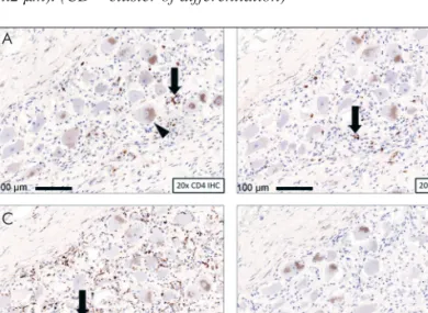
Semi-thin sections were made from the sural nerve, triceps surae muscle and parietal cortex, and stud- ied by electron microscopy. Immunohistochemistry was performed to identify helper and cytotoxic T- lymphocytes, B-lymphocytes, plasma cells and macrophages (Figure 2). Severe dorsal column and dorsal root atrophy (Figure 1.A)were found. Mild neuronal loss was observed within the dorsal root ganglia (DRG) accompanied by mixed mononu- clear cell infiltration (Figure 1.B and 2.A, B, C).
No B-lymphocytes were found in the DRG (Figure 2.D). Moderate axonal loss was identified in the
141-144 salamon-rajna_UJ ISZ TUKOR ALAP.qxd 2020. 03. 16. 15:00 Page 142
Az alábbi dokumentumot magáncélra töltötték le az eLitMed.hu webportálról. A dokumentum felhasználása a szerzôi jog szabályozása alá esik.
cross-sections of the sural nerve with no signs of acute demyelination, necrosis or vasculitis (Figure 1.C). Congo-red staining excluded local and sys- temic amyloidosis as the cause of neuropathy. No ultrastructural abnormalities were found on the myelin-sheaths. Skeletal muscles exhibited global, but not typical selective type II atrophy (Figure 1.D). Histological examination excluded neurode- generative diseases of the brain. Neither hema to - poietic or lymphoid tumor, nor systemic amyloido- sis was found.
Discussion and conclusion
Earlier described CANOMAD cases reported pa - tients with optic nerve involvement, extramem - branous glomerulonephritis, and a syndrome of inappropriate antidiuretic hormone secretion (SIADH)2, 11–14. Cases without ophthalmoplegia, fur thermore, with temporary respiratory failure associated with facial involuntary movements were also mentioned2, 11–14. We report a case of CANO - MAD syndrome with severe respiratory failure fol- lowed by neuropathological evaluation. In another case described in the literature, acute respiratory failure was resolved with IVIg treatment2. Despite the stabilization of symptoms after IVIg administra- tion in our patient, persistent severe respiratory fail- ure required additional therapeutic steps (ritux- imab). One month after the rituximab treatment, the cranial nerve symptoms were partially resolved.
The absence of B-lymphocytic infiltration in the peripheral nerves suggests that rituximab may be effective at the histological level. We assume that the patient’s death could be explained by the long disease course, and the long stay in ICU, complicat- ed with infections.
In summary, CANOMAD syndrome is a rare sensorimotor, slowly developing polyneuropathy, which predominantly affects middle-aged men and it can be fatal. In the case of a patient with immune- mediated neuropathy, cranial nerve symptoms and Raynaud phenomenon, the physician should con- sider CANOMAD syndrome. A quick, precise diagnosis allows more rapid initiation of potent immunosuppressive treatment.
ACKNOWLEDGEMENTS
Funding of the studies reported in the paper:
GINOP-2.3.2.-15.2016-00034.
CONFLICT OF INTEREST
None of the authors has any conflict of interest to disclose.
Ideggyogy Sz 2020;73(3–4):141–144.
143
Figure 1. Neuropathological features of CANOMAD syndrome.
ALight microscope image from the spinal cord. Note the general atrophy of the dorsal column (arrow) in the transverse section of the sacral spinal cord. Loss of myelinated fibers results in faint staining of the gracile fascicle, while the pyramidal tract (arrow- head) is preserved. (Luxol Fast Blue stain + cresyl-violet, magni- fication 2.5x).BMononuclear cell infiltrate (arrow) in the center of a dorsal root ganglion, accompanied by focal loss of ganglion cells. Preserved ganglion cells (arrowhead) are visible around the inflammatory infiltrate. (HE stain, 20x magnifictaion). CRegular myelin sheath (arrow) around an axon from the sural nerve. No immunocomplex deposits, amyloid fibrils or structural alterations of the myelin sheath were found (x200 nm). DElectron micro - scopy of the skeletal (intercostal) muscle – global muscle atrophy (x2 µm). (CD – cluster of differentiation)
Figure 2. Mixed mononuclear infiltration of the dorsal root gang- lia. ACD4+ immunohistochemistry reveals helper T-lymphocytes (arrow). Brown granules in the ganglion cells are intracytoplas- mic melanin and lipofuscin pigments (arrowhead). BCD8+ im mu - nohistochemistry shows cytotoxic T-lymphocytes (arrow). CMost of the mononuclear cells are CD68+ macrophages (arrow). D CD20+ B-lymphocytes were not found. (CD – cluster of differen- tiation)
A B
C D
A B
C D
141-144 salamon-rajna_UJ ISZ TUKOR ALAP.qxd 2020. 03. 16. 15:00 Page 143
Az alábbi dokumentumot magáncélra töltötték le az eLitMed.hu webportálról. A dokumentum felhasználása a szerzôi jog szabályozása alá esik.
144
Salamon: CANOMAD syndrome with respiratory failureREFERENCES
1.Garcia-Santibanez R, Zaidman CM, Sommerville RB, et al.
CANOMAD and other chronic ataxic neuropathies with disialosyl antibodies (CANDA). J Neurol 2018;265:1402-9.
https://doi.org/10.1007/s00415-018-8853-4
2.Johnson K, Malkan A, Shaffi M. Facial involuntary move- ments and respiratory failure in CANOMAD, responsive to IVIg therapy. Case Rep Med 2015;170543.
https://doi.org/10.1155/2015/170543
3.Willison HJ, O’Leary CP, Veitch J, et al. The clinical and laboratory features of chronic sensory ataxic neuropathy with anti-disialosyl IgM antibodies. Brain 2001;124:1968-77.
https://doi.org/10.1093/brain/124.10.1968
4.McKelvie PA, Gates PC, Day T. Canomad: report of a case with a 40-year history and autopsy. Is this a sensory gang- lionopathy with neuromuscular junction blockade? Muscle Nerve 2013;48:599-603.
https://doi.org/10.1002/mus.23897
5.Obi T, Murakami T, Takatsu M, et al.Clinicopathological study of an autopsy case with sensory-dominant polyradi- culoneuropathy with antiganglioside antibodies. Muscle Nerve 1999;22:1426-31.
https://doi.org/10.1002/(sici)1097-4598(199910)22:10<
1426: :aid-mus13>3.0.co;2-h
6.Krenn M, Keir G, Wieshmann UC.CANOMAD respon- ding to weekly treatment with intravenous immunoglobulin (IVIg). BMJ Case Rep 2014;2014.
https://doi.org/10.1136/bcr-2013-202545
7.Jacobs BC, O’Hanlon GM, Breedland EG, Veitch J, van Doorn PA, Willison HJ. Human IgM paraproteins de - monstrate shared reactivity between Campylobacter jejuni lipopolysaccharides and human peripherial nerve disialyla- ted gangliosides. J Neuroimmunol 1997;80:23-30.
https://doi.org/10.1016/s0165-5728(97)00130-6
8.Löscher WN, Woertz A, Wallnöfer M, Wanschitz JV, Luef G.Successful treatment of CANOMAD with IVIg and ritu- ximab. J Neurol 2013;260:1168-70.
https://doi.org/10.1007/s00415-013-6867-5
9.Delmont E, Jeandel PY, Hubert AM, Marcq L, Boucraut J, Desnuelle C. Successful treatment with rituximab of one patient with CANOMAD neuropathy. J Neurol 2010;257:
655-7. https://doi.org/10.1007/s00415-009-5412-z 10.Van den Bergh PY, Hadden RD, Bouche P, et al. European
Federation of Neurological Societies/Peripheral Nerve Society guideline on management of chronic inflammatory demyelinating polyradiculoneuropathy: report of a joint task force of the European Federation of Neurological Societies and the Peripheral Nerve Society - first revision.
Eur J Neurol 2010;17:356-63.
https://doi.org/10.1111/j.1529-8027.2010.00245.x 11.Iorio R, Capone F, Iannaccone E, et al.SIADH in a pa tient
with sensory ataxic neuropathy with anti-disialosyl anti - bodies (CANOMAD). J Neurol 2009;256:1177-9.
https://doi.org/10.1007/s00415-009-5071-0
12.Sanvito L, Rajabally YA.Optic neuropathy associated with CANOMAD: description of 2 cases. Muscle Nerves 2011;
44:451-5.
https://doi.org/10.1002/mus.22157
13.Kam C, Balaratnam MS, Purves A, et al. Canomad presen- ting without ophthalmoplegia and responding to intraveno- us immunoglobulin. Muscle Nerve 2011;44:829-33.
https://doi.org/10.1002/mus.22167
14.Delval A, Stojkovic T, Vermersch P. Relapsing sensorimo- tor neuropathy with ophthalmoplegia, antidisialosyl antibo- dies, and extramembranous glomerulonephritis. Muscle Nerve 2006;33:274-7.
https://doi.org/10.1002/mus.20452 141-144 salamon-rajna_UJ ISZ TUKOR ALAP.qxd 2020. 03. 16. 15:00 Page 144
Az alábbi dokumentumot magáncélra töltötték le az eLitMed.hu webportálról. A dokumentum felhasználása a szerzôi jog szabályozása alá esik.