HIGH INCIDENCE OF MULTIRESISTANT BACTERIAL ISOLATES FROM BLOODSTREAM
INFECTIONS IN TRAUMA EMERGENCY DEPARTMENT AND INTENSIVE CARE UNIT
IN SERBIA
OLIVERADJURIC1,2*, LJILJANAMARKOVIC-DENIC1,2, BOJANJOVANOVIC1,3and VESNABUMBASIREVIC1,3
1Faculty of Medicine, University of Belgrade, Belgrade, Serbia
2Institute of Epidemiology, Faculty of Medicine, University of Belgrade, Belgrade, Serbia
3Center for Anaesthesiology, Emergency Center, Clinical Center of Serbia, Belgrade, Serbia
(Received: 23 November 2018; accepted: 18 December 2018)
We investigated the incidence of bloodstream infections (BSIs) in trauma emergency department (ED) and intensive care unit (ICU), to assess ED- and ICU- related predictors of BSI and to describe the most common bacteria causing BSI and their antimicrobial resistance markers. A prospective study was conducted in two trauma ICUs of the ED of Clinical Center of Serbia. Overall, 62 BSIs were diagnosed in 406 patients, of which 13 were catheter-related BSI (3.0/1,000 CVC-days) and 30 BSIs of unknown origin, while 15% were attributed to ED CVC exposure. Lactate
≥2 mmol/L and SOFA score were independent ED-related predictors of BSI, while CVC in place for>7 days and mechanical ventilation>7 days were significant ICU- related predictors. The most common bacteria recovered wereAcinetobacter spp., Klebsiella spp., and Pseudomonas aeruginosa. All Staphylococcus aureus and coagulase-negative staphylococci isolates were methicillin-resistant, whereas 66% of Enterococcus spp. were vancomycin-resistant. All isolates of Enterobacteriaceae were resistant to third-generation cephalosporins, whereas 87.5% ofP. aeruginosaand 95.8% of Acinetobacterspp. isolates were resistant to carbapenems. ED BSI con- tributes substantially to overall ICU incidence of BSI. Lactate level and SOFA score can help to identify patients with higher risk of developing BSI. Better overall and CVC-specific control measures in patients with trauma are needed.
Keywords: trauma, bloodstream infections, antimicrobial resistance, emergency department, intensive care unit
*Corresponding author; E-mail:oliveradjuric87@gmail.com
First published online February 21, 2019
Introduction
Healthcare-associated bloodstream infections (HA-BSIs) are considered as major preventable threat to healthcare and patient safety. They approximately affect 4% of all critically ill patients and are the second most common healthcare- associated infection (HAI) among patients staying more than 48 h in intensive care unit (ICU) [1]. Moreover, HA-BSI is independently associated with increased risk of both short- and long-term mortality in critically ill patients [2, 3].
Patients with trauma represent considerable part of patients admitted in emergency department (ED) since the advances in initial resuscitation of critically injured patients increased survival after the injury. However, those who survive initial trauma are at particular risk of developing BSI over the course of ED and ICU treatment, which is enabled by exposure to numerous invasive devices and facilitated by disrupted barriers and altered immune response [4]. Consequently, BSI is two times more frequent in patients with trauma compared to surgical patients and after ventilator-associated pneumonia, it is the second most frequent HAI in patients with trauma [5, 6]. Yet, surveillance studies and preventative measures usually target BSI in ICU inpatients and little is known about contribution of ED in occurrence of BSI.
In Serbia, trauma represents the third cause of mortality and every tenth person dies from injury [7]. Despite this, there is neither in depth data on incidence of BSI in patients with trauma in both ED or ICU department in Serbia nor the countries of the Southeastern–European Region.
In critically ill patients, healthcare-acquired BSI is usually accompanied with sepsis, which has significant impact on patient’s clinical course, since primary bacteremia is associated with higher mortality compared with pulmonary and abdominal sources of sepsis [8]. Clinicians mostly rely on positive blood culture in early diagnosis of BSI and sepsis as it helps distinguishing infectious from non-infectious inflammatory response and allows optimization of empirical antimicrobial therapy. However, even when requested on time, blood culture requires up to 48 h to yield results, while every hour of the delay of the antibiotics initiation in patients with sepsis increases risk for unfavorable outcome [9]. Hence, search for factors available infirst hours of admission to ED could help to select patient at risk for development of BSI and sepsis later over the course of ED and ICU treatment. Moreover, choosing the adequate empirical antimicrobial therapy requires knowledge about predominant causative agents and their resistance patterns in particular group of patients and medical setting. This is allowed by constant update of emergency care providers on main antimicrobial markers of pathogens causing BSI, as it has been shown that feedback of surveillance data to clinical staff is significant tool in reducing the rates of HAI [10].
Therefore, the aims of this study were: (a) to estimate incidence of bacterial BSI in ICU patients with trauma and fraction of BSI cases attributed to ED; (b) to identify which factors among those assessed in thefirst hours in ED and those estimated later on in ICU are independent predictors of BSI in patients with trauma; and (c) to estimate most common bacteria causing BSI and their most common resistance markers in patients with trauma in Emergency Center in Serbia. To assess this, we used patients with trauma as a pilot population since they are at highest risk of acquiring HAI, and moreover, those who sustained trauma are considered not to have infection on the admission.
Materials and Methods Study setting and infection control measures
Emergency Department of Clinical Center of Serbia is a 308-bed tertiary teaching hospital and the sole referral and adult care trauma center serving the southern and central areas of Serbia. Two trauma-surgical ICUs together comprise 25 beds and have approximately 800 admissions annually. Nurse-to-patient ratio is 1:3.
Hand hygiene policy includes alcohol-based hand sanitizers placed at each bedside in each ICU room, according to the national recommendations, which are adopted from World Health Organization’s hand hygiene guidelines [11]. Central venous catheter (CVC) insertion and maintenance are done following the guidelines of American Society of Anesthesiologists for insertion and maintenance of CVC [12]. Placement of the CVC is done in aseptic conditions either by the anesthesiology specialist or by anesthesiology resident under the supervision of a specialist. Hand hygiene (using alcohol-based hand rubs), universal barrier precautions (use of a cap, mask, sterile gown, sterile gloves, and a sterile partial-body drape), and skin preparation (using chlorhexidine) are performed prior and during every CVC insertion.
The infection control team in the ED was established in 2001 within the Department for HAI at the Clinical Center of Serbia. It comprises one infection control epidemiologist and one infection control nurse who deliver general and specific infection control measures recommended by the National Infection Control Committee. No modifications in infection control measures were observed during the study period.
Study design and patients
A prospective surveillance study was conducted at the Emergency Depart- ment of Clinical Center of Serbia in a year and a half period (from November 2014
to April 2016). All consecutive adult patients with trauma were admitted to one of two trauma ICUs from ED and those who spent more than 48h in ICU were eligible for the study. Patients were transferred either from the ED directly to the ICU or via operating room if urgent surgery was indicated. Exclusion criteria were unspecified injuries, non-traumatic injures (poisoning, drowning, and suffocation), hip fractures, late effect of injury, superficial injuries, and foreign bodies. Patients with isolated brain injuries caused by low falls due to non-traumatic intracranial hemorrhage were also excluded.
The study was approved by the Ethics Committee of the Clinical Center of Serbia (no. 1358/19) and by the Ethics Committee of the Faculty of Medicine at the University of Belgrade, Serbia (no. 29/X-5).
Surveillance and case definitions
Patients were prospectively assessed for the presence of BSI by daily visits by attending physicians and infection control epidemiologists and examination of records of patients, clinical course, and microbiological results. The centers for disease and prevention definitions were used to diagnose BSI [13]. BSI was diagnosed when clinical symptoms of infection were present 48 h from admission, and laboratory-confirmed causative agent was isolated, i.e., recognized pathogen cultured from one or more blood cultures or common skin contaminant cultured from two or more blood cultures.
BSIs were reported as primary and secondary BSI. Primary BSI was defined as BSI not related to another infection site and for the purpose of the comparability of results with studies, which have used European Centre for Disease Prevention and Control criteria, these were divided into catheter-related BSI (CR-BSI) and BSI of unknown origin [14, 15]. All other cases of BSI, i.e., those related to another infection site (same organism cultured from bloodstream and primary HAI site, and the organisms exhibit same antibiogram) were considered as secondary BSI [16]. A microbiological proof of a catheter-related infection was warranted when the same organism was isolated from a peripheral blood culture and a catheter blood or as a positive semi-quantitative CVC culture (>15 CFU per catheter segment).
The extent of exposure to CVC was described as device utilization ratio, calculated by dividing the total number of CVC-days by total number of patient- days. The presence of multiple CVCs in a single patient on a single day was represented as one CVC-day. Cumulative incidence (number of BSI episodes per 100 patients) and incidence rate (number of BSI per 1,000 patient-days or 1,000 CVC-days) were calculated as outcome measures.
Definitions
Severity of trauma was assessed through the Abbreviated Injury Scale (AIS) and Injury Severity Score (ISS). Glasgow Coma Scale (GCS) was used to measure conscious level before sedation. Severe trauma brain injury (TBI) was defined as AIShead≥3 and GCS≤8. The Acute Physiology and Chronic Health Evaluation II score (APACHE II) and Sequential Organ Failure Assessment (SOFA) score are a severity-of-disease classification scores and were calculated within 24 h of admission of a patient in ED. Comorbidity polypharmacy score was defined as the sum of total number of pre-admission medications and the total number of comorbidities for each patient [17].
Microbiological assessment
Isolation and identification of bacterial strains were carried out following standard microbiological procedures. Antimicrobial susceptibility was performed using the Kirby–Bauer disk diffusion method and a Vitek2 automated system (bioMérieux, Marcy-I’Etoile, France). Zone diameter was measured and inter- preted according to the Clinical and Laboratory Standards Institute guidelines [18]. Strains that showed intermediate susceptibility and resistance to the specific antibiotic were considered resistant. Antimicrobial resistance (AMR) was pre- sented through the AMR markers as follows: forStaphylococcus aureus: markers of methicillin (oxacillin, cefoxitin, or methicillin) resistance–methicillin-resistant Staphylococcus aureus; forEnterococcusspp.: markers of glycopeptide resistance– vancomycin-resistant enterococci; forEnterobacteriaceae: third-generation cepha- losporins (ceftriaxone, cefotaxime, or ceftazidime) and carbapenems (imipenem, meropenem or doripenem, and ertapenem) resistance; and for Gram-negative rods (Pseudomonas aeruginosaandAcinetobacterspp.): antipseudomonal carbapenems (imipenem or meropenem) and colistin resistance.
Statistical analysis
For continuous variables, the Kolmogorov–Smirnov test was used to assess the assumption of normality. Continuous data are presented as the mean± standard deviation or median (interquartile range) and categorical data are presented as numbers (percentages). Univariate analysis of significant difference between groups of patients with and without BSI for continuous data was performed using Student’sttest for normally distributed data or Mann–Whitney U test for skewed data, and for categorical data using the Pearson’s χ2 test
(less than 20% of cells with expected frequencies<5 and no cell has expected frequency<1) or Fisher’s exact test (for 2×2 contingency tables where more than 20% of cells have expected frequencies<5). A multivariate logistic regression was applied to indentify independent predictors of BSI. Variables were entered into the multivariate logistic regression model based on statistical significance (p<0.25 in univariate analysis) and clinical significance, and stepwise logistic regression was performed. Odds ratios (ORs) with 95% confidence intervals (95% CIs) were computed. Hosmer–Lemeshow goodness-of-fit test and area under the receiver- operating characteristic curve (AUC) were applied to assess overall modelfit. C statistics was calculated for two distinct models (with and without predictors from ED) as a measure of discrimination of BSI and non-BSI cases and comparison of AUCs was done using DeLong’s method [19]. All statistical tests were two-sided and were performed at a 5% significance level. Statistical analyses were performed using SPSS version 20.0 software (IBM-SPSS Inc., Armonk, NY, USA).
Results Study population
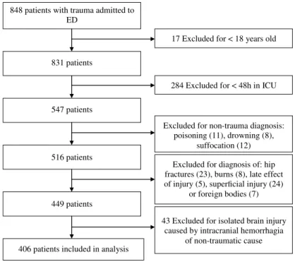
During the study period, 848 patients with trauma were admitted to ED and consequently to one of two trauma-surgical ICUs, of which 547 were adult patients who were hospitalized for more than 48 h. Thirty-one patients were excluded due to non-traumatic mechanisms of injury (poisoning, drowning, and suffocation) and 36 were excluded due to late effect of injuries, superficial injuries, or foreign bodies. In addition, 74 patients were excluded due to diagnosis of burns, hip fractures, or isolated brain injuries caused by non-traumatic injury, such as spontaneous intracranial bleeding. This led to the sample of 406 patients with trauma included in thefinal analysis. The process of patient selection is depicted in Figure1.
A cohort of 406 patients with trauma (312 males and 94 females) were followed for 5,258 patient-ICU days. Median length of stay was 9 days (range: 2–131).
Incidence of BSI in ED and ICU
Out of 406 patients with trauma, 57 patients had at least one episode of BSI (cumulative incidence 14.0%). Overall, 62 BSI episodes were diagnosed in 57 patients with the incidence rate of 11.8/1,000 patient/days. Out of 62 BSI episodes, 19 (30.6%) BSI episodes infection cite was identified and those were considered as secondary BSI. For 30 BSI episodes (48.4%), no source of infection could be
identified while 13 BSI (21.0%) were ascertained as CR-BSI, which over the 4,295 catheter-days resulted in incidence rate of 3.0/1,000 CVC-days. Out of 13 CR-BSI, 2 were attributed to ED since the CVC was placed in ED, which resulted in IR=3.7/1,000 CVC/days (532 CVC-days in ED).
Clinical characteristics of patients at admission to ED
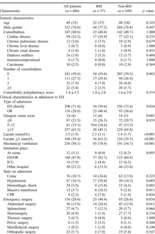
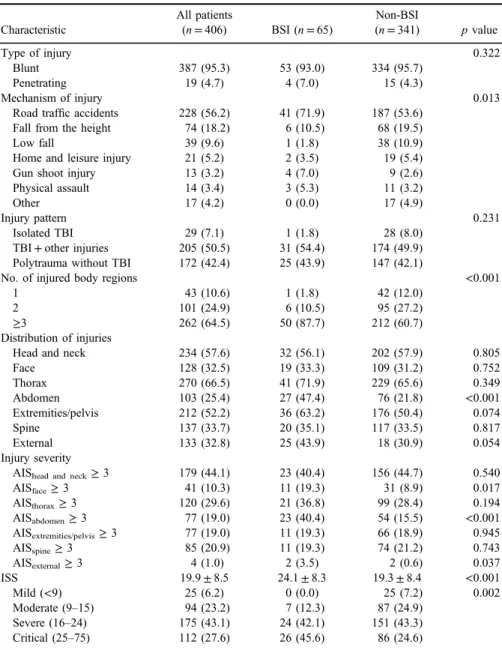
Table I provides comparison of demographic characteristics, underlying conditions, and data about severity of the disease at the admission to ED, between patient with and without BSI. There was no significant difference in demographics and comorbidities in patients with BSI and those without BSI. However, signifi- cantly more patients with BSI had received cardiopulmonary resuscitation on scene or at admission (p=0.026), had lower median GCS on admission (p=0.001), had almost twice more patients with lactate level ≥2 mmol/L (p<0.001) and in higher percent required mechanical ventilation (p<0.001) as well as massive transfusion (p=0.011) and emergency surgery (p=0.034) on admission. Patients with BSI evinced more severe medical condition on admission
848 patients with trauma admitted to ED
831 patients
284 Excluded for < 48h in ICU 547 patients
Excluded for diagnosis of: hip fractures (23), burns (8), late effect of injury (5), superficial injury (24)
or foreign bodies (7)
406 patients included in analysis
Excluded for non-trauma diagnosis:
poisoning (11), drowning (8), suffocation (12) 516 patients
17 Excluded for < 18 years old
449 patients
43 Excluded for isolated brain injury caused by intracranial hemorrhagia
of non-traumatic cause
Figure 1.Flowchart of patient selection
Table I.Characteristics of patients with trauma with and without BSI
Characteristic
All patients (n=406)
BSI (n=57)
Non-BSI
(n=349) pvalue General characteristics
Age 49 (35) 52 (37) 48 (34) 0.525
Male gender 312 (76.8) 44 (77.2) 268 (76.8) 0.947
Comorbidities 165 (40.6) 23 (40.4) 142 (40.7) 1.000
Cardiac disease 94 (23.2) 17 (29.8) 77 (22.1) 0.235
Chronic pulmonary disease 12 (3.0) 2 (3.5) 10 (2.9) 0.679
Chronic liver disease 3 (0.7) 0 (0.0) 3 (0.9) 1.000
Chronic renal disease 4 (1.0) 1 (1.8) 3 (0.9) 0.455
Diabetes mellitus 16 (3.9) 3 (5.3) 13 (3.7) 0.480
Immunocompromised 6 (1.7) 0 (0.0) 6 (1.7) 1.000
Carcinoma 10 (2.5) 0 (0.0) 10 (2.9) 0.369
Number of comorbidities
0 241 (59.4) 34 (59.6) 207 (59.3) 0.883
1 111 (27.3) 17 (29.8) 94 (26.9)
2 32 (7.9) 4 (7.0) 28 (8.0)
≥3 22 (5.4) 2 (3.5) 20 (5.7)
Comorbidity polypharmacy score 1.6±2.5 1.8±2.6 1.6±3.0 0.519 Clinical characteristics at admission to ED
Type of admission
ED directly 290 (71.4) 34 (59.6) 256 (73.4) 0.034
OR 116 (28.6) 23 (40.4) 93 (26.6)
Glasgow coma score 14 (6) 12 (6) 14 (5) 0.001
≤8 87 (21.5) 15 (26.3) 72 (20.7) 0.033
9–12 61 (15.1) 914 (24.6) 47 (13.5)
≥13 257 (63.5) 28 (49.1) 229 (65.8)
Lactate (mmol/L) 1.5 (1.8) 2.3 (3.1) 1.4 (1.7) <0.001
Lactate≥2 mmol/L 160 (39.4) 36 (63.2) 124 (35.5) <0.001
Mechanical ventilation 236 (58.1) 45 (78.9) 191 (54.7) <0.001 Intubation place
At scene 12 (5.1) 0 (0.0) 12 (6.3) 0.095
ED/OR 160 (67.8) 37 (82.2) 123 (64.4)
ICU 14 (5.9) 2 (4.4) 12 (6.3)
Other hospital 50 (21.2) 6 (13.3) 44 (23.0)
State on admission
Coma 76 (18.7) 14 (24.6) 62 (17.8) 0.223
Hypotension 67 (16.5) 17 (29.8) 50 (14.3) 0.003
Hemorrhagic shock 24 (5.9) 9 (15.8) 15 (4.3) 0.003
Massive transfusion 15 (3.7) 6 (10.5) 9 (2.6) 0.011
CPR 9 (2.2) 4 (7.0) 5 (1.4) 0.026
Emergency surgery 116 (28.6) 23 (40.4) 93 (26.6) 0.034
Abdominal surgery 56 (13.8) 14 (24.6) 42 (12.0) 0.011
Splenectomy 27 (6.7) 7 (12.3) 20 (5.7) 0.066
Neurosurgery 28 (6.9) 1 (1.8) 27 (7.7) 0.154
Thoracic surgery 3 (0.7) 0 (0.0) 3 (0.9) 1.000
Pelvic surgery 6 (1.5) 2 (3.5) 4 (1.1) 0.200
Maxillofacial surgery 1 (0.2) 1 (1.8) 0 (0.0) 0.140
Orthopedic surgery 23 (5.7) 4 (7.0) 19 (5.4) 0.547
considering higher median APACHE II and SOFA score in BSI group (p<0.001 for both). Regarding the exposure to invasive devices over the course of ED and ICU treatment, median number of days spent with CVC in place or on mechanical ventilation was significantly higher in patients who acquired BSI compared to those who did not (p<0.001 for both).
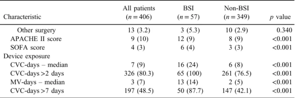
TableIIshows distribution and comparison of injury patterns and severity in patients with trauma with and without BSI. Blunt trauma was a more common type of injury in both groups of patients. Polytrauma (p<0.001) and abdominal injury (p<0.001) were more frequent injury pattern in patients with BSI. They also had significantly more severe injuries on face (p=0.017), abdomen (p<0.001), and external body surface (p=0.037) according to AIS score ≥3 for these body regions. Overall, patients with BSI had more severe injuries according to higher ISS (24.1±8.3 vs. 19.3±8.4,p<0.001) and significantly higher percent of BSI patient had critically severe injuries (45.6% vs. 24.6%, p=0.002).
Predictors of BSI
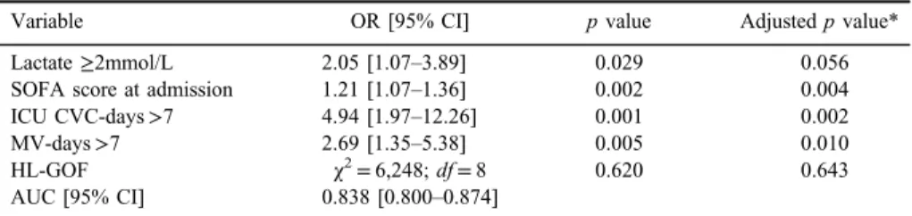
Multivariate logistic regression of factors associated with BSI was applied and model was started with 34 variables that were statistically or clinically associated with BSI in univariate analysis. Variables that remained independently associated with occurrence of BSI in multivariate logistic regression model were lactate concentration≥2 mmol/L (OR=2.05; 95% CI=1.07–43.89), SOFA score (OR=1.21; 95% CI=1.07–1.36), CVC in place for more than 7 days (OR=4.94;
95% CI=1.97–12.26), and mechanical ventilation more than 7 days (OR=2.69;
Table I.Characteristics of patients with trauma with and without BSI(Continued)
Characteristic
All patients (n=406)
BSI (n=57)
Non-BSI
(n=349) pvalue
Other surgery 13 (3.2) 3 (5.3) 10 (2.9) 0.340
APACHE II score 9 (10) 12 (9) 8 (9) <0.001
SOFA score 4 (3) 6 (4) 3 (3) <0.001
Device exposure
CVC-days–median 7 (9) 16 (24) 6 (8) <0.001
CVC-days>2 days 326 (80.3) 65 (100) 261 (76.5) <0.001
MV-days–median 3 (7) 13 (14) 2 (5) <0.001
CVC-days>7 days 197 (48.5) 50 (87.7) 147 (42.1) <0.001 Note:Values are number (%), mean±SD or median (IQR).pvalue corresponds to comparison between BSI and non-BSI. BSI: bloodstream infection; ED: emergency department; OR: operating room; CPR:
cardiopulmonary resuscitation; APACHE II score: Acute Physiology and Chronic Health Evaluation II Score; SOFA score: Sequential Organ Failure Assessment score; CVC: central venous catheter; MV:
mechanical ventilation.
Table II.Characteristics of injuries in patients with trauma with and without BSI
Characteristic
All patients
(n=406) BSI (n=65)
Non-BSI
(n=341) pvalue
Type of injury 0.322
Blunt 387 (95.3) 53 (93.0) 334 (95.7)
Penetrating 19 (4.7) 4 (7.0) 15 (4.3)
Mechanism of injury 0.013
Road traffic accidents 228 (56.2) 41 (71.9) 187 (53.6)
Fall from the height 74 (18.2) 6 (10.5) 68 (19.5)
Low fall 39 (9.6) 1 (1.8) 38 (10.9)
Home and leisure injury 21 (5.2) 2 (3.5) 19 (5.4)
Gun shoot injury 13 (3.2) 4 (7.0) 9 (2.6)
Physical assault 14 (3.4) 3 (5.3) 11 (3.2)
Other 17 (4.2) 0 (0.0) 17 (4.9)
Injury pattern 0.231
Isolated TBI 29 (7.1) 1 (1.8) 28 (8.0)
TBI+other injuries 205 (50.5) 31 (54.4) 174 (49.9) Polytrauma without TBI 172 (42.4) 25 (43.9) 147 (42.1)
No. of injured body regions <0.001
1 43 (10.6) 1 (1.8) 42 (12.0)
2 101 (24.9) 6 (10.5) 95 (27.2)
≥3 262 (64.5) 50 (87.7) 212 (60.7)
Distribution of injuries
Head and neck 234 (57.6) 32 (56.1) 202 (57.9) 0.805
Face 128 (32.5) 19 (33.3) 109 (31.2) 0.752
Thorax 270 (66.5) 41 (71.9) 229 (65.6) 0.349
Abdomen 103 (25.4) 27 (47.4) 76 (21.8) <0.001
Extremities/pelvis 212 (52.2) 36 (63.2) 176 (50.4) 0.074
Spine 137 (33.7) 20 (35.1) 117 (33.5) 0.817
External 133 (32.8) 25 (43.9) 18 (30.9) 0.054
Injury severity
AIShead and neck≥3 179 (44.1) 23 (40.4) 156 (44.7) 0.540
AISface≥3 41 (10.3) 11 (19.3) 31 (8.9) 0.017
AISthorax≥3 120 (29.6) 21 (36.8) 99 (28.4) 0.194
AISabdomen≥3 77 (19.0) 23 (40.4) 54 (15.5) <0.001
AISextremities/pelvis≥3 77 (19.0) 11 (19.3) 66 (18.9) 0.945
AISspine≥3 85 (20.9) 11 (19.3) 74 (21.2) 0.743
AISexternal≥3 4 (1.0) 2 (3.5) 2 (0.6) 0.037
ISS 19.9±8.5 24.1±8.3 19.3±8.4 <0.001
Mild (<9) 25 (6.2) 0 (0.0) 25 (7.2) 0.002
Moderate (9–15) 94 (23.2) 7 (12.3) 87 (24.9)
Severe (16–24) 175 (43.1) 24 (42.1) 151 (43.3)
Critical (25–75) 112 (27.6) 26 (45.6) 86 (24.6)
Note: pvalue corresponds to comparison between BSI and non-BSI. BSI: bloodstream infection; TBI:
trauma brain injury; AIS: Abbreviated Injury Scale; ISS: Injury Severity Score.
95% CI=1.35–5.38; TableIII). The modelfit the data properly since the Hosmer– Lemeshow test was insignificant (p=0.620). The discrimination of the model was good according to AUC=0.838 (95% CI=0.800–0.874). However, after con- trolling for patients’ characteristics (age, gender, and comorbidities), injury severity (severe TBI and ISS) and presence of hemorrhagic shock on admission, lactate concentration≥2 mmol/L did not remain statistically significant predictor (OR=1.93; 95% CI=0.984–3.795).
In order to estimate the impact of ED predictors in the model, we compared AUCs of models with only ICU variables [CVC in place for more than 7 days and mechanical ventilation (MV) more than 7 days] with full model with ED variables added (lactate ≥2 mmol/L and SOFA score). Inclusion of variables from ED significantly improved discriminative accuracy of the model since AUC increased from fair (AUC=0.785; 95% CI=0.741–0.824) to good accuracy (AUC= 0.839; 95% CI=0.800–0.874), respectively (p=0.0004; Figure2).
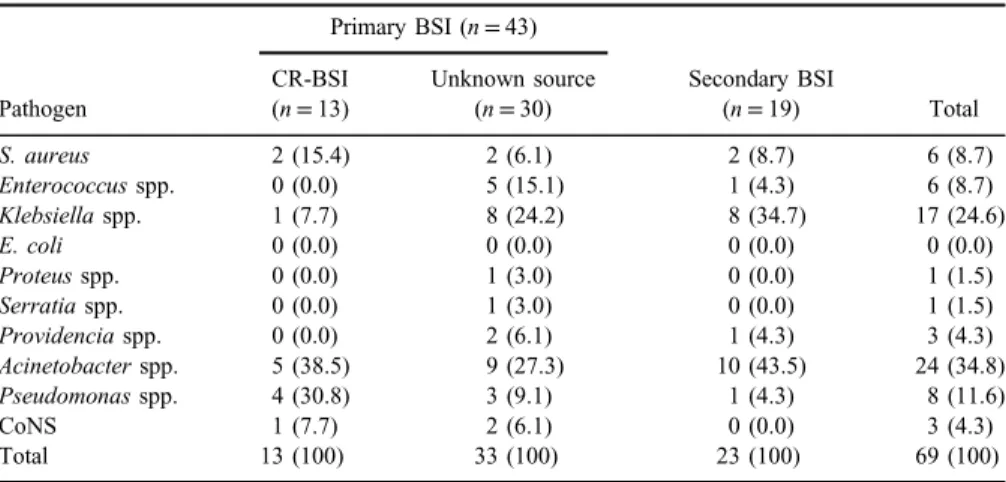
Causative agents and AMR
Sixty-nine pathogens were obtained from 62 BSI episodes, of which 6 were polymicrobial (5 isolates with 2 bacteria and 1 with 3 bacteria). The most common pathogens recovered were Gram-negative bacteria (Acinetobacterspp.,Klebsiella pneumoniae, Pseudomonas spp., andKlebsiellaspp.) accounting for 71% of all isolates (TableIV). AMR markers in bacteria causing BSI in patients with trauma are presented in TableV. While all isolates ofS. aureus and coagulase-negative staphylococci (CoNS) were methicillin-resistant, 66% ofEnterococcusspp. were resistant to vancomycin. RegardingEnterobacteriaceae,all isolates ofKlebsiella spp., K. pneumoniae, Proteus spp., Serratia spp., and Providentia spp. were
Table III.Multivariate logistic regression of predictors of BSI in patients with trauma
Variable OR [95% CI] pvalue Adjustedpvalue*
Lactate≥2mmol/L 2.05 [1.07–3.89] 0.029 0.056
SOFA score at admission 1.21 [1.07–1.36] 0.002 0.004
ICU CVC-days>7 4.94 [1.97–12.26] 0.001 0.002
MV-days>7 2.69 [1.35–5.38] 0.005 0.010
HL-GOF χ2=6,248;df=8 0.620 0.643
AUC [95% CI] 0.838 [0.800–0.874]
Note:SOFA score: sequential organ failure assessment score; OR: odds ratio; ICU: intensive care unit;
CVC: central venous catheter; HL-GOF: Hosmer and Lemeshow goodness of fit; MV: mechanical ventilation; BSI: bloodstream infection; AUC: area under the receiver-operator characteristic curve;
CI: confidence interval.
*Adjusted for age, gender, comorbidities, severe trauma brain injury (AIShead≥3 and GCS≤8), trauma severity (ISS), and hemorrhagic shock.
Figure 2.Comparison of AUCs of models with and without ED predictors. AUC difference=0.055;
[95% CI=0.0.3–0.08];p=0.0004
Table IV.Distribution of bacteria causing BSI by type of source
Pathogen
Primary BSI (n=43)
Secondary BSI
(n=19) Total
CR-BSI (n=13)
Unknown source (n=30)
S. aureus 2 (15.4) 2 (6.1) 2 (8.7) 6 (8.7)
Enterococcusspp. 0 (0.0) 5 (15.1) 1 (4.3) 6 (8.7)
Klebsiellaspp. 1 (7.7) 8 (24.2) 8 (34.7) 17 (24.6)
E. coli 0 (0.0) 0 (0.0) 0 (0.0) 0 (0.0)
Proteusspp. 0 (0.0) 1 (3.0) 0 (0.0) 1 (1.5)
Serratiaspp. 0 (0.0) 1 (3.0) 0 (0.0) 1 (1.5)
Providenciaspp. 0 (0.0) 2 (6.1) 1 (4.3) 3 (4.3)
Acinetobacterspp. 5 (38.5) 9 (27.3) 10 (43.5) 24 (34.8)
Pseudomonasspp. 4 (30.8) 3 (9.1) 1 (4.3) 8 (11.6)
CoNS 1 (7.7) 2 (6.1) 0 (0.0) 3 (4.3)
Total 13 (100) 33 (100) 23 (100) 69 (100)
Note:BSI: bloodstream infection; CR-BSI: catheter-related BSI; CoNS: coagulase-negative staphylococci.
resistant to third-generation cephalosporins. Considering carbapenem resistance of these bacteria, majority of isolates of Klebsiella spp. (85.7%), K. pneumoniae (70%), and Providentia spp. (66.7%) were denoted as carbapenem resistant, whereas no isolates ofProteusspp. andSeratia spp. were resistant to this class of antimicrobial agents. Gram-negative rods,Pseudomonasspp. andAcinetobac- ter spp., were highly resistant to carbapenems, as 87.5% and 95.8% of their isolates, respectively, were not susceptible to this antibiotic. Of 24 isolates of Acinetobacter spp., 4 (16.7%) were also resistant to colistin.
Discussion
In this study, we assessed the incidence of BSI in patients with trauma in ICU of a major referral academic Emergency Center in Belgrade, Serbia, as well as the fraction of BSI, which can be attributed to CVC exposure initiated in ED. In addition, we sought to estimate which of the ED- and ICU-derived variables are independent predictors of BSI and whether ED predictors significantly improve that prediction. At least, we delineated distribution of causative agents of BSI and the major AMR markers of these pathogens.
Table V.AMR markers in bacteria causing BSI in patients with trauma
Bacteria AMR marker n n(%)R
S. aureus MRSA 6 6 (100)
CoNS MRCoNS 3 3 (100)
Enterococcusspp. VRE 6 4 (66.7)
Klebsiellaspp. 3GC-NS 17 17 (100)
CAR-NS 17 13 (76.5)
Proteusspp. 3GC-NS 1 1 (100)
CAR-NS 1 0 (0.0)
Serratiaspp. 3GC-NS 1 1 (100)
CAR-NS 1 0 (0.0)
Providentiaspp. 3GC-NS 3 3 (100)
CAR-NS 3 2 (66.7)
Pseudomonasspp. CAR-NS 8 7 (87.5)
CST-NS 8 0 (0.0)
Acinetobacterspp. CAR-NS 24 23 (95.8)
CST-NS 24 4 (16.7)
Note:AMR: antimicrobial resistance; BSI: bloodstream infection; MRSA: methicillin-resistantStaphylo- coccus aureus; CoNS: coagulase-negative staphylococci; MRCoNS: Methicillin-resistant coagulase- negative staphylococci; VRE: vancomycin-resistant enterococci; 3GC: third-generation cephalosporin;
CAR: carbapenem; CST: colistin; NS: non-susceptible.
Cumulative incidence of BSI in this study was 14% and the incidence rate was 11.8/1,000 patient/days. Similar to these findings, cumulative incidence in studies that reported BSI as a primary outcome in patients with trauma was 15.2%
in American trauma ICU [20]. However, diverse results were reported within wards and countries where BSI was analyzed as a fraction of all HAIs [21–26].
Regardless of heterogeneity of results, most of the studies focused on incidence and risk factors for BSI acquired in ICU. The reason is that majority of infections present upon admission to ED are community-acquired, whereas those that occur later are usually, due to delayed diagnostic, identified upon admission in ICU. Moreover, according to protocols and guidelines for surveil- lance studies, if invasive device, such as vascular catheter, is inserted in ED and signs and symptoms of infection occur in ICU, these are reported as ICU-acquired BSI since ED is not considered as inpatient location and for which no denominator data are collected [27]. The consequence is the attribution of ED-acquired HAI to the ICU and underestimation of ED as a significant source of HAI. LeMaster et al.
[28] showed that in a study conducted in large urban academic hospital in Boston, based on cases with vascular catheters inserted in ED, incidence of ED BSI is similar to the rates of BSI in ICU. Moreover, Theodoro et al. [29] showed that although ED contributes only 7.7% of total institutional catheter-days, incidence of central line-associated BSI (CLABSI) in ED is similar to incidence in medical and composite ICUs and it is higher compared to surgical ICU. No such study was conducted in patients with trauma to date, especially not in low or middle income countries. In this study, 15% of CR-BSIs were acquired in ED with the incidence 3.7/1,000 CVC-days, which is similar to BSI incidence in ICU (3.0/1,000 CVC-days). This can be partially explained by smaller number of CVC-days in ED used as a denominator for incidence calculation. However, the results of this study confirm findings from two previously mentioned studies conducted in unselected population of critically ill patients in terms of considerable fraction of BSI that can be attributed to ED and the similar incidence of BSI acquired in ED and ICU. This is particularly important as it shows that exposure to CVC in ED is significant in developing BSI and necessity of the adaptation of future preventative strategies to include ED in CVC maintenance and hand hygiene guidelines.
We also sought to determine which variables regarding the patient’s and trauma characteristic assessed in veryfirst hours of ED admission are associated with later occurrence of BSI together with variables from ICU. Factors identified to independently predict occurrence of BSI in patients with trauma in this study were lactate level≥2 mmol/L and SOFA score at the admission to ED and CVC in place for more than 7 days and mechanical ventilation for more than 7 days in ICU.
This is supported by results of previous studies, which also suggest that severity of disease scores such as APACHE II and SOFA as well as days spent on mechanical
ventilation and CVC in place is independently associated with HAI and BSI in patients with trauma [22, 25,26].
Lactate is widely accepted as marker of hypoperfusion, and its diagnostic value in risk stratification for adverse outcomes has been frequently investigated in diverse subpopulations of critically ill patients [30]. However, the studies mostly focused on unselected ICU patients with suspected or confirmed infection or septic shock and evaluated mortality as an outcome [31]. Several studies addressed this issue within the ED patients and only few with respect to infection prediction. In a study of prediction of bacteremia in adult patients admitted to Swedish ED, Ljungström et al. [32] showed that lactate was significantly higher in patients with bacteremia compared to those without infection (2.10 vs. 1.66 mmol/L) and that it has as good predictive ability as a single biomarker as well as in composition with other biomarkers. However, Lin et al. [33] showed that lactate level 19.9 mg/dl (2 mmol/L) has slightly better diagnostic accuracy in prediction of Gram-positive bacteremia than procalcitonin (AUC 0.66 vs. 0.61) in adult ED patients, but lower accuracy in predicting Gram-negative bacteremia (AUC 0.71 vs. 0.79) and overall positive blood culture (AUC 0.69 vs. 0.72). In this study, however, we do not have in-depth information on other inflammation markers as lactate was selected as marker of tissue damage following trauma and not as an infection diagnostic marker.
In this study, elevated lactate did not remain significant after adjustment for patients’characteristics, trauma severity, hemorrhagic shock, and severe TBI. This result may suggest that increased lactate is only reflecting the severity of the underlying condition motivating the use of CVC, which maintained the strongest association with BSI in adjusted model. Although, in Serbia, CVCs are inserted following CVC guidelines, and CVC management includes change of femoral CVC after 2 days and subclavian and jugular CVC after 7 days of insertion [12], high BSI incidence in ED and ICU can be attributed to possible low compliance to these policies and lack of hygiene measures. Although such measures represent a challenge in life-saving medical setting, they should not be neglected, considering unfavorable outcomes of infections related to CVC use and the fact that great part of CLABSI can be prevented just by adequate adherence to the components of the CL bundles [34].
Considering AMR markers in causative pathogens of BSI, we found alarmingly high rates of Enterobacteriaceaestrains resistant to third-generation cephalosporins (100% of strains) and carbapenems (66.7%–85.7%) as well as high percentage ofPseudomonasspp. andAcinetobacterspp. resistant to carbapenems (87.5% and 95.8% of non-susceptible strains, respectively). On the contrary, National Healthcare Safety Network and International Nosocomial Infection Control Consortium reported considerably lower rates of isolates resistant to common AMR markers [35,36]. Although there is no data available on resistance
to common AMR markers of pathogens causing BSI in patients with trauma from countries close to Serbia, our results are similar to AMR data of invasive isolates in acute care hospitals in countries of Southeastern–European Region [37]. At clinical level, this data is highly important since the choice of empirical therapy of BSI/CLABSI and its duration depend on understanding the distribution and resistance patterns of their causative agents in specific setting or patient popula- tion. At national level, this imposes the necessity for implementation of measures for antibiotic consumption restriction together with general and pathogen-specific measures for disruption of transmission of resistant clones.
Several limitations of the study should be addressed. Collecting data from one academic trauma center can be subject to selection bias due to inclusion of mostly severe trauma cases. We did not have data on previous antibiotic consumption due to the urgent type of referral. However, this is thefirst study in Southeastern–European Region to outline epidemiology of BSI and to estimate contribution of both ED and ICU incidence of BSI as well as contribution of ED variables as risk factors of BSI in population of patients with trauma. Strength of the study is prospective daily collection of numerous clinical and disease severity variables, as well as assessment of signs of infections and the exposure to risk factors with low possibility of misclassification of both exposure and outcome.
In conclusion, BSI is a common HAI in trauma ICU affecting 14% of all inpatients in ICU. Considerable part of BSI in ICU arises in ED, resulting in similar incidence of BSI in ED compared to ICU. In this study, combination of predictors for BSI assessed in ED and those measured in ICU indicates that constant monitoring of patients on MV and those with CVC for reassessing the need for their continuation may be useful in order to detect primary BSI and pneumonia-related secondary BSI, especially in patients who had hypoperfusion and more severe condition on admission, as estimated by lactate level and SOFA score. Predominance of primary BSI is caused by Gram-negative bacteria highly resistant to commonly used antibiotics, stress necessity for more effective overall, and CVC-specific infection control measures in critically ill patients with trauma.
Acknowledgements
This work was supported by the Ministry of Education, Science and Technological Development of Serbia, contract no. 175046, 2011–2018.
Conflict of Interest
The authors declare no conflict of interest related to this work.
References
1. European Centre for Disease Prevention and Control: Annual Epidemiological Report 2016 – Healthcare-Associated Infections Acquired In Intensive Care Units [Internet].
ECDC, Stockholm, 2016. Available athttps://ecdc.europa.eu/sites/portal/files/documents/
AER-HCAI_ICU_3_0.pdf
2. Wong, S. W., Gantner, D., McGloughlin, S., Leong, T., Worth, L. J., Klintworth, G., Scheinkestel, C., Pilcher, D., Cheng, A. C., Udy, A.: The influence of intensive care unit- acquired central line-associated bloodstream infection on in-hospital mortality: A single- center risk-adjusted analysis. Am J Infect Control44, 587–592 (2016).
3. Czaja, A. S., Rivara, F. P., Wang, J., Koepsell, T., Nathens, A. B., Jurkovich, G. J., Mackenzie, E.: Late outcomes of trauma patients with infections during index hospitaliza- tion. J Trauma67, 805–814 (2009).
4. Claridge, J. A., Crabtree, T. D., Pelletier, S. J., Butler, K., Sawyer, R. G., Young, J. S.:
Persistent occult hypoperfusion is associated with a significant increase in infection rate and mortality in major trauma patients. J Trauma48, 8–14 (2000).
5. Wallace, W. C., Cinat, M., Gornick, W. B., Lekawa, M. E., Wilson, S. E.: Nosocomial infections in the surgical intensive care unit: A difference between trauma and surgical patients. Am Surg65, 987–990 (1999).
6. Glance, L. G., Stone, P. W., Mukamel, D. B., Dick, A. W.: Increases in mortality, length of stay, and cost associated with hospital-acquired infections in trauma patients. Arch Surg 146, 794–801 (2011).
7. Institute of Public Health of Serbia: Health Statistical Yearbook of Republic of Serbia, 2015. Available athttp://www.batut.org.rs/download/publikacije/pub2015.pdf
8. Mansur, A., Klee, Y., Popov, A. F., Erlenwein, J., Ghadimi, M., Beissbarth, T., Bauer, M., Hinz, J.: Primary bacteraemia is associated with a higher mortality risk compared with pulmonary and intra-abdominal infections in patients with sepsis: A prospective observa- tional cohort study. BMJ Open5, e006616 (2015).
9. Liesenfeld, O., Lehman, L., Hunfeld, K. P., Kost, G.: Molecular diagnosis of sepsis: New aspects and recent developments. Eur J Microbiol Immunol (Bp)4, 1–25 (2014).
10. Gaynes, R., Richards, C., Edwards, J., Emori, T. G., Horan, T., Alonso-Echanove, J., Fridkin, S., Lawton, R., Peavy, G., Tolson, J.: Feeding back surveillance data to prevent hospital-acquired infections. Emerg Infect Dis7, 295–298 (2001).
11. World Health Organization: WHO Guidelines on Hand Hygiene in Health Care. WHO, Geneva, 2009. Available athttp://www.who.int/gpsc/5may/tools/9789241597906/en 12. American Society of Anesthesiologists Task Force on Central Venous Access, Rupp, S. M.,
Apfelbaum, J. L., Blitt, C., Caplan, R. A., Connis, R. T., Domino, K. B., Fleisher, L. A., Grant, S., Mark, J. B., Morray, J. P., Nickinovich, D. G., Tung, A.: Practice guidelines for central venous access. Anesthesiology116, 539–573 (2012).
13. Horan, T. C., Andrus, M., Dudeck, M. A.: CDC/NHSN surveillance definition of health care-associated infection and criteria for specific types of infections in the acute care setting.
Am J Infect Control36, 309–32 (2008).
14. Hansen, S., Sohr, D., Geffers, C., Astagneau, P., Blacky, A., Koller, W., Morales, I., Moro, M. L., Palomar, M., Szilagyi, E., Suetens, C., Gastmeier, P.: Concordance between European and US case definitions of healthcare-associated infections. Antimicrob Resist Infect Control1, 28 (2012).
15. Djuric, O., Markovic-Denic, L., Jovanovic, B., Bumbasirevic, V.: Agreement between CDC/NHSN surveillance definitions and ECDC criteria in diagnosis of healthcare- associated infections in Serbian trauma patients. PLoS One13, e0204893 (2018).
16. Horan, T. C., Emori, T. G.: Definitions of key terms used in the NNIS System. Am J Infect Control25, 112–116 (1997).
17. Justiniano, C. F., Evans, D. C., Cook, C. H., Eiferman, D. S., Gerlach, A. T., Beery, P. R., Lindsey, D. E., Saum, G. E., Murphy, C. V., Miller, S. F., Papadimos, T. J., Steinberg, S. M., Stawicki, S. P.: Comorbidity-polypharmacy score: A novel adjunct in post- emergency department trauma triage. J Surg Res181, 16–19 (2013).
18. Clinical and Laboratory Standards Institute: Performance Standards for Antimicrobial Susceptibility Testing: 24th Informational Supplement, M100-S24. CLSI, Wayne, PA, 2014.
19. DeLong, E. R., DeLong, D. M., Clarke-Pearson, D. L.: Comparing the areas under two or more correlated receiver operating characteristic curves: A nonparametric approach.
Biometrics 44, 837–845 (1988).
20. El-Masri, M. M., Hammad, T. A., McLeskey, S. W., Joshi, M., Korniewicz, D. M.:
Predictors of nosocomial bloodstream infections among critically ill adult trauma patients.
Infect Control Hosp Epidemiol25, 656–663 (2004).
21. Mathur, P., Varghese, P., Tak, V., Gunjiyal, J., Lalwani, S., Kumar, S., Misra, M. C.:
Epidemiology of blood stream infections at a level-1 trauma care center of India. J Lab Physicians6, 22–27 (2014).
22. Mitharwal, S. M., Yaddanapudi, S., Bhardwaj, N., Gautam, V., Biswal, M., Yaddanapudi, L.: Intensive care unit-acquired infections in a tertiary care hospital: An epidemiologic survey and influence on patient outcomes. Am J Infect Control44, e113–e117 (2016).
23. Lazarus, H. M., Fox, J., Burke, J. P., Lloyd, J. F., Snow, G. L., Mehta, R. R., Evans, R. S., Abouzelof, R., Taylor, C., Stevens, M. H.: Trauma patient hospital-associated infections:
Risks and outcomes. J Trauma59, 188–194 (2005).
24. Lazarus, H. M., Fox, J., Lloyd, J. F., Evans, R. S., Abouzelof, R., Taylor, C., Pombo, D. J., Stevens, M. H., Mehta, R., Burke, J. P.: A six-year descriptive study of hospital-associated infection in trauma patients: Demographics, injury features, and infection patterns. Surg Infect (Larchmt) 8, 463–473 (2007).
25. Giamberardino, H. I., Cesário, E. P., Carmes, E. R., Mulinari, R. A.: Risk factors for nosocomial infection in trauma patients. Braz J Infect Dis11, 285–289 (2007).
26. Papia, G., McLellan, B. A., El-Helou, P., Louie, M., Rachlis, A., Szalai, J. P., Simor, A. E.:
Infection in hospitalized trauma patients: Incidence, risk factors, and complications. J Trauma 47, 923–927 (1999).
27. Centers for Disease Control and Prevention: National Healthcare Safety Network Device Associated Module: Central Line Associated Bloodstream Infection Event, 2010. Available athttp://www.cdc.gov/nhsn/PDFs/pscManual/4PSC_CLABScurrent.pdf
28. LeMaster, C. H., Schuur, J. D., Pandya, D., Pallin, D. J., Silvia, J., Yokoe, D., Agrawal, A., Hou, P. C.: Infection and natural history of emergency department-placed central venous catheters. Ann Emerg Med56, 492–497 (2010).
29. Theodoro, D., Olsen, M. A., Warren, D. K., McMullen, K. M., Asaro, P., Henderson, A., Tozier, M., Fraser, V.: Emergency department central line-associated bloodstream infec- tions (CLABSI) incidence in the era of prevention practices. Acad Emerg Med22, 1048– 1055 (2015).
30. Baron, B. J., Nguyen, A., Stefanov, D., Shetty, A., Zehtabchi, S.: Clinical value of triage lactate in risk stratifying trauma patients using interval likelihood ratios. Am J Emerg Med 36, 784–788 (2018).
31. del Portal, D. A., Shofer, F., Mikkelsen, M. E., Dorsey, P. J., Gaieski, D. F., Goyal, M., Synnestvedt, M., Weiner, M. G., Pines, J. M.: Emergency department lactate is associated with mortality in older adults admitted with and without infections. Acad Emerg Med17, 260–268 (2010).
32. Ljungström, L., Pernestig, A. K., Jacobsson, G., Andersson, R., Usener, B., Tilevik, D.:
Diagnostic accuracy of procalcitonin, neutrophil-lymphocyte count ratio, C-reactive pro- tein, and lactate in patients with suspected bacterial sepsis. PLoS One12, e0181704 (2017).
33. Lin, C. T., Lu, J. J., Chen, Y. C., Kok, V. C., Horng, J. T.: Diagnostic value of serum procalcitonin, lactate, and high-sensitivity C-reactive protein for predicting bacteremia in adult patients in the emergency department. PeerJ5, e4094 (2017).
34. Morales-Cartagena, A., Fernández-Ruiz, M., Lalueza, A., Lora-Tamayo, J., San Juan, R., L´opez-Medrano, F., Origüen, J., Chaves, F., María Aguado, J.: Impact on mortality of adherence to evidence-based interventions in patients with catheter-related bloodstream infection due to methicillin-sensitiveStaphylococcus aureus. Infect Dis (Lond)16, 1–10 (2018).
35. Weiner, L. M., Webb, A. K., Limbago, B., Dudeck, M. A., Patel, J., Kallen, A. J., Edwards, J. R., Sievert, D. M.: Antimicrobial-resistant pathogens associated with healthcare- associated infections: Summary of data reported to the National Healthcare Safety Network at the centers for disease control and prevention, 2011–2014. Infect Control Hosp Epidemiol37, 1288–1301 (2016).
36. Rosenthal, V. D., Maki, D. G., Mehta, Y., Leblebicioglu, H., Memish, Z. A., Al-Mousa, H. H., Balkhy, H., Hu, B., Alvarez-Moreno, C., Medeiros, E. A., Apisarnthanarak, A., Raka, L., Cuellar, L. E., Ahmed, A., Navoa-Ng, J. A., El-Kholy, A. A., Kanj, S. S., Bat-Erdene, I., Duszynska, W., Van Truong, N., Pazmino, L. N., See-Lum, L. C., Fernández-Hidalgo, R., Di-Silvestre, G., Zand, F., Hlinkova, S., Belskiy, V., Al-Rahma, H., Luque-Torres, M. T., Bayraktar, N., Mitrev, Z., Gurskis, V., Fisher, D., Abu-Khader, I. B., Berechid, K., Rodríguez-Sánchez, A., Horhat, F. G., Requejo-Pino, O., Hadjieva, N., Ben-Jaballah, N., García-Mayorca, E., Kushner-Dávalos, L., Pasic, S., Pedrozo-Ortiz, L. E., Apostolopoulou, E., Mejía, N., Gamar-Elanbya, M. O., Jayatilleke, K., de Lourdes-Duenas,˜ M., Aguirre-Avalos, G., International Nosocomial Infection Control Consortium: Interna- tional Nosocomial Infection Control Consortium (INICC) report, data summary of 43 countries for 2007–2012. Device-associated module. Am J Infect Control 42, 942–956 (2014).
37. WHO Regional Office for Europe: Central Asian and Eastern European Surveillance of Antimicrobial Resistance. Annual report 2017. Denmark, 2018. Available athttp://
www.euro.who.int/__data/assets/pdf_file/0005/354434/WHO_CAESAR_AnnualReport_
2017.pdf?ua=1