Original Research Article
HIP
Early detection of developmental dysplasia of hip by ultrasound
HIP International 1-6
© The Author(s) 2019 Article reuse guidelines:
sagepub.com/journals-permissions D Ó I: 10.1 177/1 120700019879687 journals.sagepub.com/home/hpi
®SAGE
Z ita G yu rko vits', G e llé rt So h ár2, A nna B aricsa2, G á b o r N é m e th 1, H ajnalka O r v o s1 and B eát D u b s1
A b s t r a c t
O b je c t iv e : T o assess the effectiveness o f early universal ultrasound (U S ) screening o f developm ental dysplasia of the hip (D D H ).
S tu d y d e sig n : A prospective study of universal hip screening of all m ature neonates w as conducted from 2012 to 2013, at the D ep artm ent of O b ste trics and Gynaecology, U n ive rsity of Szeged; 1636 new bo rns (3272 hips) had clinical exam inations and hip ultrasound by the G ra f method w ithin the 1st 3 days of life. Prevalence of D D H , risk factors, sensitivity and specificity of clinical exam inations w e re evaluated.
R e su lts: A t the 1st US, 70 of the exam ined 3272 hips (2.14% ) w e re found to be positive. A cco rd in g to G ra f categories, the following distribution was observed: type II C , 21 hips (30.0% ); D , 24 hips (3 4.28% ); III, 24 hips (3 4.28% ); IV, I hip (1.44% ). Regarding the risk factors, female gender, breech presentation and positive fam ily h isto ry proved to be significant. Interestingly, 28 (50.90% ) o f the 55 new borns w ith D D H had neither positive physical signs n o r any risk facto rs, e xce p t being female. T h e physical exam ination w as calculated fo r sensitivity (20.0% ) and specificity (98.34% ).
C o n c lu s io n s : In o u r I-year period study, 50.9% o f the new borns with D D H had n eith er any positive physical signs n o r any risk factors, excep t being a fem ale. In co ntrast, early universal US screening of the hip facilitated to diagnose all cases w ith hip dysplasia. Hip sonography is an effective mode of prevention in orthopaedics, h o w e ve r fu rth e r studies are needed to com pare the rates of operative procedures in selective versus universal screening m odels.
K e y w o r d s
D evelopm ental dysplasia of the hip, neonatal screening, ultrasonography Date received: 23 July 2 0 18; accepted: I I September 2 0 19
Introduction
Developmental dysplasia of the hip (DDH), if untreated, is of the main causes of disability in childhood. It can lead to higher than normal load and shearing forces of the hip with the potential risk of hip replacement in adult life.1--’
Introduction of early detection and management of DDH has given the chance for faster improvement with mainly non-invasive treatments. However, the number of late cases requiring surgery has still not decreased substantially.4 Stiil no general agreement exists neither on the type of screening (how and when), nor on treatment, and a widely accepted definition of pathological dysplasia is still not established.
Surprisingly, as Graf first stated in 1980, the diagnosis by- ultrasound (US) has changed the clinical view of the dis
ease;5 it has been shown that morphological abnormalities
may not be associated with Ortolani and Barlow signs.6 Ultrasound screening can be universal for all newborns or selective for risk groups only. In Hungary, newborns are screened by clinical examinations within 72 hours of birth and at 3 weeks of age by paediatricians and at 6-8 weeks of age by orthopaedic or paediatric surgeon specialists.
Selective ultrasound screening is performed only for infants
'Department of Obstetrics and Gynaecology, University of Szeged, Hungary
d e p artm en t of Orthopaedics, University of Szeged, Hungary
Corresponding author:
Zita Gyurkovits, Department of Obstetrics and Gynaecology, University of Szeged, Semmelweis St I, Szeged 6722, Hungary.
Email: gyurkovits2000@yahoo.com
T able i. Sonographic hip types.
Type Maturity Bony
roof
Bony angel
Bony rim Cartilage roof Beta-angel Age Therapy
Type 1 Mature Good a 3* 60° Sharp Good coverage
femoral head
la = ß < 55°
ib = ß > 5 5 °
All No
Type 11 a f immature but appropriate for age
Adequate 50-59° Blunt Coverage femoral head
< 12 weeks No
Type II a- Immature and inappropriate for age
Deficient 50-59° Rounded Coverage femoral head
< 12 weeks Needed with controls Type II b Delay in
ossification
Deficient 50-59° Rounded Coverage femoral head
> 12 weeks Needed with controls Type II c Stable or
unstable
Severely deficient
43-49° Rounde/flat Still coverage femoral head
ß < 77° All Needed with
controls Type D Decentring hip Severely
deficient
43-49° Rounde/flat Displaced ß > 77° All Needed with
controls
Type III Eccentric hip Poor *s43° Flat Labrum pressed
upwards
All Needed with
controls
Type IV Eccentric hip Poor =£43° Flat Labrum pressed
downwards
All Needed with
controls
with positive clinical findings and for the ones who were considered to belong to the following risk groups: positive family history for DDH; breech presentation; macrosomia;
or other persisting foot deformities.
The purpose of this study was to evaluate a universal newborn screening hip US model, performed on the 3rd day, followed on the 3rd and 6th weeks o f life, combined with clinical tests. Incidence of DDH, risk factors, sensi
tivity, specificity o f clinical examination, effectiveness of different kind o f treatments, number of late-diagnosed cases and complications o f the universal US screening model were evaluated.
Methods
A prospective study o f a universal hip screening and treat
ment of all mature newborns (gestational age 2=37 weeks), born between 0 January 2012 and 31 December 2012 at the Department of Obstetrics and Gynaecology, University of Szeged was performed. During this period, 2529 newborns were born, out of which 1636 (64.68%) administered to the Neonatology Ward were checked (3272 hips). The other 35.5% were excluded from the study due to various reasons, e.g. low birth weight, signs of congenital malformations, intrauterine infection or transfer to Neonatal Intensive Care Unit. These newborns were examined later when their gen
eral condition had improved. Graf method ultrasound exam
ination was performed usually on the 3rd day (range day 1-4) by the same trained professional orthopaedic specialist on the Neonatology Ward. The precise standard technique introduced by Graf was used. The Graf method of ultra
sound classification for developmental dysplasia o f the hip is a standardised examination technique with appropriate
equipment. During the ultrasound examination, the infant is is placed in lateral position with the hips slightly flexed, adducted and medially rotated. The coronal sonogram is obtained with a high-resolution (5-10 MHz) linear trans
ducer on a standard section through the mid-portion of the acetabulum. After the anatomical identification, the bony roof angle (alfa-angle) and the cartilage roof angle (beta- angle) arc determined. Measurements arc done in the stand
ard plane, and the classification of the basic types and subtypes arc made by objective parameters. Separation of pathological movements (instability) front harmless move
ments (elastic whipping) is important. In certain cases, stress test is carried out to decide whether the joint is stable or unstable.7
The hips were assessed in a coronal plane by morphol
ogy and angular measurements according to Graf (Tabic 1)}
In our study, DDH includes all the types worse than Ila hips, which arc considered physiologically immature.
Ultrasound positivity was declared if a angel was <50°
and (3 angel >55°. Newborns having hip type lie, D, III, IV were controlled at age of 3 and 6 weeks by physical examination and US. Afterwards, follow-up was continued until 1 year o f age. All newborns with abnormal US and clinical findings received treatment according to the national protocol followed in our university: physiother
apy, and broad diapering were applied from diagnosis and depending on the grade o f DDH, Pavlik harness or fixed abduction brace treatments were added on, after the 2nd examination at 3 weeks o f age.
All neonates with hips worse than Ila on 1st measure
ment were started on treatment. Mothers were trained to conduct physiotherapy, apart from the single newborn with type IV hip, and to use broad diapering until 1st control on
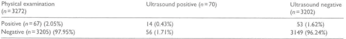
T able 2. The results of physical examination, including false positive and false negative cases.
Physical examination (n = 3272)
Ultrasound positive (o = 70) Ultrasound negative
(n = 3202)
Positive (n = 67) (2.05%) 14 (0.43%) 53 (1.62%)
Negative (n = 3205) (97.95%) 56(1.71%) 3149 (96.24%)
Table 3. The distribution of ultrasound positive hip types, the percentage of physically negative hips and the gender ratio.
Sonographic hip types Number of hips (n = 70) Physically negative Male/female ratio
II c 21 (30.0%) 14 (66.6%) 4/17
D 24 (34.28%) 16 (66.6%) 6/18
III 24 (34.28%) 15 (62.5%) 3/21
IV 1 (1.44%) 1 (100.0%) 0/1
week 3 o f life. The 4 newborns having hip dislocation type III and D were treated with Pavlik harness or fixed abduc
tion brace started on week 3 until complete normalisation.
Parallel with the US screening, the usual physical examinations, Ortolani manoeuvre and Barlow tests were performed on every neonate.
Results were collected and recorded; sensitivity and specificity of clinical examination were calculated.
Statistical analysis was performed using the chi-square test; p < 0.05 was considered to be statistically significant.
Macrosomia was declared when birthweight was >4000 g.
Study protocol was approved by Ethical Committee al University o f Szeged, Hungary (140/2017-SZTE).
Results
The overall presence o f DDH was 21.4 per 1000. Out of 55 newborns, 45 were female (81.8%) and 10 were male (18.2%); the difference is significant (p < 0 .0 1).
Independent of US screening, all newborns had physi
cal examination: 67 of 3272 hips (2.05%) were Barlow or Ortolani positive, but among these 67 hips only 14 (20.8%) were US positive, the remaining 53 hips (79.2%) were declared negative according to G raf method by US.
Important to emphasise that with physical examination, 14 hips of 70 US positive cases (20.0%) were positive, while 56 US positive hips (80.0%) had no physical alteration at all.
Sensitivity and specificity of clinical examinations were cal
culated: 20.0% (95% confidence interval [Cl], 11.39- 31.27%) and 98.34% (95% Cl, 97.84-98.76%) respectively.
Accuracy, namely overall probability that a patient will be correctly classified with physical examination was 96.67% (95% Cl, 96.00-97.26%). Positive predictive value of physical examination was 20.90% (95% Cl, 13.35-31.17%), negative predictive value 98.25% (95%
Cl, 98.04-98.44%) (Table 2).
Different types of positive hips on US according to Graf classification, results of physical examinations and the male/fcmalc ratio are shown on Table 3.
Regarding the risk factors, 79 among 1636 infants (4.82%) were born in breech presentation and 7 of them (12.72%) were in the DDH group, breech presentation proved to be a significant risk factor (/; < 0.05). Regarding macrosomia, 119 neonates of 1636 (7.27%) were affected, among them 5 (9.09%) were in the DDH group, difference was not signifi
cant (jp=0.61). Family history was positive in 49 cases (2.99%), and 6 of them (10.90%) were infants with DDH, which also showed significant difference {p < 0.05). No new
borns had serious foot defects. All twin newborns (46) and triplet neonates (3), were physical and US negative (Tabic 4).
Taking into consideration only the proven significant risk factors, 13 o f 55 involved newborns (23.64%) had either positive family history or were born in breech pres
entation. In addition, 28 o f 55 newborns with DDH (50.90%) had neither any positive physical signs nor any risk factors except being female.
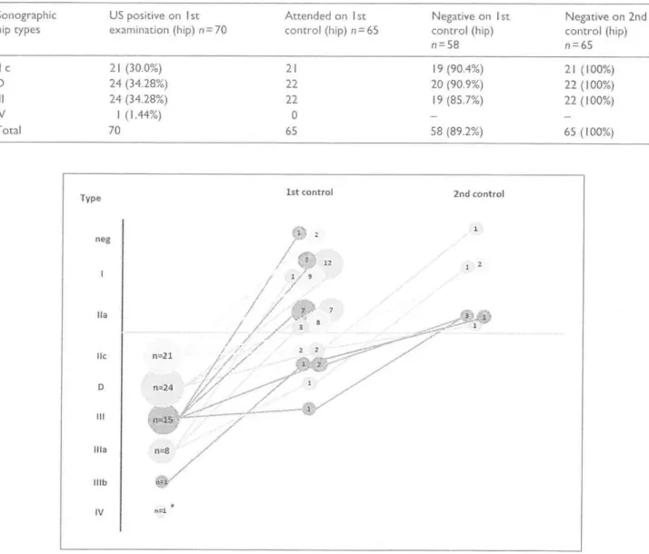
On the 3rd week of life, 50 of 55 US positive newborns were controlled; the remaining 5 did not appear for follow
up examination. At this 1st control. 58 o f 65 hips (89.2%) had already negative US (Table 5) and all o f them (50 new
borns) attending the nd control, on the 6th week of life, were both on US and physical examination negative (Figure 1). In our study, patients were followed up for I year, during this time, we encountered no complications related to treatment and no cases of late diagnosis.
The single patient with type IV dislocated hip was referred straight to an orthopaedic surgeon specialist but the family left the country and returned when the child was 10 months of age. Surgical treatment was offered and per
formed. In her case, only female gender but neither risk factors nor any physical examination signs were positive.
Discussion
The prevalence o f DDH in our universal US screening study was 2.14% which is consistent with the earlier reported data, 0.5-4% according to ethnicity, method of ascertainment and age.4 0
T able 4. Risk factors of developmental dysplasia of the hip (DD H).
Risk factors DDH positive DDH negative p-value
Female gender n = 820 45 (5.48%) 775 (94.52%) <0.05*
Macrosomia n= 1 19 5 (9.09%) 1 14 (90.91%) 0.61
Breech presentation n = 79 7(12.72%) 72 (87.28) <0.05'
Positive family history n = 49 6 (10.9%) 43 (83.1%) <0.05'
Multiple gestations n = 49 0 49 (100%) -
Significance at p < 0.05 is indicated by'
Table 5. Ultrasound (US) positive hips and control examinations.
Sonographic US positive on 1 st Attended on 1st Negative on 1st Negative on 2nd
hip types examination (hip) n = 70 control (hip) n = 65 control (hip) control (hip)
COLOilc
n = 65
lie 21(30.0%) 21 19 (90.4%) 21 (100%)
D 24 (34.28%) 22 20 (90.9%) 22 (100%)
III 24 (34.28%) 22 19 (85.7%) 22 (100%)
IV 1 (1.44%) 0 - -
Total 70 65 58 (89.2%) 65 (100%)
Figure I. Grades of ultrasound positive hips on the 1st and 2nd control examinations.
Prior to this study, selective US screening model had been used in our university, namely newborns in risk groups and with positive physical signs had US examina
tion. In our study, we showed that more than half o f the neonates (50.90%) diagnosed with DDH had neither posi
tive physical signs nor any risk factors except being
female. According to our previous selective screening protocol, all these neonates would have been missed and diagnosed late.
The effectiveness of the standard physical examination method, consisting of Ortolani manoeuvre and Barlow tests, was carefully analysed. We also demonstrated that physical
examination has a poor sensitivity (20.0%) compared to the Graf US method; using physical examination only is an inac
curate and ineffective way of screening DDH. Several previ
ous studies have also reported that selective screening protocol would have missed large percentage o f DDH cases, and the sensitivity of the Ortolani and Barlow manoeuvres was similarly low.10 Discrepancies between clinical and ultra
sound examinations were present even if both examinations had been performed by the same orthopaedic specialist.11
“G raf technique” of hip sonography no longer uses the original clinical and x-ray classification of normal, dys- plastic, subluxated and dislocated hip, but classifies according to the exact anatomical pathology that must be identified and treated appropriately. Not only the bony structures but also the hyaline cartilaginous preformed parts can be identified in a sonogram: the bony angle alpha quantifies the bony socket, and the cartilage angle beta quantifies the cartilaginous acetabular roof. In order to be reproducible, the same sonographic section through the hip joint must be always used, the standard plane. The accurate diagnosis can be achieved only after the proper anatomical identifications and objective measurements of hip joint with considering the age of the patient as well.
Potential risk factors o f DDH are under continuous inves
tigations.12 Some of them like female gender and positive family history, have genetic predisposition and others are related to intrauterine circumstances, like breech presenta
tion and macrosomia. Macrosomia and twin pregnancy, generally considered as risk factors, did not show signifi
cant correlation with DDH in our study.13
Immediately after the 1st positive US result, physio
therapy and wide diapering for spreading were started fol
lowing our treatment protocol. We observed that 89.2% of positive US cases, mainly those with minimal anatomic abnormalities, showed significant improvement on the 3rd week. Benefit of early-age spreading has been known, however the effect of physiotherapy and broad diapering versus spontaneous improvement cannot be judged own
ing to the lack of randomisation.7
Adjusted rate of 1st operative procedure that we calcu
lated, 0.61 per 1000 live births, showed similarity to many other studies with general US screening.14 Furthermore, the only I operative intervention in our study could have been avoided if after the correct diagnosis of DDH, recom
mendations had been followed by the parents. These observations are in accordance with the earlier published research findings. Marks et al.15 found no late detected cases in a group of 14,050 neonates screened by US. Even the randomised controlled trial o f Holen et al.1'’ showed that US screening could have the potential to eradicate the late presenting cases. Reduction of operative procedures was observed in a nationwide survey in Germany as well;
data collected over 5 years showed that incidence of 1st operative procedures was 0.26 per 1000.14 Previous reports from the period of selective screening had shown higher,
relatively stable incidence o f late presenting DDH of approximately 2-3 per 1000, including frank dislocation, subluxation, and acetabular dysplasia.17
Limitations o f our study are its nature of being non- randomised, the relatively small sample size and the lack of long-term follow-up.
The role of DDH screening is known to be essential; how
ever, the exact methodology and mode, whether universal or selective, has not been fully established yet.18 Taking into consideration that DDH can potentially lead to lifelong dis
ability, beneficial effect of universal US has been recognised and its application was recommended more than half a cen
tury ago.19 General US screening was introduced in some European countries, like Austria (1991), Switzerland (1995) and Germany (1996) resulting in a dramatic fall in the rate of open reduction and osteotomies and with Graf technique there was no overtreatment.20'21 On the other hand, there arc opinions that the general screening can lead to overdiagnosis and overtreatrnent, therefore, according to these opinions, considering cost-benefit-equation, selective screening is more favourable.22 In many of these earlier studies, however, the different sonographic techniques were not properly dis
tinct. There are few randomised controlled trials and meta
analysis comparing effectiveness of general versus selective US screening, where absence of conclusive evidence for improved outcomes were shown.23
Rosendahl et al.24-25 stated that the effect of US screening in reducing prevalence of late DDH was at best marginal despite a considerable increase in diagnostic and therapeutic efforts. Woolacott et al.18 in their systematic review based on 23 medical databases, concluded that general US com
pared with clinical screening in newborns may increase overall treatment rates, but US screening seemed to be asso
ciated with shorter and less intrusive treatment. Similarly, in our study, an increased treatment rate was observed com
pared to previous selective US era, but we concluded that all diagnostic and therapeutic works used in our study were noninvasive, cheap and tolerable without any long-term consequences, in contrast to surgical procedures.
Diagnosis and the treatment were both conducted by specially trained orthopaedical and physiotherapeutic spe
cialists. Exact diagnosis is crucial, to make sonograms reproducible and comparable only the standard plane is allowed with standardised examination technique, to avoid typical mistakes like tilting effect.26 In experienced hands with correct technique, the anatomical structures can be visualised and classified according to G raf in seconds with
out any harmful radiation, leading to proper diagnosis.
In our study no operative procedure was needed, except on the patient who did not appear in time for control exam
inations. Clinical screening combined with universal US examination was effective and early detection o f DDH was useful to reduce the need of surgical interventions. Patients without any clinical signs but proven US abnormalities were given the chance for better long-term outcome.
We found the universal screening model with G raf tech
nique a valuable tool for identifying infants with DDH. With careful follow-up and treatment, including physiotherapy, broad diapering, Pavlik harness or fixed abduction brace, it proves to be a simple, non-invasive way to restore a cen
tered hip within the 1st year o f life.
These results should be taken into consideration when new screening models and clinical guidelines are discussed. Early diagnosis is extremely important; every newborn, not only the risk group, should have technically correctly performed US examination. Follow-up studies over more decades would be useful to assess benefits o f US screening in preventing impaired hip function and degenerative joint diseases in adulthood which could be a subject o f further investigation.
D e c la r a tio n o f co n flictin g in t e r e s ts
The aulhor(s) declared no potential conflicts of interest with respect to the research, authorship, and/or publication of this article.
Fu n d in g
The author(s) received no financial support for the research, authorship and/or publication of this article.
R e fe r e n c e s
1. Barlow TO. Early diagnosis and treatment of congenital dis
location of the hip. J Bone Joint Surg 1962; 44: 292-301.
2. Pauwels F. Biomechanics o f the normal and diseased hip.
New York: Springer-Verlag Berlin Heidelberg, 1976.
3. Ganz R, Leunig M, Leunig-Ganz K, et al The etiology of osteoarthritis of the hip: an integrated mechanical concept.
Clin Orthop Relat Res 2008; 466: 264-272.
4. Dezateux C and Rosendahl K. Developmental dysplasia of the hip. Lancet 2007; 5: 1541-1552.
5. Graf R. The diagnosis of congenital hip-joint dislocation by the ultrasonic-combound treatment. Arch Orthop Trauma Surg 1980;97:117-133.
6. Tonnis D. Congenital dysplasia and dislocation o f the hip in children and adults. New York: Springer-Verlag Berlin Heidelberg, 1987.
7. Graf R. Fundamentals of sonographic diagnosis of infant hip dysplasia. JPediatr Orthop 1984; 4: 735-740.
8. Graf R. Guide to sonography o f the infant hip. Stuttgart:
Georg Thiernc Verlag, 2010.
9. Eastwood DM Neonatal hip screening. Lancet 2003; 361:
595-597.
10. Jimenez C, Delgado-Rodriguez M, Lopez-Moratalla M, el al. Validity and diagnostic bias in the clinical screening for congenital dysplasia of the hip. Acta Orthop Belg 1994;
60: 315-321.
11. Kyung BS, Lee SIl. Jeong WK, et al. Disparity between clinical and ultrasound examinations in neonatal hip screen
ing. Clin Orthop Surg 2016; 8: 203-209.
12. Kolb A, Schweiger N, Mailath-Pokorny M, et al. Low incidence of early developmental dysplasia of the hip in universal ultrasonographic screening o f newborns; analy
sis and evaluation of risk factors. Int Orthop 2016; 40:
123-127.
13. American Academy of Pediatrics. Clinical practice guide
line: early detection of developmental dysplasia of the hip. Committee on Quality Improvement, Subcommittee on Developmental Dysplasia of the Hip. Pediatrics 2000;
105fPt 1): 896-905.
14. Von Kries R, ihme N, Oberle D, et al. Effect of ultrasound screening on the rate of first operative procedures for devel
opmental hip dysplasia in Germany. Lancer 2003; 362;
1883-1887.
15. Marks DS, Clegg J and al-Chalabi AN. Routine ultrasound screening for neonatal hip instability. Can it abolish late- presenting congenital dislocation of the hip? ./ Bone Joint Surg Br 1994; 76: 534-538.
16. Holen KJ, Tegnander A, Brcdland T, et al. Universal or selective screening of the neonatal hip using ultrasound? A prospective, randomised trial of 15.529 newborn infants../
Bone Joint Sing Br 2002; 84: 886-890.
17. Terjesen T. Bredland T and Berg V. Ultrasound for hip assessment in the newborn. J Bone Joint Surg Br 1989; 71:
767-773.
18. Woolacott NF. Puhan MA, Steiner J. el al. Ultrasonography in screening for developmental dysplasia of the hip in new
borns; systematic review. BM.J2005; 330: 1413.
19. Ortolani M. A little known sign and its importance for the diagnosis of early subluxation in congenital hip. La Pediatri
1937;45:129-134.
20. Thallinger C, Pospischill R. Ganger R. et al. Long-term results of a nationwide general ultrasound screening sys
tem for developmental disorders of the hip: the Austrian hip screening program../ Child Orthop 2014; 8: 3-10.
21. Graf R. Hip sonography: 20 years experience and results.
Hip Int 2007; I7(Suppl. 5): S8-S14.
22. Shipman SA, Helfand M. Moyer VA, ct al. Screening for developmental dysplasia of the hip: a systematic lit
erature review for the US Preventive Services Task Force.
Pediatrics 2006; 117: e557-e576.
23. Olsen SF, Blom HC and Rosendahl K. Introducing univer
sal ultrasound screening for developmental dysplasia of the hip doubled the treatment rate. Acta Paediatrica 2018; 107:
255-261.
24. Rosendahl K. Markestad T and Lie RT. Ultrasound screen
ing for developmental dysplasia of the hip in the neonate:
the effect on treatment rate and prevalence of late cases.
Pediatrics 1994; 94: 47-52.
25. Rosendahl K and Toma P. Ultrasound in the diagnosis of developmental dysplasia of the hip in newborns. The European approach. A review of methods, accuracy and clinical validity. Fair Radiol 2007; 17: 1960-1967.
26. Graf R. Hip sonography: background; technique and com
mon mistakes; results; debate and politics; challenges. Hip Int 2017; 27: 215-219.