SEXUAL R E P R O D U C T I O N
MARIE-AGNES LETROUIT-GALINOU
I. Ascolichens 59 A. The Basic Cycle of Sexual Reproduction in Ascolichens and
Ascomycetes 59 B. Reproductive Apparatus of the Gametophyte 60
C. Problem of Fertilization of the Ascogones 63
D. The Sporophytic Apparatus 64
E. The Ascus 68 F. Morphology and Anatomy of the Ascocarp 73
G. Basic Sterile Structures of the Ascocarp 75 H. Basic Types of Perithecia and Apothecia 77 I. Special Types of Ascocarps in Pyrenolichens and Discolichens . . . 82
J. Summary 85 II. Basidiolichens 87 References 87
I. Ascolichens
The sexual reproduction of ascolichens is basically similar to that in the other Ascomycetes and will be discussed with reference to those.
A. The Basic Cycle of Sexual Reproduction in Ascolichens and Ascomycetes (Fig. 1)
According to Chadefaud (1953, 1960), the basic reproductive cycle in Ascomycetes is not digenetic, as often claimed, but is in principle trigenetic, as in the algal family Floridaceae or, among the Basidiomycetes, the Uredinales with alternation of (1) a gametophyte producing male and female sexual elements; (2) sporophyte I (= prosporophyte), siphonaceous and mictohaploid, that is, with cells containing several nuclei, some male and the others female but still immature and not coupled; and (3) sporophyte II (= ascosporophyte), dikaryotic and giving rise to asci. Sporophytes I
59
60 MARIE-AGNES LETROUIT-GALINOU
ο 0
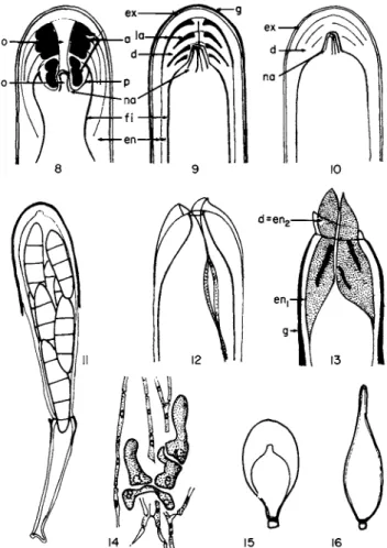
Δ Β C D FIG. 1. Basic scheme of sexual reproduction in ascolichens. A , Haploid spore (ascospore or conidiospore) producing a thallus; B, pycnidium in which spores arise, some considered to be direct spores (left arrow), others spermatia (right arrow); C, ascogonial apparatus (as) included in the gametophytic primordium, trichogamy may possibly occur; D, sporophytic generating asci(a) apparatus which develops on the ascogonial apparatus and is included in an ascocarp derived from the primordium. Chromosomal reduction (R!) takes place in the asci (S, and S2, prosporophyte and ascosporophyte).
and II together constitute the sporophytic apparatus and develop parasiti- cally on the gametophyte in the interior of fruiting bodies that are called ascocarps.
B. Reproductive Apparatus of the Gametophyte
In the nonlichenized Ascomycetes, the gametophyte consists of mycelia and appendages, sclerotia and stromas, and it produces three types of re
productive filaments: (1) asexual reproductive filaments, or conidiogenous filaments, directly producing nonmotile spores called conidial spores or conidia; (2) male filaments which are either spermatogenous filaments producing nonmotile male gametes (called spermatia) or parascogonial filaments associated with ascogonial filaments and which ensure fertilization directly; and (3) female or ascogonial filaments, certain cells of which (ascogonial cells) have a role as female elements. According to Chadefaud (1960), the existence of spermatia to which parascogonial filaments have not been substituted might be a primitive character indicative of an early stage of evolution.
In ascolichens, the gametophyte consists of the thallus which produces or can produce only two types of reproductive structures: microsporogenous filaments and ascogonial filaments. Microsporogenous filaments produce microspores, the nature of which (whether conidial or spermatial) is under discussion. They are generally confined in fructifications that resemble pycnidia. Ascogonial filaments which are lodged in the primordia of the ascocarps are never accompanied by fertilizing parascogonial filaments.
They are formed in the primordia or these secondarily develop around them, depending on the species.
1. THE MICROSPOROGENOUS FILAMENTS (Fig. 2)
Microspores of various shapes and forms are produced by more or less differentiated sporogenous cells. These spores are always exogenous, that is, arising from buds. They are formed on a sporogenous cell, quite often at the tip of a style, never at the bottom of a neck or a ring collar as is seen in certain nonlichenized Ascomycetes.
FIGS. 2 AND 3. Reproductive apparatus of the gametophyte. Fig. 2, microsporogenous filaments. A, endobasidial filament of Buellia canescens; B, part of the endobasidial micro- sporogenous plexus in Lobaria laetevirens; C, exobasidial filament of Roccella montagnei (c, microspore). Fig. 3, ascogonial filaments. A, complex ascogonial apparatus formed of a large number of ascogonial filaments of the Collema type in Lecidea elaeochroma; B, ascogonial filament of Collema sp. (after Stahl, 1877); C, vesiculate multinucleate cells (a) of the ascogonial apparatus of Peltigera rufescens (a, ascogonial cells; c, microspore; tr, trichogyne).
62 MARIE-AGNES LETROUIT-GALINOU
The sporogenous cells, in general, belong to differentiated sporogenous filaments. Always multicellular, they are simple or branched and free or anastomosed, depending on the species. In this case, they can form a loose network or a kind of lacunose paraplectenchyma (Fig. 2B).
Two types of sporogenous filaments may be distinguished (Gluck, 1899).
In the exobasidial type (Fig. 2C), the sporogenous cells, in this case called
"sterigmata," arise singly on lateral or subterminal sporophores. They are homologous to lateral branches and, as a rule, are elongated and morpholog- ically distinct from sterile cells. In the endobasidial type (Fig. 2A, B) the sporogenous cells are generally less distinct from sterile cells and by contrast lie in an intercalary or terminal position. The resulting chainlike arrange- ment is similar to that in some nonlichenized Ascomy cetes such as Neurospora sitophila.
The sporogenous filaments are joined into pycnidia except in some poorly known groups (Ozenda and Clauzade, 1970). Pycnidia are spherical or flask-shaped, opening at the apex with an ostiole. Rarely projecting or stalked {Cladonia), they are often included in the thallus. They have a wall, sometimes dark. The sporogenous filaments can fill the entire cavity in a lacunose mass when anastomosed. They very often form a fertile layer, sometimes plicate {Cladonia, Ramalina, etc.), covering the inner surface of the wall.
According to some authors (Moller, 1888), these microspores are asexual spores capable of germinating, but according to others they behave as spermatia, functional or not, for one can observe them adhering to tricho- gynes of ascogenous filaments (Stahl, 1877; Baur, 1898; Ahmadjian, 1966;
Jahns, 1970). If they are indeed spermatia, the pycnidia from which they emerge must be called spermogonia.
2. THE ASCOGONIAL FILAMENTS (Fig. 3)
The female reproductive organ in the Ascomycetes as a group is made up of differentiated filaments that are always multicellular, ascogonial filaments apparently homologous to the male sporogenous filaments. They are formed mainly of three parts, going from the base to the apex (but not all the parts will always be present): a foot composed of sterile cells; theascogone, the essential part composed of the fertile cell, sometimes several (= asco- gonial cells); and the trichogyne, slender and erect, sterile, and when there is trichogamy attached either with spermatia or a fertilizing cell of parasco- gonial filaments. The ascogonial filaments do not give rise to free gametes.
It is the ascogonial cells which play the role of fertile elements. Each of these can be considered to beanongametogenousgametocyst(Chadefaud, 1960).
Ascogonial filaments were first observed in lichens by Fuisting (1868) for Lecidea fumosa and later by Stahl (1877). Stahl interpreted them as female
elements and carefully described their structure and evolution in various species of Collema. The filaments were eventually observed in all species examined, with the exception of Phlyctis agelaea.
The ascogonial filaments in lichens are always composed of a large number of cells, contrary to many of the Ascomycetes. This applies especially to the fertile part that presents a clearly filamentous aspect not unlike that of thalline hyphae. For the most part the foot is generally little differentiated.
Moreau and Moreau (1928) distinguished two kinds of ascogonial fila- ments. The Collema type (Fig. 3B), by far the most common, has uninucleate, cylindrical, and isodiametric ascogonial cells; the Peltigera type (Fig. 3C).
found in Peltigera and Solorina, has ellipsoidal and multinucleate ascogonial cells. Trichogynes in both types are differentiated slowly and often branched.
The ascogonial filaments may be simple and originate singly. More often they are branched and their parts together makeup the ascogonial apparatus.
At times one finds an arbuscular type where each branch remains free. At other times we have a more or less glomerular type where the branches are intertwined in a more or less dense cluster or glomerule, sometimes giving a paraplectenchymatous aspect (Fig. 3A), from which trichogynes arise. In such a case only the central elements in the glomerule are fertile. Another type of ascogonial apparatus has been found in Pertusariapertusa (Letrouit- Galinou, 1966), Baeomyces and Icmadophila ericetorum (Nienburg, 1908), and various Cladoniae (Krabbe, 1891). This is the spreading type, composed of a branched system of sterile supporting hyphae on which fertile hyphae develop from place to place, and contain ascogonial cells.
Nothing is known about the basis of differentiation of the reproductive apparatus. There are no reports in lichens of either heterothallism, so common in nonlichenized Ascomycetes, or of a dioecious condition.
C Problem of Fertilization of the Ascogones
There has been heated discussion on the question of whether fertilization of the ascogone is involved in the developmental stages. According to several authors microspores of lichens are spermatia which do in some cases become attached to the trichogyne and fertilize the ascogone. Consequently, spermatial trichogamy would be a basic phenomenon. In support of this thesis one should note that (1) the asexual nature of these microspores is not well established, (2) spermatia have often been found adhering to the apex of trichogynes (Stahl, 1877; Baur, 1898; Ahmadjian, 1966) and they can result in structural modifications of the trichogyne and ascogonial cells, and (3) according to Ahmadjian (1966), the great variability in monospore cultures of Cladonia cristatella can be explained only by genetic factors in- compatible with apogamy.
64 MARIE-AGNES LETROUIT-GALINOU
The opposite theory, stressed by Moreau and Moreau (1928), holds apogamy to be the rule. This is clearly the case when microspores are lacking;
when present it is stated that they are always either asexual spores or non- functional spermatia. One of the more serious arguments for partisans of this theory is that the initial hyphae of the ascosporophyte in a number of lichens (especially many of those cited as examples of trichogamy) are uninucleate, not dikaryotic. This is easily explained by apogamy, and with greater difficulty by trichogamy. In this last instance one must allow for a hypothesis referred to as double fertilization (Gwynne-Vaughan and Williamson, 1931), according to which the fertilized ascogone and the sporophytic apparatus derived from it would have diploid nuclei (conjugated or not) and in no case conjugated haploid nuclei. This hypothesis, however, was rejected some time ago (see Martens, 1946; Chadefaud, 1960).
Until better informed, we will take the following point of view:
1. Apogamy is probable if primary ascosporophytic hyphae with uninu- cleate cells are formed in the developmental stages. It is then always accompanied either by a fusion between cells of the ascosporophyte (= perittogamy) or by other phenomena leading to the formation of dikaryo- tic cells.
2. One could admit fertilization of the ascogone, resulting in the forma- tion of a dikaryon, if the hyphae of the ascosporophyte are dikaryotic from the outset. According to the species, this fertilization would be classified as spermatial trichogamy (basic case); somatogamy, the conjugated nuclei being issued from normal gametophytic cells [for example, Lecidea elaeo-
chroma (Letrouit-Galinou, 1966)]; or autogamy, in other words the transfor- mation of nuclei of the ascogone, some into male nuclei, others into female nuclei (that seen perhaps in Peltigera).
D. The Sporophytic Apparatus 1. ASCOMYCETES IN GENERAL
The sporophytic apparatus is normally formed by the combination of two phases of the reproductive cycle: that of sporophyte I (the prosporophyte) and that of sporophyte II (the ascosporophyte).
a. SPOROPHYTE I. This phase of the trigenetic cycle, often reduced and appearing absent, is intercalated between the ascogonial cell, fertilized or apogamous, and the dikaryotic ascogenous filaments described by Claussen (1912). Unsuspected for a long time (Martens, 1946), it was first defined and interpreted by Chadefaud (1953, 1960).
Basically the fertilized ascogonial cell, which takes the place of the zygote, gives rise to some vesicles or tubes (cf. Pyronema confluens and Peltigera
spp.) that are, as a rule, siphonaceous, multinucleate, and mictohaploid, that is, provided with separate haploid nuclei, some male and some female but still immature and not conjugated. These vesicles and tubes constitute the prosporophyte which develops as a parasite on the gametophyte. Later, in certainly the most primitive case (Dothidea puccinioides; Luttrell, 1951b), they are transformed into sporocysts, in the interior of which small spores with dikaryons are differentiated. The two included conjugated nuclei are one male and one female, semimature, so that they are coupled without fusing. These spores (= Chadefaud's carpospores) are not liberated; they give rise to the ascosporophyte at this point.
The prosporophyte is, in fact, never this typical since (1) it is often little developed, sometimes reduced to the ascogonial cells alone; (2) the spores are only very rarely individualized: sometimes their formation is only present in outline; sporulation is often replaced by a simple division into spore cells
(Pertusaria pertusa). There is also often complete apospory and then the prosporophytic vesicle gives rise directly to sporophyte II. In some of the Discomycetes, especially Pyronema confluens, and perhaps also in Peltigera rufescens, sporulation is replaced by the emission of dikaryotic carposporous tubes on which the ascosporophyte then arises.
b. SPOROPHYTE II. Sporophyte II or the ascosporophyte develops from dikaryotic spores, the spore cells, or the carpospore tubes produced by sporophyte I, or directly from sporophyte I. As a parasite on the gametophyte it is formed by "ascogenous filaments" with dikaryons, each dikaryon being composed of two coupled haploid nuclei, one male and one female, which divide by conjugated mitoses. These filaments generally consist of hyphae with clamp connections comparable to those of sporophytes in the Basi- diomycetes and, like them, composed of successive dangeardia. The type of dangeardium involved here is the well-known croziers. They are made up of a group of cells which arise by division of one cell with a dikaryon. They are most often formed with two dikaryotic cells, but there are other rarer types (Chadefaud, 1943, 1960). Certain of them, situated at tips of hyphae, are ascogenous dangeardia: their dikaryotic cells give rise to an ascus in which ascospores are formed, and these will produce new gametophytes when liberated.
c. THE PERITTOGAMOUS SPOROPHYTE (Fig. 6). In certain apogamous species, especially with many lichens, the ascosporophyte is not initially dikaryotic. It consists of primary ascosporophytic filaments with uninu- cleate, haploid cells, connected to dikaryotic secondary ascosporophytic filaments. Each secondary filament begins either with one cell of a primary filament becoming binucleate by a mitosis, not followed by septation, or by two primary filaments, fusing without karyogamy. Such a fusion has been
66 MARIE-AGNES LETROUIT-GALINOU
FIGS. 4 - 7 . Sporophytic apparatus of ascolichens. Fig. 4, evolution of the sporophytic apparatus in Buellia canescens. A, ascogonial apparatus; B, vesiculate, uninucleate prosporo- phytic cells; C , primary part formed of uninucleate cells of the perittogamous ascosporophyte;
D, secondary dikaryotic part of the ascosporophyte. Fig. 5, evolution of the sporophytic apparatus in Pertusaria pertusa. A, mictohaploid prosporophytic apparatus; B, mass of uninucleate cells coming after the prosporophyte (carposporal cells?); C , uninucleate cells of the primary part of the ascosporophyte; D, dikaryotic, ascogenous secondary part of the ascosporophyte. Fig. 6, perittogamy in Collema pulposum (after Moreau and Moreau, 1928).
Fig. 7, Lecanora subfuscata: matured part of the ascosporophyte with secondarily uninucleate cells, (a, ascogonial filament; asp,, primary part of the ascosporophyte; asp2, secondary part of the ascosporophyte; cs, carposporal cells; psp, prosporophyte.)
described by Killian (1938) in the pyrenomycete Lasiobotrys lonicerae under the term perittogamy. According to drawings by Moreau and Moreau (1928), it also occurs in the lichen Collema pulposum and, according to Erbisch (1969), in Pertusaria pertusa.
2. THE SPOROPHYTIC APPARATUS IN LICHENS
The development of this apparatus is known in only about 15 discolichens.
Sporophyte I appears to be more or less reduced to the ascogonial cells alone
except in Peltigera where it gives rise apparently to carposporic tubes.
Formation of the sporophytic apparatus at the expense of the carpospore cells has been observed in Pertusariapertusa, and this phenomenon may also occur in Lobaria laetevirens (Letrouit-Galinou, 1966, 1971). The ascosporo- phyte in all other cases arises directly from prosporophytic vesicles.
Sporophyte II begins as a primary part of uninucleate cells in almost all species that have been studied. To this a dikaryotic secondary part is next added by perittogamy or otherwise. This secondary part with a few excep- tions (perhaps certain Baeomycetaceae and Peltigeraceae) is formed of hyphae with clamp connections or with lateral hooks (Moreau and Moreau, 1925) and is then "dangeardian."
This sequence can be illustrated with the five examples that follow. The ascosporophyte in the first three examples comprises a primary part with uninucleate cells.
a. Collema sp. (Baur, 1898; Moreau and Moreau, 1928). The asco- gonial apparatus is an "archicarp" coiled in a helix and terminated by a long multicellular trichogyne, the tip of which projects above the thallus.
The ascogonial cells in this archicarp enlarge and become vesiculate and multinucleate and so are transformed in prosporophytic vesicles. The vesicles give rise to branched hyphae which form the primary part of the ascosporophyte and are made of uninucleate cells. One would be led to be- lieve that there is apogamy in accordance with the views of Moreau and Moreau. Next, hyphae of the secondary part are developed; these are dikaryotic and provided with lateral clamps and give rise to asci at their tips.
The first dikaryotic cells can originate from the fusion of two adjacent uninucleate cells, and this would then be considered perittogamy (Fig. 6).
b. Pertusariapertusa (Baur, 1901; Letrouit-Galinou, 1966; Erbisch, 1969) (Fig. 5). The ascogonial apparatus is complex and branched with asco- gonial filaments of the Collema type (Fig. 5A). Certain cells evolve into multinucleate prosporophytic vesicles which later become masses of uninu- cleate carposporal cells (Fig. 5B). From these cells come primary ascosporo- phytic hyphae with uninucleate cells (Fig. 5C). Next, through perittogamy, dikaryotic cells are formed (Fig. 5D) which give rise to secondary asco- sporophytic filaments with lateral clamps, producing asci. According to Erbisch the development varies from one species to the next in Pertusaria.
c. Buellia canescens (Letrouit-Galinou, 1966) (Fig. 4). The ascogonial apparatus is formed of isolated filaments of the Collema type (Fig. 4A).
Certain of these elements consists of ascogonial cells which enlarge and are transformed into apparently prosporophytic vesicles; these nevertheless
68 MARIE-AGNfeS LETROUIT-GALINOU
remain uninucleate (Fig. 4B), contrary to the situation in the preceding case and reminiscent of apogamy. The resulting ascosporophyte is formed of a primary part with uninucleate cells (Fig. 4C) and a secondary ascogenous dikaryotic part with lateral clamps (Fig. 4D).
d. Lecidea elaeochroma (Letrouit-Galinou, 1966). The ascogonial ap- paratus with a glomerular aspect consists of ascogonial filaments of the
Collema type in which the ascogonial cells become binucleate, perhaps as a consequence of somatogamy. They enlarge and become transformed into a vesicular prosporophytic apparatus, probably siphonaceous and multinucleate. From this apparatus come the ascosporophytic hyphae which are at first dikaryotic and provided with lateral clamps. The asci form slowly on the hyphae.
e. Peltigera (Moreau and Moreau, 1928; Letrouit-Galinou and Lallemant, 1971). The ascogonial cells are initially multinucleate. They give rise immediately, or after transformation into prosporophytic vesicles, to long siphonaceous tubes which are apparently carposporous tubes. The hyphae of the ascosporophyte, dikaryotic ascogenous hyphae with lateral clamps, arise from these tubes either directly or after division into dikaryotic cells.
The oldest ascosporophytic cells in Buellia canescens, Lecidea elaeochroma,
and Lecanora subfuscata seem to become secondarily uninucleate (Fig. 7) but the reason for this phenomenon is unknown.
E. The Ascus
Asci in lichens are always derived from dikaryotic proascal cells so that karyogamy takes place at the beginning of their development and the same if the ascogonial apparatus is apogamic. The ascus originates most often from a fertile cell of a dangeardium with formation of a lateral clamp or a hook;
in that case, one can see at the base of the ascus traces of the two septa which separated it from the basal cell of this dangeardium and from its clamp or hook. A single ascus can be formed initially from an ascogenous dangeardium, but many are often formed because the clamp of the dangear- dium produces a secondary ascogenous dangeardium of which the clamp in turn produces a third, etc.
The three elements of the ascogenous dangeardium in Icmadophila erice-
torum (Chadefaud, 1960)—the basal cell, fertile cell, and hook—are oriented in a nearly straight line; the ascus rises laterally on the fertile cell in this case.
At other times,, as in Pertusaria pertusa according to Erbisch, there is no ascogenous dangeardium. The ascus comes directly from a dikaryotic cell without producing such a dangeardium at first.
Asci are generally claviform, a character that could be interpreted as primitive according to Chadefaud (1960), but they may also have more characteristic shapes in certain families: subglobose or pear-shaped in the Arthoniaceae and Cryptotheciaceae (Fig. 15), flask-shaped in the Thelocar- paceae (Fig. 16), or cylindrical in the Caliciaceae.
FIGS. 8 - 1 6 . Asci of lichens. Fig. 8 , basic scheme of the apical apparatus of the ascus (adapted from Parguey-Leduc and Chadefaud, 1 9 6 3 ) ; Fig. 9 , the archaeasceous type; Fig. 1 0 , the nassasceous type; Fig. 1 1 , jack-in-the-box-type dehiscence in Roccella montagnei; Fig. 1 2 , bivalve dehiscence in Pertusaria pertusa; Fig. 1 3 ; rostrate dehiscence of Parmelia caperata;
Fig. 1 4 , young asci at the extremity of ascosporophytic hyphae; Fig. 1 5 , ascus of Arthonia;
Fig. 1 6 , flask-shaped ascus of Thelocarpon. (a, amyloid ring; co, ocular chamber; d, apical dome; en, endoascus; en,, external layer of the endoascus; e n2, internal layer of the endo- ascus; ex, exoascus; fi, internal film; g, amyloid gelatin; la, annular amyloid lamellae; na, apical nasse; o, oculus of the dome obscured here by the apical cushion and its manubrium;
p, pendant of the dome.)
70 MARIE-AGNES LETROUIT-GALINOU 1. THE ASCAL WALL, APICAL APPARATUS, AND METHOD
OF DEHISCENCE
According to observations with the light microscope, which are almost the only ones available, juvenile asci have a thin wall which is covered in discolichens with an I + blue gelatin (= amyloid gelatin) at a very early stage.
Later on, this wall thickens so that the asci in lichens look more like those of bitunicate nonlichenized Ascomycetes than those of unitunicate nonlichen- ized Ascomycetes.
The ascal wall in lichens, fundamentally conforming to the general type (Chadefaud, 1942, 1960, 1969), consists of two layers, both complex: (1) an external firm layer, the exoascus, often covered with the gelatin mentioned above, and (2) an internal layer, the endoascus, sometimes thick and basically double. An internal membrane can be added to these layers, in connection with the plasmalemma of the ascus.
The structure or staining qualities of these layers are often modified at the apex of the ascus. Various formations that are differentiated make up the apical apparatus of the asci as studied by Chadefaud (1942, 1960, 1969, Fig. 8). This apparatus can include the following three parts:
1. An apical calotte derived from the exoascus.
2. An apical dome, an essential part of the apical apparatus. Laminate, it arises from the endoascus, particularly from the inner layer. Its sides extend far down toward the base of the ascus. The summit is thickened, and its axis is often hollow with one ocular chamber open at the base or with the oculus open at both ends. If there is one oculus, it is generally plugged apically by an apical cushion elongated into the oculus by a manubrium. The dome can produce around the lower orifice of the ocular chamber or oculus a tubular pendant projecting toward the base in the ascal cavity. Various parts of a more or less complex apical ring are differentiated in the dome and in its pendant. These are always amyloid in lichens.
3. An apical nasse (trap), characteristic of the nassasceous type. This is composed of straight or coiled longitudinal rods lodged in the ocular chamber, lying against the walls of this chamber and joined at the summit.
These rods seem to be in connection with the epiplasm.
All lichens are inoperculate as far as the type of dehiscence is concerned (Ziegenspeck, 1926; Richardson and Morgan-Jones, 1964; Letrouit-Galinou,
1966; Richardson, 1970), with the following main variations:
1. Bitunicate jack-in-the-box dehiscence (Fig. 11) (pyrenolichens, some discolichens): the exoascus ruptures at the tip or in a circle at a distance from the tip; the endoascus is ejected and elongates carrying along the ascal contents.
2. Bivalvate dehiscence (Fig. 12) (e.g.,Pertusariaa.ndBaeomyces; Calopla-
caceae, Gyalectaceae): the exoascus splits longitudinally into two valves.
The endoascus does not come out.
3. Rostrate dehiscence (Fig. 13) (most discolichens): the exoascus and the external layer of the endoascus split only at the tip; the apical dome bursts out, sometimes with eversion.
4. Poricidal dehiscence: dehiscence by a more or less well-defined apical pore (Graphis elegans; Richardson, 1970).
5. Rupture, disarticulation, or gelification of the ascal wall (Caliciaceae).
Some asci seem to be indehiscent (e.g., Phlyctis).
Particulars of the asci, their walls, apical apparatus, and manner of dehiscence may be defined under several broad ascal types (Chadefaud, 1942, 1969; Luttrell, 1951a). Most lichens fall under two types, and these may in fact be connected: the nassaceous bitunicate and the archaeasceous types:
1. The bitunicate nassasceous type (Fig. 10) is found in nearly all pyreno- lichens and in certain groups of discolichens (e.g., Arthonia, Opegrapha, Chiodecton, etc.). It is the type known in the dothideaceous pyrenomycetes and some nonlichenized Discomycetes, in particular Patellaria atrata which is classified in the Lecanorales. The exoascus is thin and the endoascus thick. The apical dome is nonamyloid and hollowed with an ocular cham- ber containing a more or less distinct apical nasse with dehiscence of the jack-in-the-box type.
2. The archaeasceous type (Fig. 9) is characteristic of lichens in the order Lecanorales, apart from some of the Patellariaceae. The exoascus is covered with a very characteristic amyloid gelatin; the endoascus, also amyloid, is made up of two layers. The apical dome arises from the innermost layer which may even be reduced to this dome, a thick, laminate, and usually amyloid structure. It is often hollow with an ocular chamber. This chamber can include an apical nasse which leads to the nassasceous type. It can be surrounded by an amyloid ring differentiated in the dome which also leads into the nassasceous type. Dehiscence is rostrate or bivalvate. Several variants are distinguishable by the fine structure of the dome (Chadefaud et al., 1963; Letrouit-Galinou, 1973).
2. CYTOLOGY OF THE Ascus: SPOROGENESIS AND ASCOSPORES
We know very little of the cytoplasmic features of the ascus and its inclu- sions, nature of reserves (sometimes lipids, other times glycogens), or evolu- tion. Some authors (Maire, 1905; Moreau and Moreau, .1919; Stevens, 1941;
Letrouit-Galinou, 1966) have observed various configurations in nuclear division, confirming in the species investigated that the first mitosis is indeed reductional.
Some aspects of spore delimitation in Roccella montagnei resemble stages
72 MARIE-AGNES LETROUIT-GALINOU
described by Harper (Fig. 19), but one should realize that these stages are not recognized today, and it appears rather certain that they would not be found in all species. In Physcia aipolia (Fig. 17) (Rudolph and Giesy, 1966) with brown biseptate spores, the mechanism of ascospore formation is in effect one described by Carroll (1969) for Ascodesmis and Ascobolus. A very thin, flexible double membrane, related to the endoplasmic reticulum, com- pletely surrounds the sporoplasma with the eight spore nuclei at first. The sac subsequently formed by invagination subdivides into eight sections, each containing a nucleus and forming the primordial envelope of each spore. The primary membrane of the spore (Fig. 17A) is first laid down between the two layers of this envelope, next becoming the epispore. Later the secondary membrane with radial structure is laid down between this and the external layer and becomes theexospore(Fig. 17B).Theintercellular septum appears as an annular fold of the primary membrane which extends
FIGS. 17-19. Ascospores. Fig. 17, differentiation of the spore wall in Physcia aipolia (based on photographs of Rudolph and Giesy, 1966). A, deposition of the primary membrane (i) ( = epispore); B, formation of the secondary membrane (exs) ( = exospore); C, differentiated spore (cu, epilocular cupules; ens, endospore; exs, exospore; i, epispore). Fig. 18, examples of ascospores. A, Caloplaca cerina; B, Roccella montagnei; C, Phlyctis agelaea; D , Lecanora subfuscata\ E, Acarospora; F, Pertusaria pertusa (half-spore). Fig. 19, beginning of spore differentiation in Roccella montagnei.
centripetally. This septum contains a very thin interlocular lamina (Fig.
17C). Moreover, a thin endospore forms around the cytoplasm of each cell, and a lamellar deposit around this endospore thickens the epispore con- siderably. The endospore of each cell seems to be what Chadefaud (1960) calls a locula and the layers contributing to a thickening of the epispore might then be the epilocular cupules.
There are generally eight spores per ascus, but there may be more (several hundred in the Acarosporales) or less, either because some of the nuclei abort or because the spore membrane includes two or more nuclei per spore.
Spores (Fig. 18) are sometimes unicellular (and these uni-, bi-, or multi- nucleate), uniseptate, or multiseptate and muriform or not. Size varies in length from a few microns (Acarosporales) to several hundred microns (some Pertusaria and Varicellaria species).
F. Morphology and Anatomy of the Ascocarp
The ascosporophyte and asci of lichens develop in the interior of the specific gametophyte formations, the ascocarps, comparable to those of nonlichenized Ascomycetes, with the exception of those in the Cryptothe- ciaceae which seem directly immersed in the thallus. We will examine in turn the morphology, anatomy, and ontogeny of lichen ascocarps.
1. MORPHOLOGY OF THE ASCOCARP
Two broad types of ascocarps, the perithecium and apothecium, are traditionally recognized in lichens, just as in the nonlichenized Ascomycetes.
The asci of perithecia are immersed in a perithecial cavity which is bounded by an exciple distinct from the thallus. This cavity opens at the top through a narrow pore, which is sometimes located at the end of a more or less elongated neck.
With apothecia the asci are by contrast directly exposed to the exterior.
Apothecia are as a rule round but they can also be lirelline, that is, elongated and sometimes branched. They may or may not have an exciple.
2. ANATOMY OF THE ASCOCARP
The ascocarps of lichens, as in other ascomycetes, are made up of three parts: the hymenium, the subhymenium, and the exciple (Fig. 20):
a. The hymenium consists of the asci, among which are very often inter- mingled sterile interascal filaments, uni-or multicellular, simple or branched, and free or anastomosed. There are three kinds of filaments depending on their origin: (1) true paraphyses, free filaments produced from the sub-
74 MARIE-AGNkS LETROUIT-GALINOU
FIGS. 20 AND 21. Basic structure of ascocarps. Fig. 20, basic structure of a mature ascocarp, (in this case Lecanora subfuscatd). Fig. 21, basic developmental scheme of ascocarps of ascolichens. A, the primordium; B, primary corpus; C, formation of a secondary preparathecial envelope; D, mature ascocarp provided with a typical parathecial apparatus, (am, amphi- thecium; cl, epihymenial cupola which gives rise to pseudoparaphyses; cpa, parathecial crown;
es, secondary parathecioid envelope; exc, exciple, often double; h, hymenium; pa, para- thecium; pc, circumcentral plexus; pi, pericentral base; q, paraphysoid apparatus; sh, sub- hymenial meniscus; sh,, primary part of the subhymenium; sh2, secondary part of the subhymenium; t, pericentral tegment.)
hymenium and extending upward in the same direction as the asci; (2) pseudoparaphyses, free or anastomosed filaments produced from the epi- hymenial cupola and growing out in directions opposite to the asci. They are sometimes attached to the subhymenium and break off apically, a condition that is capable of mimicking true paraphyses; and (3) paraphysoids, attached from the start at both ends and formed by stretching and intercalary elonga- tion of carpocentral elements to be described below.
A pulverulent mazaedium containing spores is formed in the Caliciales by transformation of the hymenium after breakdown of the ascal walls and fragmentation of the paraphyses.
b. The subhymenium contains the sporophytic apparatus. In addition it consists of sterile filaments, the primary ones being part of the carpocenter (see below), the secondary ones consisting of the bases of the interascal filaments.
c. The exciple, sometimes lacking (Arthoniaceae, Phlyctidiaceae), con- sists solely of sterile hyphae. It more or less completely surrounds the hymenium and subhymenium. It is called lecideine when no algae are present and is clearly distinguished from the thallus by texture and color. Lecanorine is in the alternative case where algae are incorporated in the exciple. As a matter of fact, the nature of the exciple varies considerably from case to case depending on its ontogeny.
G. Basic Sterile Structures of the Ascocarp
Development of ascocarps is a complex phenomenon which involves many structures of which some are transitory, ones being primary, the others secondary (Letrouit-Galinou, 1968). There are many types and variations which will be discussed below.
1. PRIMARY STRUCTURES
These consist of the primordium and the primary corpus derived from it.
a. The primordium (Fig. 21 A) (= generative Gewebe of Henssen, 1968) is a small gametophytic mass of homogeneous texture, containing the asco- gonial apparatus. The constituent hyphae arise at the foot of the ascogone in the Collemataceae (Stahl, 1877; Henssen, 1965). It is derived directly from the thalline hyphae in all other cases, often preserving their structure (e.g.,
Nephroma, Peltigera) or are ramifications of them.
As a rule the primordium is formed before ascogonial filaments, more rarely later (e.g., Collema, Buellia, Physcia). It is paraplectenchymatous in some Arthopyreniae (Janex-Favre, 1970), as the structure called an asco- stroma in some of the nonlichenized ascomycetes. It is arbuscular in some cases.
b. The primary corpus (Fig. 21B) is derived directly from the primordium, from which are differentiated a central mass or carpocenter (containing the ascogonial apparatus, then the sporophytic apparatus produced by it) and a pericentral envelope. This includes, generally speaking, a tectal part or tegment, a basal part or floor. It may be lacking when the primordium is totally transformed into a carpocenter (e.g., Lecanora).
From the carpocenter are formed (1) the subhymenial apparatus which
76 MARIE-AGNES LETROUIT-GALINOU
contains the sporophytic apparatus and is sometimes paraphysogenous and gives rise to true paraphyses; (2) an epihymenial apparatus often in the form of a bell; it can give rise to descending filaments which remain short or develop into definite pseudoparaphyses; and (3) a paraphysoid apparatus composed of paraphysoidal filaments which are often anastomosed and joined with the other two apparatus described above and which may or may
not persist in the form of paraphysoidal interascal filaments.
The primary corpus can enlarge by adding on elements of the same nature at its periphery, with this aggregate making up the peribase. It is formed at the expense of the circumcentral plexus, lateral and annular, probably in nature similar to that of the primordium.
The primary corpus in Lobaria laetevirens is probably differentiated at the expense of the thallus.
2. SECONDARY STRUCTURES
In contrast to the primary structures these are not derived directly from the primordium. They are added secondarily to the primary elements. Some of these are preparathecial sensu lato (Bellemere, 1967); others comprise the parathecial apparatus sensu stricto.
a. Secondary preparathecial structures (Fig. 21C), still poorly known, develop after the differentiation of the carpocenter and before that of the parathecial apparatus when this is present. They could be derived from the carpocenter but possibly also from the thallus. These structures are complete if they entirely surround the carpocenter and incomplete otherwise. They are usually formed of erect, parallel, or contiguous filaments which gives them a parathecioid appearance. They may be provided with amphithecioid or amphithecial filaments on the exterior face and with secondary para- physes on the inner face. The tip may be provided with a parathecial ring composed of divaricate filaments.
In this way the following are (or could be) secondary formations: the proparathecium of Buellia canescens; the lateral wall and inner lining of the lirellae in the Graphidales; the periphyses-invested wall of the Thelotrema- taceae; the secondary envelope (and perhaps the carpocentral envelope as well) of certain pyrenolichens; the perihy menial envelope of Pertusaria; that in Lichina; and, finally, the parathecioid envelope of Peltigera and Lobaria.
These formations are of special interest because their structure is inter- mediate between that of the pericentral envelope, to which it is added, and that of the parathecial envelope which follows.
b. The parathecial apparatus (Figs. 21D and 32D) was first carefully described by Corner (1929-1930) and Dughi (1952, 1954). It comprises (l)a parathecium, often cup-shaped, flaring, and composed of filaments which,
oriented directly obliquely toward the top and exterior, are elongated and branched; (2) an amphithecium, lecideine or lecanorine, formed of hyphae that arise at the outer face of the parathecium and are oriented toward the exterior; this makes up part of the exciple; ( 3 ) a system of secondary paraphyses which, by contrast, arise at the inner face of the parathecium and encircle the primary part of the hymenium; these remain extrahymenial in some of the ascohymenial nonlichenized Pyrenomycetes; (4) a more or less distinct parathecial ring at the summit of the parathecium composed of short filaments intercalated between those of the amphithecium and the secondary paraphyses.
The parathecial ring forms first. It elongates and branches, its hyphae yielding the parathecium, the amphithecium, and the secondary paraphyses.
The hyphae reform at the same time, in proportion to structures derived.
H. Basic Types of Perithecia and Apothecia
The basic types are differentiated by the arrangement and degree of devel- opment of the ontogenetic structures which were described above. Unfor- tunately, we still know the sequence of ascocarp development for only a small number of species and actually, from this limited information, can gain no more than a partial insight into the problem.
I. PERITHECIAL TYPES IN THE PYRENOLICHENS (FIGS. 2 2 - 2 6 )
Doppelbaur (1959) and Janex-Favre ( 1 9 7 0 ) have studied perithecial devel- opment in various species in the Arthopyreniaceae, Pyrenulaceae, and Verrucariaceae. Janex-Favre has concluded the following in these lichens (Fig. 22): (a) the primordium is normally plexiform, rarely paraplectenchy- matous; (b) the evolution of the carpocenter conforms to a type which has been called advanced; when the epihymenial apparatus and the subhymen- ium coexist, they constitute a perilocular lining homologous to that in some nonlichenized ascolocular pyrenomycetes (Wehmeyer, 1955; Moreau and Moreau, 1956; Chadefaud, 1960; Parguey-Leduc, 1966); (c) four envelopes of distinct origin and structure may contribute to the formation of the exciple, the thalline envelope derived from the thallus, the primary peri- central envelope derived from the primordium at the same time as the carpo- center, the secondary envelope, developed after differentiation of the carpocenter and comparable to a parathecioid envelope since it is formed of contiguous hyphae parallel to the meridian of the perithecium and is some- times provided with an apical ring, and a carpocentral envelope derived from the carpocenter as the primary envelope but very slowly and at the outside of the perilocular lining; and (d) an ostiolar apparatus at the top of the perithecium in various species may include an inner formation coming
78 MARIE-AGNES LETROUIT-GALINOU
FIGS. 22-26. Types of perithecia in ascolichens (after Janex-Favre, 1970). Fig. 22, scheme;
Fig. 23, ascostromatic type with pseudoparaphyses: two developmental stages; Fig. 24, Artho- pyrenia submicans: ascostromatic type perithecium in stage A of development (the subhymenial
meniscus is lacking in this species); Fig. 25, ascolocular type with noninterascal pseudo- paraphyses of the Verrucariaceae; Fig. 26, ascohymenial type with true paraphyses and secondary envelope, (as, ascostroma; cl, epihymenial cupola; ec, carpocentral envelope; ep, primary envelope; es, secondary envelope; et, thalline envelope; O,, inner structure of the ostiolar apparatus; 02, intermediate structure of the ostiolar apparatus; q, paraphysoid apparatus; sh, subhymenial meniscus.)
from the carpocenter, an intermediate formation derived from the inner layers of the wall, and an outer formation related to the outer layers of it.
Janex-Favre (1970), then, distinguishes three perithecial types among the species she examined:
1. An ascolocular type with a paraplectenchymatous primordium and interascal pseudoparaphyses, comparable to that in the dothideal ascolo- cular pyrenomycetes (Arthopyrenia fallax, A. submicans) (Figs. 23 and 24).
This is the primordium called a stromatic sphere or pyrenosphere by Chade- faud (1960). There is no paraphysoid apparatus. The carpocenter may be reduced to the epihymenial apparatus, producing long hymenial pseudo- paraphyses. The perithecial wall derived from the primordium is para- plectenchymatous and lacks an ostiolar apparatus.
2. An ascolocular type with a plexiform primordium and short extra- hymenial pseudoparaphyses (several species in the Verrucariaceae sensu
stricto) (Fig. 2 5 ) . The perilocular lining is complete; the epihymenial appa- ratus gives rise to short pseudoparaphyses which resemble periphyses. The paraphysoidal apparatus is generally lacking, and the subhymenial appa- ratus is only very rarely paraphysogenous. The perithecial wall includes an inner, generally carpocentral envelope and an outer either pericentral or thalline envelope. An ostiolar apparatus is present but usually reduced to its inner formation.
3. An ascohymenial type with true paraphyses and a secondary envelope present {Dermatocarpon miniatum, Pyrenula nitida, Porina sp., Arthopyrenia sublittoralis, A. conoided) (Fig. 2 6 ) . There is no epihymenial apparatus and the carpocenter is reduced to a subhymenial apparatus that is always para- physogenous and to which a transitory paraphysoidal apparatus may or may not be added. The perithecial wall always includes a secondary envelope.
Derived from this envelope, the ostiolar apparatus is reduced to its inter- mediate formation. A number of variants have been discovered.
2. BASIC APOTHECIAL TYPES IN DISCOLICHENS
These include the graphidian type, the prelecanorian types and the leca- norian type.
a. THE GRAPHIDIAN TYPE. (Figs. 2 7 - 2 9 ) . The graphidian type is charac- teristic of the Graphidales, especially Arthonia (Zogg, 1944), Opegrapha
(Letrouit-Galinou 1966), Graphis (Janex-Favre, 1964), Roccella (Letrouit- Galinou, 1966), as well as Phlyctis (Letrouit-Galinou, 1966). The mature round or lirelliform ascocarp is derived directly from the primary corpus which enlarges by adding on a peribase. A parathecial apparatus is lack- ing. The primordium is plexiform and the carpocenter composed of a frequently paraphysogenous subhymenial apparatus, persistent or not persistent. Consequently, the interascal filaments could be true paraphyses, paraphysoids, or a combination of the two. The pericentral envelope is some- times well developed and carbonized (Graphidaceae), incomplete or absent at other times (Arthoniaceae). The primary corpus and afterwards the mature ascocarp grows at the expense of the circumcentral plexus, which is continuous and circular in the case of round apothecia {Phlyctis, Roccella),
discontinuous and localized at some points in the periphery in lirelline species. In the latter case growth is localized at points where the plexus is present. Thus, the apothecium will elongate later if there are just one or two growth points but branch if there are more than two. Between these points a lateral wall is formed, homologous perhaps to a secondary preparathecial formation.
b. THE PRELECANORIAN TYPES (Figs. 3 0 and 31). We group here, perhaps artificially, the types known for Lichina (Henssen, 1963; Janex-Favre, 1967),
80 MARIE-AGNES LETROUIT-GALINOU
FIGS. 2 7 - 3 1 . Graphidian and prelecanorian types. Fig. 27, extremity of alirellaof Graphis scripta (graphidian type) (after Janex-Favre, 1964); Fig. 28, growth of a circular graphidian ascocarp; Fig. 29, growth of a lirella; Fig. 30, young secondary ascocarps of Pertusaria pertusa (prelecanorian type); Fig. 31, detail of the secondary envelope of a mature ascocarp of Thelotrema lepadinum (prelecanorian type), (es, secondary envelope; f, periphysoid filaments of the secondary envelope; h, hymenium; p, paraphyses; pc, circumcentral plexus; pi, pericentral base; q, paraphysoid apparatus; t, pericentral tegment; th, thallus.)
Pertusaria, and Thelotrema (Letrouit-Galinou, 1966). These types are diffi- cult to interpret. All three cited have in common a secondary preparathecial envelope but lack a parathecial apparatus sensu stricto. Furthermore the primary corpus is reduced because the circumcentral plexus is lacking and the apothecia are more or less perithecioid.
The primary corpus in Lichina is reduced to the carpocenter, consisting of the subhymenial base and a persistent paraphysoidal apparatus. The carpo- center is later surrounded by a secondary envelope.
Secondary carpocenters are formed in Pertusaria pertusa (Fig. 30) in the abortive primary corpus. These are transformed into mature perithecioid, coalescent ascocarps, each composed of a nonparaphysogenous subhy- menial base with a persistent paraphysoid apparatus and of a perihymenial envelope provided on the outer face with amphithecioid filaments, giving a thalline aspect.
In Thelotrema lepadinwn (Fig. 3 1 ) the mature ascocarp is made up of a paraphysogenous subhymenial base to which is added a lateral wall provided with extrahymenial filaments (= periphyses), reminiscent of the nonlichen- ized discomycetes in the order Ostropales (Bellemere, 1967).
c. THE LECANORIAN TYPE (Figs. 3 2 - 3 4 ) . This is the commonest type, characterized by the formation of a parathecial apparatus in the strict
FIGS. 32-34. Lecanorian type. Fig. 32, development of ascocarps in Buellia canescens.
A, differentiation of the primary corpus; B, placement of the secondary envelope and the parathecial corona; C , mature ascocarp; D, detail of the margin of the parathecial apparatus.
Fig. 33, immature ascocarp of Peltigera rufescens, further provided with an epicentral velum (t). Fig. 34, Parmelia caperata: very young ascocarp at the time of differentiation of the paraplectenchymatous husk (right, explanatory diagram), (am, amphithecium; cpa, parathecial corona; co, paraplectenchymatous husk; es, secondary parathecioid envelope; f, descending filaments; h, hymenium; p, paraphyses; pa, parathecium; q, paraphysoid apparatus; sh,,
carpocentral subhymenium; sh2, secondary subhymenium; t, pericentral tegment.)
8 2 MARIE-AGNES LETROUIT-GALINOU
sense and true paraphyses as interascal filaments. Different variants are exemplified by the following three species:
1. Buellia canescens (Fig. 32): The primary corpus (A) is derived from a plexiform primordium. It consists of a pericentral envelope, a paraphysogen- ous subhymenial apparatus, and a transitory paraphysoid apparatus. Next, a proparathecium (B), probably homologous to a preparathecial formation, is added to the primary corpus. It has an apical parathecial ring from which is derived the parathecial apparatus sensu stricto. The much reduced primary corpus in Lecanora subfuscata and Lecidea elaeochroma produces the para- thecial apparatus directly.
2. Peltigera rufescens (Letrouit-Galinou and Lallemant, 1 9 7 1 ) (Fig. 33):
The primordium consists of a palisade of branched erect thalline filaments.
It gives rise to a primary corpus reduced to the carpocenter, including a paraphysogenous subhymenial base and an epicentral palisade, itself divided into a paraphysoid apparatus and an epihymenial apparatus. This has descending filaments. Next, a secondary preparathecial envelope of remark- able structure is formed consisting of erect filaments much as a real para- thecial apparatus, and with lateral branches. Filaments oriented toward the interior, however, are not transformed into paraphyses; instead they add to different parts of the primary corpus, much as circumcentral hyphae would do. A parathecial apparatus sensu stricto is derived next from this envelope. The primary corpus and the parathecioid envelope are consider- ably reduced in Nephroma resupinatum (Letrouit-Galinou and Lallemant,
1970). That in Lobaria laetevirens (Letrouit-Galinou, 1 9 7 1 ) is formed directly at the expense of the thallus.
3. Parmelia (Fig. 3 4 ; Baur, 1904; Moreau and Moreau, 1925; Letrouit- Galinou, 1970) as well as Usnea (Nienburg, 1908). The development in these genera obviously differs from the above types. In effect, an envelope is organized around the primordium, at least partly at the expense of the thalline hyphae. This envelope is at the same time comparable to the para- thecioid envelope of Lobaria laetevirens and to a true parathecial apparatus but whose internal ramifications are not transformed into paraphyses. The ramifications form a paraplectenchymatous husk (proper rim of the asco- carp) and a paraphysogenous subhymenial plexus. The outer ramifications are thalloid and are incorporated at the thalline rim, as well as the para- thecioid envelope itself.
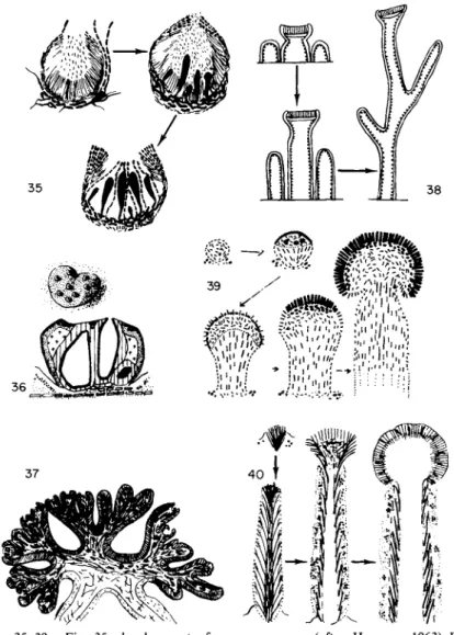
/. Special Types of Ascocarps in Pyrenolichens and Discolichens 1. PYCNOASCOCARPS (Fig. 3 5 )
The ascogonial filaments in Physma (Stahl, 1877) and various Ephebeaceae (Henssen, 1963) form beneath the pycnidia. Asci later develop in these,
FIGS. 35-39. Fig. 35, development of pycnoascocarps (after Henssen, 1963) Fig. 36, stromatoid structure in Laurera sanguinaria: general aspect and transverse section; Fig. 37, Umbilicaria proboscidea: transverse section of an old apothecium (after Henssen, 1970); Fig. 38, schematic representation of the ontogenetic and phylogenetic development of pseudopodetia in different Stereocaulon (according to Lamb, 1951); Fig. 39, development of podetia in Baeomyces rufus; Fig. 40, development of podetia in Cladonia floerkeana.
between the conidiophores that are sometimes still functional, and even- tually true paraphyses are born.
2. PSEUDOSTROMATA (Fig. 36)
Perithecia in some of the pyrenolichens in the Trypetheliaceae are aggre- gated in specific structures often designated as stromata. This stroma is, in
84 MARIE-AGNES LETROUIT-GALINOU
fact, an accessory structure, slowly differentiated around the ascocarps that were already well developed and at the expense of the thallus or perithecial envelope (Johnson, 1940; Letrouit-Galinou, 1957). This structure is, there- fore, not comparable to the stroma of mycologists which forms early in development before the appearance of ascogonial filaments.
3. GYROPHORINE APOTHECIA (Fig. 37)
The formation of asci in the hymenium of the Umbilicariaceae is most often confined to fertile zones that often project in an annulate or stellate manner (= gyri). This arrangement results from the repeated formation of sterile zones (Scholander, 1934; Frey, 1936; Henssen, 1970) and dichotom- ously dividing fertile zones. These gyri, according to Henssen, project be- cause their growth is arrested in the central part of the sterile portion while they continue to grow actively at their periphery in the fertile zones.
4. PODETIA (Figs. 38-40)
The ascocarps in some discolichens formerly united in the family Cladon- iaceae develop on special structures that are erect, often branched, and thalline in appearance. These are classified as podetia, of which three types may be distinguished:
a. In Stereocaulon and related genera they are actually pseudopodetia (Fig. 38; Wolff, 1905; Lamb, 1951; Jahns, 1970), purely thalline structures formed by the vertical growth of some of the thalline granules. The apothecia that develop later seem to be lecanorian at least in Stereocaulon, with a plexiform primordium and a well-developed primary corpus.
b. The podetium of Baeomyces (Fig. 39) differs fundamentally from that in Stereocaulon in the sense that it becomes the ascocarp (Nienburg, 1908;
Letrouit-Galinou, 1966; Jahns, 1970). The primary corpus is derived from a superficial plexiform primordium. It consists of a carpocenter, which con- tains the ascogonial apparatus and extends marginally, and a pericentral envelope consisting of a thin transitory cover and a thick foot. The foot elongates and becomes the podetium. The carpocenter is later covered by epicentral filaments, some of which become paraphyses while others at the periphery remain extrahymenial. The podetium of Baeomyces, derived from the lower part of a primary corpus, is the same as the structure called a pseudodiscopodium by Bellemere (1967).
c. The podetium of Cladonia, as in Baeomyces, is part of the ascocarp but its structure and mode of development are different (Krabbe, 1891; Letrouit- Galinou, 1966; Jahns, 1970). It terminates in a point or a cup and is often branched, being derived from a bundle-shaped primordium composed of divaricate hyphae that elongate and branch and within which the ascogonial
filaments arise, as in the primary corpus of a typical apothecium. These filaments are often differentiated quite early, in the initial bundle itself, but most frequently appear only after the podetia develop at branch tips or along the margin of the cup. Hyphae are drawn aside around them, form- ing a parathecioid or parathecial envelope. These produce paraphyses, the bases of which form a paraphysogenous subhymenial plexus.
/. Summary
Lichenized Ascomycetes are characterized as follows in comparison with nonlichenized Ascomycetes.
1. THE SPOROPHYTIC APPARATUS
a. The ascogonial apparatus is often complex and always composed of long multicellular filaments, in general with uninucleate cells (the Collema
type) but rarely multinucleate (the Peltigera type).
b. This apparatus produces a reduced but still recognizable prosporo- phyte.
c. Next, in many species, the ascosporophyte formed from the prosporo- phyte consists of a primary part with uninucleate cells and later, after perit- togamy or otherwise, a secondary dikaryotic ascogenous part with lateral clamps.
d. As a rule the asci are bitunicate-nassasceous or archaeasceous.
2. THE ASCOCARP
a. The lichenized fungi do not differ basically from nonlichenized Asco- mycetes as far as ontogeny and organization of the ascocarps is concerned.
In particular, the interascal filaments may be represented by paraphysoids (as in Opegrapha), pseudoparaphyses (as in Arthopyrenia fallax and A. sub- micans), or true paraphyses (the majority of species). However, it is only in some pyrenolichens (Arthopyrenia fallax and A. submicans with pseudopara- physes) that the ascocarp is derived from an ascostroma, though reduced to a pyrenosphere, whereas this happens in many of the ascolocular pyrenomy- cetes (Pleospora, etc.). No true stroma is present in other lichens, and one could believe in this case either the thallus itself replaces it or the primordium is a particular type of ascostroma.
b. In discolichens, (1) it is possible (cf. Chadefaud et al., 1968) that the ascocarp in its primitive form is comparable to that of the less evolved discomycetes (cf. Bellemere, 1967), ascostromatic, lenticular, and with para- physoids, but the ascostroma replaced by a plexiform primordium; (2) the
86 MARIE-AGNES LETROUIT-GALINOU
graphidian type would be, therefore, the closest relative to this primitive hypothetical form; (3) for the more highly evolved types we can say that the one in Thelotrema is reminiscent of the Ostropales; the podetial types in
Baeomyces and Cladonia correspond to those of Cudonia (Duff, 1922) and
Mitrula (Corner, 1929-1930), respectively; the lecanorian type (= para- thecial ; Chadefaud, 1965) is by far the most common type in lichens but very poorly represented among the nonlichenized Discomycetes; and finally, the eudiscopodian type, so very common in these latter, is lacking in lichens.
FIGS. 4 1 - 4 6 . Sexual reproduction in basidiolichens. Fig. 41, basic reproductive cycle in Basidiomycetes (after Chadefaud, 1960); Fig. 42, Omphalina ericetorum (Agaricales): general aspect of the carpophore and the thalline lobes (after Poelt and Oberwinkler, 1964); Fig.
43, basidia and basidiospores of Omphalina ericetorum; Fig. 44, columnar carpophore of Clavulinopsis septentrionalis (Aphyllophorales) (after Poelt, 1959); Fig. 45, Cora pavonia:
transverse section of the carpophore on the level with a fertile papilla (after Grassi, 1950);
Fig. 46, lower surface of the carpophore with fertile papillae in Cora pavonia (after Mattirolo, 1881). (b, basidia; m „ primary mycelium; m2, secondary mycelium; s, spore; v, multinucleate vesicle.)
II. Basidiolichens
There are very few basidiolichens—according to recent accounts, only about 20 species and 10 genera. They all are Basidiomycetes with carpo
phores and neobasidiates with typical basidia (Fig. 43).
The reproductive cycle has not been studied in any of the species. One would judge, however, that it should resemble that in nonlichenized species (Fig. 41), where in principle a haploid basidiospore gives rise to a haploid primary mycelium. A secondary dikaryotic mycelium is then produced by intercellular fusions (= perittogamies), often with lateral clamps. The mycelial hyphae become aggregated in the form of carpophores. The mycelial elements of the fertile layer produce the basidia, and these give rise to basidiospores after nuclear fusion and meiosis.
The primary and secondary mycelia, according to Chadefaud (1944, 1960), are homologous with primary and secondary sporophytes of perit- togamous Ascomycetes; there would be no gametophytic phase.
The carpophores of basidiolichens are only rarely of the Agaricales type where the hymenium develops simultaneously and is located on lamellae
(Omphalina, Fig. 42). More frequently we find the Aphyllophorales type with a continuously developing hymenium. In addition, they do not have a vallicular hymenium (not localized in the pits of particular podia). They are in columns (Clavaria and Clavulinopsis; Fig. 44), in sheets (Stereum),
or multiple ridges or segments (Cora). In Cora the brackets are formed of intertwined mycelia, in the center of which a gonidial layer is lodged. The lower surface (Fig. 46) bears fertile papillae (Fig. 45). According to Tomaselli and Caretta (1969) the branched hyphae in an umbell comprising these papillae have their origin in the gonidial layer.
References
Ahmadjian, V. (1966). Artificial reestablishment of the lichen Cladonia cristatella. Science 151, 199-201.
Baur, E. (1898). Zur Frage nach der Sexualitat der Collemaceen. Ber. Deut. Bot. Ges. 16, 363-367.
Baur, E. (1901). Uber Anlage und Entwicklungsgeschichte einer Flechtenapothecien. Flora (Jena) 88, 319-332.
Baur, E. (1904). Untersuchungen uber die Entwicklungsgeschichte der Flechtenaphothecien.
Bot. Z. 62, 21^14.
Bellemere, A. (1967). Contribution a Ρ etude du developpement de Papothecie chez les Disco- mycetes Inopercules. Bull. Soc. Mycol. Fr. 83, 393-640, 755-931.
Carroll, G. (1969). A study of the fine structure of ascosporogenesis in Saccobolus kerverni and Ascodesmis sphaerospora. Arch. Mikrobiol. 66, 321-339.
Chadefaud, M. (1942). Etudes d'asques. II. Structure etanatomiecompareedel'appareil apical des asques chez divers Disco et Pyrenomycetes. Rev. Mycol. 7, 57-88.