Isolation of persistent left superior vena cava during atrial fibrillation ablation
Emin E. Ozcan, Gabor Szeplaki, Bela Merkely, Laszlo Geller
*Semmelweis University, Heart Center, Budapest, Hungary
a r t i c l e i n f o
Article history:
Available online 29 July 2015
Keywords:
Persistent left superior vena cava Paroxysmal atrial fibrillation Pulmonary vein isolation Ablation
a b s t r a c t
Persistent left superior vena cava is a rarely seen anomaly but it may be an arrhythmogenic source for paroxysmal atrial fibrillation. Furthermore, the complex anatomicregion be- tween the left superior vena cava and the pulmonary veins may leads to misinterpretation of the pulmonary vein recordings during atrial fibrillation ablation. Approaches that might be helpful to overcome these problems are discussed in this case report.
Copyright©2015, Indian Heart Rhythm Society. Production and hosting by Elsevier B.V.
This is an open access article under the CC BY-NC-ND license (http://creativecommons.
org/licenses/by-nc-nd/4.0/).
Introduction
Triggers of paroxysmal atrial fibrillation (AF) do not originate only from the pulmonary veins. Several structures such as coronary sinus (CS), superior vena cava and ligament of Marshall which is the developmental remnant of left superior vena cava (LSVC) may be substrates for the triggers[1]. In some rare cases, LVSC may persist and become a source of the AF episodes[2,3]. The diagnosis of LSVC is often made when a pacemaker is implanted or during insertion of CS catheter[4].
Misinterpretation of intracardiac electrograms due to persis- tent LSVC and the approaches that might be used during pul- monary vein isolation (PVI) are discussed in the present paper.
Case report
A 61 years old male patient with history of PVI due to parox- ysmal AF one year ago was referred to our unit for repeat
ablation because of drug-resistant episodes. During insertion of CS catheter, it stepped out of the heart shadow and the electrical signals disappeared. The course of catheter was consistent with persistent LSVC. The diagnosis was confirmed with contrast injection. After insertion of CS catheter, we continued standard PVI protocol by using electroanatomic mapping (Ensite NavX mapping system, St. Jude Medical, Minneapolis, MN, USA). Since the position of CS catheter was not stable, the surface patch was chosen as the reference.
Pulmonary vein potentials were observed to delay and disappear in the recordings of the left superior pulmonary vein (LSPV) during the wide area circumferential ablation of the left sided veins. However, localized sharp potentials just behind the far-field signals were notable and they became prominent with advancing the circular multielectrode cath- eter (Inquiry Optima, St. Jude Medical Inc., St. Paul MN, USA) from the ablation line toward distal part of the vein (Fig. 1A).
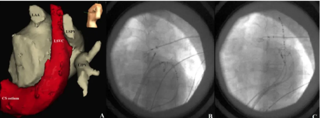
These potentials thought to be far-field signals resulted from the anatomical neighborhood of LSVC and LSPV (Fig. 2A, B).
Pacing from the LSPV at the lowest output was capturing the
*Corresponding author. Semmelweis University, Heart Center, Gaal Jozsef street 9, 1122, Budapest, Hungary. Tel.:þ36 20 3658330; fax:
þ36 1 4586842.
E-mail address:laszlo.geller@gmail.com(L. Geller).
Peer review under responsibility of Indian Heart Rhythm Society.
H O S T E D BY Available online atwww.sciencedirect.com
ScienceDirect
journa l home page: www.elsevier.com /locate/IPEJ
i n d i a n p a c i n g a n d e l e c t r o p h y s i o l o g y j o u r n a l 1 5 ( 2 0 1 5 ) 1 3 0e1 3 2
http://dx.doi.org/10.1016/j.ipej.2015.07.011
0972-6292/Copyright©2015, Indian Heart Rhythm Society. Production and hosting by Elsevier B.V. This is an open access article under the CC BY-NC-ND license (http://creativecommons.org/licenses/by-nc-nd/4.0/).
vein, and therefore exit block could not be demonstrated (Fig. 1B). When output was increased to 10 mA LSVC was captured directly and sharp potentials became simultaneous with the pace spike (Fig. 1B). Pacing from the CS catheter localized in the LSVC demonstrated the same findings sug- gesting that these sharp potentials were far-field signals from LSVC. Timing of the signals was not altered by pacing from left atrial appendage. Failure of exit block might be a result of conduction through a gap at the ablation line or a far-field capture of LSVC. Moreover, this might also be caused by the connections between left atrium (LA) or LSPV and persistent LSVC which may show propagation similar to the far-field capture[2]. Therefore, we decided to map LSVC.
The multielectrode catheter was retrogradely introduced into LSVC through CS (Fig. 2C). The mapping catheter was introduced from the CS junction and the catheter was advanced into the LSVC up to the level without electrical activity. Besides local sharp LSVC potentials following the far-field LA potentials (Fig. 1D) premature ectopic beats with the earliest activation in LSVC were observed during the mapping (Fig. 1E). Thereupon, LSVC was isolated, applying circumferential ablation within the CS. Local sharp poten- tials disappeared and the ectopic beats were not conducting to LA (Fig. 1F). The pacing performed from LSVC with maximal energy did not capture LA. Then, ablation catheter was advanced into the LA again and exit block was Fig. 1eA,B,C are recordings from LSPV and D,E,F are from LSVC. (A) Sharp far-field signals resulted from the anatomical contiguity of LSVC. (B) Pacing from the LSPV at the lowest output capturing the vein, exit block could not be demonstrated.
When output was increased to 10 mA LSVC was captured directly and far-field signals disappeared. (C) After isolation of LSVC sharp potentials disappeared. CS catheter is in the distal LSVC. LSVC recordings during sinus rhythm (D) and ectopic beats (E). Note both ectopic beats (#) and left atrial far-field signals are preceding surface p wave (E). Following isolation LSVC potentials (*) abolished and only far-field LA signals remained (D and F).
i n d i a n p a c i n g a n d e l e c t r o p h y s i o l o g y j o u r n a l 1 5 ( 2 0 1 5 ) 1 3 0e1 3 2
131
demonstrated with pacing from LSPV. The circular catheter was inserted into LSPV through the same long sheath and the potentials persisting following PVI were observed to disappear (Fig. 1C). The intervention was successfully completed without any complication. Patient did not develop AF episodes during the follow-up period over 3 months.
Discussion
This paper emphasizes three important points regarding the association of persistent LSVC and AF.
1. Persistent LSVC may be source of the arrhythmogenic foci [1e3]. Moreover, its connections with CS and LA may lead the AF episodes to continue even the pulmonary veins are isolated.
2. The neighborhood between LSVC and LSPV may cause misinterpretations during the pulmonary vein recordings and pacing maneuvers. Using circular catheters and care- ful observation of pulmonary vein signals during ablation helps to overcome this problem.
3. There may be electrical connections between LA and LSVC, and in this case complete isolation of the pulmonary veins may not be achieved[2]. Ectopies resulted from the pul- monary veins may stimulate LA through LSVC and CS and, may trigger AF episodes. Exit block as well as entrance block must be definitely demonstrated.
Due to above conditions, electrical isolation of LSVC seems to be necessary particularly in the patients with AF recur- rence. However, evidence on the isolation of persistent LSVC are not sufficient to recommend this approach as an initial procedure[2,5]. An additional issue is what to do in the pa- tients without ectopy during the mapping of LSVC. A patient-
tailored approach is appropriate given the limited number of these patient that will likely be encountered.
Conflict of interest
None declared.
Acknowledgment
The study was supported by grants from the National Devel- opment Agency of Hungary (Semmelweis Egyetem Magiszter Program, TAMOP-4.2.2/B-10/1-2010-0013) and the J anos Bolyai Research Scholarship of the Hungarian Academy of Sciences (GSZ, LG).
r e f e r e n c e s
[1] Chen PS, Wu TJ, Hwang C, Zhou S, Okuyama Y, Hamabe A, et al. Thoracic veins and the mechanisms of non- paroxysmal atrial fibrillation. Cardiovasc Res 2002;54:295e301.
[2] Hsu LF, Jaı¨s P, Keane D, Wharton JM, Deisenhofer I, Hocini M, et al. Atrial fibrillation originating from persistent left superior vena cava. Circulation 2004;109:828e32.
[3] Liu H, Lim KT, Murray C, Weerasooriya R. Electrogram-guided isolation of the left superior vena cava for treatment of atrial fibrillation. Europace 2007;9:775e80.
[4] Mora G. A novel method of placing right ventricular leads in patients with persistent left superior vena cava using a conventional j stylet. Indian Pacing Electrophysiol J 2014;14:65e74.
[5] Wissner E, Tilz R, Konstantinidou M, Metzner A, Schmidt B, Chun KR, et al. Catheter ablation of atrial fibrillation in patients with persistent left superior vena cava is associated with major intraprocedural complications. Heart Rhythm 2010;7:1755e60.
Fig. 2e(A) Computed tomography image integrated to NavX map. Note the anatomical contiguity between LSVC and LSPV.
Fluoroscopic images in left anterior oblique projection demonstrating catheter positions. (B) Ablation and circular multielectrode catheters are in LSPV and CS catheter distal is in the LSVC-CS junction. (C) Ablation and circular multielectrode catheters are in LSVC and CS catheter is in the distal LSVC.
i n d i a n p a c i n g a n d e l e c t r o p h y s i o l o g y j o u r n a l 1 5 ( 2 0 1 5 ) 1 3 0e1 3 2