Full Terms & Conditions of access and use can be found at
https://www.tandfonline.com/action/journalInformation?journalCode=irnf20
Renal Failure
ISSN: 0886-022X (Print) 1525-6049 (Online) Journal homepage: https://www.tandfonline.com/loi/irnf20
The association of overhydration with megafistulas in hemodialysis patients
Mihály Tapolyai, Mária Faludi, Klára Berta, Melinda Forró, Lajos Zsom, Ákos G. Pethő, László Rosivall & Tibor Fülöp
To cite this article: Mihály Tapolyai, Mária Faludi, Klára Berta, Melinda Forró, Lajos Zsom, Ákos G. Pethő, László Rosivall & Tibor Fülöp (2019) The association of overhydration with megafistulas in hemodialysis patients, Renal Failure, 41:1, 440-445, DOI: 10.1080/0886022X.2019.1614954 To link to this article: https://doi.org/10.1080/0886022X.2019.1614954
© 2019 The Author(s). Published by Informa UK Limited, trading as Taylor & Francis Group.
Published online: 04 Jun 2019.
Submit your article to this journal
Article views: 159
View Crossmark data
CLINICAL STUDY
The association of overhydration with megafistulas in hemodialysis patients
Mihaly Tapolyaia,b,c, Maria Faludia,b, Klara Bertaa,b, Melinda Forrod, Lajos Zsome, Akos G. Peth}of , Laszlo Rosivallgand Tibor F€ul€opc,h
aSemmelweis University, Budapest, Hungary;bHemodialysis Unit, Fresenius Medical Care, Budapest, Hungary;cMedical Services, Ralph H. Johnson VA Medical Center, Charleston, SC, USA;dHemodialysis Unit, Fresenius Medical Care Hungary, Hatvan, Hungary;
eHemodialysis Unit, Fresenius Medical Care Hungary, Cegled, Hungary;f1st Department of Internal Medicine, Faculty of Medicine, Semmelweis University, Budapest, Hungary;gDepartment of Pathophysiology, International Nephrology Research and Training Center, Semmelweis University, Budapest, Hungary;hDepartment of Medicine, Division of Nephrology, Medical University of South Carolina, Charleston, SC, USA
ABSTRACT
Objectives: Diffuse enlargements of arteriovenous dialysis fistulas customarily attributed to either excessive arterial inflow or central outflow stenosis. The relationship between volume status and clinically enlarged (arteriovenous) fistula (CEF) formation in end-stage renal disease (ESRD) patients is not well understood.
Methods: We assessed the pre-dialysis bioimpedance spectroscopy-measured percentage of overhydration (OH%) in 13 prevalent dialysis patients with CEF development and negative angi- ography and compared the results with those of 52 control dialysis patients (CONTR). All patients were prevalent ESRD patients receiving thrice-weekly maintenance hemodiafiltration at an academic outpatient dialysis unit.
Results:10/13 CEF patients had OH%15% as compared to 20/52 control patients (Chi squarep: .02). The degree of OH% was 20.2 ± 7.4% among the CEF vs. 14.4 ± 7.1% in the control group (Student’s t-test p: .01), representing 4.2 ± 3.2 vs. 2.8 ± 1.6 L of excess fluid pre-dialysis (p: .03).
Patients with CEF development took an average of 1.7 ± 1.4 vs. 0.8 ± 0.8 (p: .002) antihypertensive medications compared to the CONTR patients, yet their blood pressure was higher: 156/91 vs. 141/
78 mmHg (systolic/diastolic p: .03<.0001). We found no difference in fistula vintage, body mass index, age, diabetes status, or diuretic use. The odds ratio of having a CEF in patients with15%
OH status was 5.3 (95% CI: 1.3–21.7;p: .01), the Number Needed to Harm with overhydration was 4.
Conclusions: There is an association between bioimpedance spectroscopy-measured overhy- drated clinical state and the presence of CEF; either as an increased volume capacitance or as a potential cause.
ARTICLE HISTORY Received 29 January 2019 Revised 21 March 2019 Accepted 27 April 2019 KEYWORDS
Bioimpedance spectroscopy;
blood pressure; body composition; end-stage renal disease; megafistula;
volume overload
Introduction and background
For decades, arterio-venous fistulas (AVFs) have been deemed the primary and desired dialysis access for hemodialysis [1], with the successful creation and main- tenance of AVFs remaining Achilles’heel of hemodialy- sis. Most dialysis fistulas fail by stenosis and clotting, representing the principal cause of access loss in hemo- dialyzed patients, more commonly occurring in dia- betics [2] or those with higher hemoglobin [3]. Early failure of arteriovenous dialysis access occurs at a rate of about 17.5% [4] to 23% [5] and the overall one-year primary patency rate is about 60%. Most efforts to maintain and salvage fistulas have focused so far on the preservation of blood flow in the arterial inflow or venous outflow tracts.
Another, albeit much less common mechanism of hemodialysis access failure is the development of fistu- las with excessively high flow and large diameters, also known as megafistulas [6], most commonly origi- nating from the brachio-cephalic location. Established risk factors for megafistulas are wide arterio-venous anastomoses with large blood flow rates or a relative narrowing of the draining vein in the venous circula- tion. Megafistula formation is most commonly recog- nized by an excessive blood flow and clinically confirmed by the Nicoladoni-Branham maneuver. The suggested criteria include an increased blood flow greater than 2.2 L/min, increased cardiac output and index, increased (>20%) cardio-pulmonary recircula- tion and hypertrophied feeding artery [6,7].
CONTACTMihaly Tapolyai mtapolyai@aol.com 2120 Dunakeszi, Szabadka u. 24/a, Budapest, Hungary ß2019 The Author(s). Published by Informa UK Limited, trading as Taylor & Francis Group.
This is an Open Access article distributed under the terms of the Creative Commons Attribution License (http://creativecommons.org/licenses/by/4.0/), which permits unrestricted use, distribution, and reproduction in any medium, provided the original work is properly cited.
https://doi.org/10.1080/0886022X.2019.1614954
Another working definition of megafistula simply refers to the ultrasonically measured blood flow rate within the fistula. ‘Although it is uncertain as to why some patients develop a megafistula, altered hoop stress (circumferential stress) almost certainly plays a role’[8]. The exact relationship between volume status and enlarged fistula formation is, nonetheless, cur- rently insufficiently understood. Objective assessment of volume status in end-stage renal disease (ESRD) remains challenging and persisting chronic volume overload may contribute to poor BP control [9,10], vas- cular stiffness [10,11], and decreased survival [12–15].
Bioimpedance monitoring is an emerging gold stand- ard methodology to assess not only body composition [16–18] but to assess the exact degree of volume sta- tus in dialysis patients [14–16,19]. As the relationship between volume status, antihypertensive drug use sta- tus and an enlargement of AVF is not well understood, we investigated whether volume overload may repre- sent an additional risk factor for these redundant vas- cular enlargements.
Methods
This was a case-control study approved by the Ethics Committee of the Hungarian Health Ministry (equiva- lent for an Independent Review Board) (TUKEB 3032-1/
2015/EKU) and Fresenius Medical Care as the dialysis provider in Hungary. The study conformed to the Helsinki Declaration as developed by the World Medical Association. The data used for the study were obtained during routine care of maintenance dialysis at the Fresenius Medical Care facilities, within the premises of and affiliated with Semmelweis University in Budapest, Hungary. All patients received thrice weekly maintenance hemodiafiltration using a post- dilution fluid replacement method, aiming at a single- session Kt/V of 1.4 and a replacement fluid volume of 21 L [20].
All of our patients routinely undergo bimonthly pre-dialysis fluid status evaluations at the designated medical care facility by using a multi-channel bioim- pedance spectroscopy apparatus (BCM – Body Composition Monitor, Software version 3.2; Fresenius Medical Care, Bad Homburg, Germany) as part of our standard clinical practice. The etiology of the proband group’s ESRD was heterogeneous including PCKD (1), urological anomalies (1), nephrectomy because of renal cell carcinoma (2), presumably hypertension (1), diabetes (3), glomerulonephritis (2), and unknown. We have previously described the measurement method [16]; however, it shall be added that we measured the
patients’ fluid compartments by positioning them flat on their backs and placing two conductive electrodes on their hands and ankles on the same side at the same time. With this method, we measured their total body water (TBW), extracellular water (ECW), intracel- lular water (ICW), and overhydration (OH) levels in lit- ers and acquired the percentage (OH%) of the excess fluid that is greater than the anticipated ECW. The val- idation of the bioimpedance methods for measuring fluid spaces has been done historically with various isotopes (of potassium, bromide, hydrogen), tagged albumin and DEXA scans and reviewed earlier in details [21].
Our goal was to define pre-dialysis BCM-measured OH% in 13 prevalent chronic dialysis patients with CEF. The diagnosis of CEF was established on clinical exam, with excessive and torturous dilatations of arteriovenous fistula observed throughout its course in the extremity; these were typically easy to palpate and traceable to the shoulder. We did not have the means to measure blood flow in the fistulas; consequently, the diagnosis of ultrasonography-determined megafis- tula could not be made with certainty and the diagno- sis of clinically enlarged AVF was made purely on clinical grounds, by the consensus of the nephrologists involved in the clinical care of the cohort. Due to this lack of diagnostic rigor, we refer to these fistulas as clinically enlarged fistulas (CEFs). All CEF subjects had negative angiographies performed earlier by a dedi- cated interventional radiologist and definitively ruled out for proximal outflow stenosis. We compared these patients with 52 consecutive control dialysis patients without enlarged fistula formation on clinical grounds but no further testing (Doppler ultrasound or angiog- raphy) was performed. We tabulated the patients’ demographic characteristics as well as their blood pressures and blood pressure medication numbers.
For the comparison of continuous data, we utilized Student’s t-tests and for categorical data, we explored statistical significance with two-tailed Fisher’s exact test statistics.
Results
The characteristics of the patients are depicted in Table 1. The CEF as well as the control patients’ data passed the normality test for the Gaussian distribu- tion when tested by the D’Agostino and Pearson omnibus normality test, the Shapiro normality test, and the Kolmogorov–Smirnov test for normality.
There was a statistically significant male predomin- ance (84.6% vs. 51.9%; p: .03) among the subjects
RENAL FAILURE 441
with CEFs, but they did not differ in age, dialysis vin- tage, prevalence of diabetes mellitus or even the amount of residual urine volume from controls.
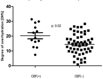
Importantly, however, the CEF patients had higher blood pressure, took more antihypertensive medica- tions and were significantly more overhydrated (p<.01 for all). The degree of overhydration, when expressed in liters showed 1.4 L of excess extracellular fluid (Student’s t-test p: .03; Mann–Whitney p: .06) among the subjects with CEF. Moreover, when the degree of volume overload was expressed as a % of OH, the difference became striking (Student’st-test p: .01; Mann–Whitney p: .01) with a clear dichotomy at 15% OH, an established cutoff point for future adverse events [12,22–24]. This is also illustrated by Figure 1’s scatter plots, demonstrating the medians and standard deviation intervals for both groups. Our
data were also examined in a categorical fashion rela- tive to the 15% OH cutoff (Table 2). The majority of CEF patients were in the >15% OH category with the two-tailed Fisher’s exact test of association significant (p: .02). The unadjusted odds ratio of those with a CEF being in fluid excess was 5.3 (95% CI: 1.3–21.7;
p: .01; the Number Needed to Harm was 4).
Discussion
The presence of a high-flow hemodialysis fistula is associated with multiple adverse outcomes. These include steal syndrome [25], exotic complications such as hemothorax [26] or pulmonary embolism [27], and the impairment of cardiac function with high-output heart failure resulting from a large proportion a large proportion of the cardiac output recirculated through the enlarged fistulas [22,28,29]. While our working def- inition of CEF in our study was not harmonized to the diagnosis of ultrasonography-defined megafistula with high overall flow nor did we perform the Nicoladoni- Branham maneuver, our study entertains yet another striking possibility contributing to enlargement of arteriovenous accesses; the one of chronic salt-water overload. Only recently, exact technology with bioim- pedance analysis become available for the daily rou- tine care of ESRD patients, to offer reliable and repeated measures of volume status with a minimized Table 1. Demographics and results comparing hemodialysis patients with and without a clinically enlarged fistula.
Clinically enlarged fistulas (n: 13) Control (n: 52) p
Gender (M:F) 84.6% 51.9% .03
Age (years) 51.1 ± 14.7 59.0 ± 14.7 .09
Dialysis vintage (years) 5.3 ± 2.4 4.8 ± 3.8 .62
Diabetes (%) 8 23 .22
Residual urine volume (mL/day) 274 ± 218 169 ± 295 .23
Number of antihypertensive medications 1.77 ± 1.4 0.80 ± 0.8 .002
Diuretics use (%) 15.3 9.6 .56
Body mass index (kg/m2) 25.1 ± 6.5 26.7 ± 5.7 .40
Systolic blood pressure (mmHg) 156 ± 24.5 141 ± 20.8 .003
Diastolic blood pressure (mmHg) 91 ± 10.9 78 ± 9.7 <.0001
OH (L) 4.2 ± 3.2 2.8 ± 1.6 .03
OH% 20.2 ± 7.4 14.4 ± 7.1 .01
EC (L) 19.7 ± 6.0 20.2 ± 18.7 .92
OH: overhydration: OH%: percent overhydration: EC: extracellular fluid volume.
When continuous data are shown, the means ± standard deviation is displayed.
Figure 1. Comparison of those with and without a clinically enlarged arteriovenous fistula (CEF) with respect to the bioim- pedance-measured fluid overhydration. The two populations’ percent-overhydration (n: 13 andn: 52) mean ± standard devi- ation is depicted by the horizontal lines.
Table 2. Results comparing those with and without a CEF in a 22 table for Fisher’s exact test (p: .02).
Data analyzed CEFþ CEF– Total
OHþ 10 20 30
OH– 3 32 35
Total 13 52 65
CEF: clinically enlarged (arteriovenous) fistula; OH: overhydration; overhy- dration (OH) was dichotomized at a 15% excess of extracellular fluid;
CEFþ: patients with a CEF; CEF–: patients without a CEF; OHþ: overhy- drated; OH–: not overhydrated.
burden and cost for the dialysis networks [9,14,15].
Our study shows that patients with a CEF have excess fluid of about 1.4 L over the control group. While this quantity does seem small, it is important to note that 10 out of 13 patients with a CEF had severe pre-dialy- sis volume overload as compared to only 20 of the 52 patients without CEF. We deemed fluid overload as
‘severe’ when the degree of overhydration (OH%) was greater than 15% [12,19]. Expressing fluid overload in liters is important to help individual patients under- stand target weight goals during renal replacement therapy, whereas expressing OH in percent is import- ant for prognosis and securing comparability among clinical studies. As Wizemann et al. [12] showed, hemodialysis patients’ mortality is strongly affected when this threshold is met or exceeded. Interestingly, Wizemann’s study also showed that the presence of hypertension may be somewhat protective (hazard ratio: 0.98, p: .01) from mortality. Thus, we used the 15% OH cutoff to define the tolerance for fluid excess.
The sustained fluid excess was not related to interdia- lytic weight gain–it hardly ever is –but rather to the inadequate determination and failure to reach the esti- mated dry weight [15]. In this particular cohort, we routinely observed that patients with CEF have stead- fastly refused physician-recommended dry weight reduction and insisted that their weight be maintained at the level they deemed appropriate for themselves.
There could be many reasons why these patients refused dry weight reduction; one possible explan- ation is the relatively long vintage (5.3 years) with the patients’sense of empowerment of‘knowing it better’. Also, the long vintage, could have contributed merely to the development of CEF, given so many cannula- tions over the years. In the past, we have demon- strated that one possible reason for fluid overload in dialysis patients is due to the difficulties reaching tar- get net ultrafiltration goals while taking multiple anti- hypertensive drug medications [16]. In our current study, the linear regression analysis coefficient was 0.673 (p<.0001), between the number of antihyper- tensive medications and the amount of excess fluid (in liters), as measured by bioimpedance. Further, there was a strong correlation (r¼0.54, p<.0001) between the number of antihypertensive medications and the degree of fluid overload. Antihypertensive medications pose a risk of intradialytic hypotension, cramping and does appear to interfere with adequate ultrafiltration [9,16,30,31]. Indeed, this CEF patient cohort was taking a significantly larger number of antihypertensive medi- cations (1.7 vs. 0.8; p: .002) and they were – perhaps consequently – more fluid overloaded. The use of
diuretics did not appear to be protective. Our study reinforces the importance of validated technologies in determining the optimal target weight in ESRD patients as they are at risk for both hypo- and hyper- volemia during treatment, if to rely on clinical assess- ment alone [9,16,30,31].
The question of interest raised by our study is whether this measured fluid excess is simply a measure- ment capacitance (the cumulative volume of enlarged fistulas) or the cause or exacerbating factor for the development of the CEF. The fact that these patients were taking more antihypertensive medications indi- cates a strong possibility of long-standing fluid over- load, in keeping with the slow speed of enlarged fistula formation. As shown in Table 1, prescribing more anti- hypertensive drugs may have been the rule for these subjects, further aggravating the tendency to both fluid overload and potential CEF formation. While conven- tional treatment for these high-flow accesses is inflow restriction [6,32], the treatment may also involve opti- mizing fluid status.
Our study has several limitations. As CEF formation is a rare event, we compensated for the low number of index patients by ensuring that the comparator control group had plenty of patients, four times as many as the group with the condition. The lack of ultrasound-measured fistula flow determination was an obvious limitation of our study. We also did not perform cardiac ultrasound investigations as the focus of the study was fluid excess and its association with the presence of fistula enlargement, even though per- sisting fluid excess does have cardiac consequences as well. We have no information on previous endovas- cular interventions. Most importantly, the largest limi- tation of this study was the case-control design and observational nature of the project; therefore, a defini- tive conclusion cannot be drawn at this time. On the other hand, it would have been ethically impossible to perform a prospective study of fluid overloaded patients. Future studies need to explore this relation- ship further and extend these investigations to include measurements of cardiovascular health and cardiac output among these patients.
Conclusions
While the observational nature of this study does not permit to imply causality, we observed a striking associ- ation between CEF formation and OH in a prevalent cohort of otherwise stable ESRD patients. This novel association may represent yet another consequence of poorly controlled volume status in ESRD and additional
RENAL FAILURE 443
studies needed to provide independent verification.
Future trials need to consider the presence of fistula enlargement when prospectively evaluating bioimpe- dance analysis and volume status among those being started on or receiving maintenance dialysis.
Acknowledgements
We sincerely appreciated the assistance of Mr. Attila Lenart- Muszka during editing and grammar review. The views and opinions expressed in the paper do not reflect the official views or opinion or endorsed by the United States Veteran Health Administrations.
Disclosure statement
MT, MF, KB, MF, LZ, AP, and TF are current or former employees of Fresenius Medical Care Kft., Hungary. MT and TF are also affiliated with the United States Veteran Health Administrations. However, the views and opinions expressed herewith do not reflect the official views or opinion or endorsed by the Fresenius Medical Care Hungary and United States Veteran Health Administrations.
ORCID
Akos G. Petho} http://orcid.org/0000-0001-9776-9841
References
[1] Burkhart HM, Cikrit DF. Arteriovenous fistulae for hemodialysis. Semin Vasc Surg. 1997;10:162–165.
[2] Yan Y, Ye D, Yang L, et al. A meta-analysis of the asso- ciation between diabetic patients and AVF failure in dialysis. Ren Fail. 2018;40:379–383.
[3] Ye Y, Liu H, Chen Y, et al. Hemoglobin targets for the anemia in patients with dialysis-dependent chronic kidney disease: a meta-analysis of randomized, con- trolled trials. Ren Fail. 2018;40:671–679.
[4] Eslami MH, Zhu CK, Rybin D, et al. Simple predictive model of early failure among patients undergoing first-time arteriovenous fistula creation. Ann Vasc Surg. 2016;35:46–52.
[5] Al-Jaishi AA, Oliver MJ, Thomas SM, et al. Patency rates of the arteriovenous fistula for hemodialysis: a systematic review and meta-analysis. Am J Kidney Dis.
2014;63:464–478.
[6] Miller GA, Hwang WW. Challenges and management of high-flow arteriovenous fistulae. Semin Nephrol.
2012;32:545–550.
[7] Dix F, Khan Y, Al-Khaffaf H. The brachial artery-basilic vein arterio-venous fistula in vascular access for haemodialysis—a review paper. Eur J Vasc Endovasc Surg. 2006;31:70–79.
[8] Work J. Dialysis vascular access: introduction. Semin Nephrol. 2012;32:517–518.
[9] Dekker M, Konings C, Canaud B, et al. Pre-dialysis fluid status, pre-dialysis systolic blood pressure and out- come in prevalent haemodialysis patients: results of
an international cohort study on behalf of the MONDO initiative. Nephrol Dial Transplant. 2018;33:
2027–2034.
[10] Hur E, Usta M, Toz H, et al. Effect of fluid manage- ment guided by bioimpedance spectroscopy on car- diovascular parameters in hemodialysis patients: a randomized controlled trial. Am J Kidney Dis. 2013;61:
957–965.
[11] Czyzewski_ Ł, Wyzgał J, Czyzewska_ E, et al.
Contribution of volume overload to the arterial stiff- ness of hemodialysis patients. Ren Fail. 2017;39:
333–339.
[12] Wizemann V, Wabel P, Chamney P, et al. The mortality risk of overhydration in haemodialysis patients.
Nephrol Dial Transplant. 2009;24:1574–1579.
[13] Onofriescu M, Siriopol D, Voroneanu L, et al.
Overhydration, cardiac function and survival in hemo- dialysis patients. PLoS One. 2015;10:e0135691.
[14] Zoccali C, Moissl U, Chazot C, et al. Chronic fluid over- load and mortality in ESRD. J Am Soc Nephrol. 2017;
28:2491–2497.
[15] Hecking M, Moissl U, Genser B, et al. Greater fluid overload and lower interdialytic weight gain are independently associated with mortality in a large international hemodialysis population. Nephrol Dial Transplant. 2018;33:1832–1842.
[16] Tapolyai M, Faludi M, Reti V, et al. Dialysis patients’ fluid overload, antihypertensive medications, and obesity. ASAIO J. 2011;57:511–515.
[17] Marcelli D, Usvyat LA, Kotanko P, et al. Body compos- ition and survival in dialysis patients: results from an international cohort study. CJASN. 2015;10:
1192–1200.
[18] Rymarz A, Gibinska J, Zajbt M, et al. Low lean tissue mass can be a predictor of one-year survival in hemo- dialysis patients. Ren Fail. 2018;40:231–237.
[19] Tapolyai M, Faludi M, Reti V, et al. Volume estimation in dialysis patients: the concordance of brain-type natriuretic peptide measurements and bioimpedance values. Hemodial Int. 2013;17:406–412.
[20] F€ul€op T, Tapolyai MB, Zsom L, et al. Successful prac- tice transitioning between hemodialysis and hemodia- filtration in outpatient units: ten key issues for physicians to remember. Artif Org. 2018;42:925–932.
[21] Tapolyai MB, Faludi M, F€ul€op T, et al. Which fluid space is affected by ultrafiltration during hemodiafil- tration? Hemodial Int. 2014;18:384–390.
[22] Pandeya S, Lindsay RM. The relationship between car- diac output and access flow during hemodialysis.
ASAIO J. 1999;45:135–138.
[23] Machek P, Jirka T, Moissl U, et al. Guided optimization of fluid status in haemodialysis patients. Nephrol Dial Transplant. 2010;25:538–544.
[24] Chazot C, Wabel P, Chamney P, et al. Importance of normohydration for the long-term survival of haemo- dialysis patients. Nephrol Dial Transplant. 2012;27:
2404–2410.
[25] Schenk WG 3rd. Subclavian steal syndrome from high-output brachiocephalic arteriovenous fistula: a previously undescribed complication of dialysis access.
J Vasc Surg. 2001;33:883–885.
[26] Salim S, Ganeshram P, Patel AD, et al. Unilateral hemothorax in a 46 year old South Indian male due to a giant arteriovenous hemodialysis fistula: a case report. Cases J. 2008;1:225.
[27] Beathard GA. (2014). Pulmonary embolism associated with dialysis access procedure. In: Yevzlin A, Asif A, Salman L. editors. Interventional nephrology. New York (NY): Springer.
[28] Basile C, Lomonte C, Vernaglione L, et al. The relation- ship between the flow of arteriovenous fistula and cardiac output in haemodialysis patients. Nephrol Dial Transplant. 2007;23:282–287.
[29] Letachowicz K, Kusztal M, GołeRbiowski T, et al.
External dilator-assisted banding for high-flow
hemodialysis arteriovenous fistula. Ren Fail. 2016;38:
1067–1070.
[30] Keane DF, Baxter P, Lindley E, et al. Time to reconsider he role of relative blood volume monitoring for fluid management in hemodialysis. ASAIO J. 2018;64:
812–818.
[31] Keane DF, Bowra K, Kearney K, et al. Use of the body composition monitor for fluid status measurements in elderly malnourished subjects. ASAIO J. 2017;63:
507–511.
[32] Chemla ES, Morsy M, Anderson L, et al. Inflow reduc- tion by distalization of anastomosis treats efficiently high-inflow high-cardiac output vascular access for hemodialysis. Semin Dial. 2007;20:68–72.
RENAL FAILURE 445