in fl uenza viruses in Iranian industrial poultry farms
MOHSEN BASHASHATI
1, ZOHREH MOJAHEDI
2,
ALI AMEGHI ROUDSARI
3, MORTEZA TAGHIZADEH
3, AIDIN MOLOUKI
1, NAJMEH MOTAMED
4,
FERESHTEH SABOURI
1and
MOHAMMAD HOSSEIN FALLAH MEHRABADI
1p1Department of Avian Disease Research and Diagnostic, Razi Vaccine and Serum Research Institute, Agricultural Research Education and Extension Organization (AREEO), Karaj, Iran
2Department of Viral Vaccine Quality Control, Razi Vaccine and Serum Research Institute, Agricultural Research Education and Extension Organization (AREEO), Karaj, Iran
3Department of Research and Development, Razi Vaccine and Serum Research Institute, Agricultural Research Education and Extension Organization (AREEO), Karaj, Iran
4Department of Poultry Vaccine Research and Production, Razi Vaccine and Serum Research Institute, Agricultural Research Education and Extension Organization (AREEO), Karaj, Iran
Received: December 19, 2019 • Accepted: September 10, 2020 Published online: November 12, 2020
ABSTRACT
Despite the use of wide-scale vaccination programmes against the H9N2 virus, enzootic outbreaks of H9N2 avian influenza (AI) have often occurred and caused serious nationwide economic losses, particularly in broiler chickens. In this study, the haemagglutinin (HA) and neuraminidase (NA) genes of nine recent H9N2s and a common vaccine strain were fully sequenced and compared with other representative Iranian viruses. Phylogenetic analysis revealed that all Iranian viruses were grouped into the G1 sub-lineage with different clusters in which recent isolates (2014–2017) formed a distinct cluster compared to the vaccine group (1998–2004). All Iranian H9N2s exhibited low pathogenicity AI connecting peptide feature with an R/KSSR motif. Amino acid 226, located in the 220 loop of the receptor binding site, was leucine among the recent Iranian viruses, a characteristic of human influenza viruses. With an overall gradual increase in the genetic diversity of H9N2s, Bayesian skyline plots of Iranian HA and NA genes depicted a fluctuation and a relative stable situation, respectively. It is recommended to apply constant surveillance to assess any increase in viral human adaptation and evolutionary changes in circulating field H9N2s. Moreover, antigenic characterisation of the prevailing H9N2 viruses seems to be necessary for evaluating the possible antigenic drift from the vaccine strain.
KEYWORDS
H9N2 avian influenza virus, HA gene, NA gene, phylogenetic analysis, evolution, chicken, Iran
INTRODUCTION
Avian influenza virus (AIV) subtype H9N2 is known as one of the most dominant low- pathogenicity (LP) AIVs in the poultry industry all over the world (Song et al., 2011). This virus was first isolated from turkeys in Wisconsin in the mid-1960s (Homme and East- erday, 1970). Subsequently, in the 1990s, extensive spread of the H9N2 virus was reported from various countries of Europe, Asia, Africa and the Middle East (Song et al., 2011).
Regardless of broad vaccination in those days, nowadays subtype H9N2 has become
Acta Veterinaria Hungarica
68 (2020) 3, 328–335 DOI:
10.1556/004.2020.00048
© 2020 Akademiai Kiado, Budapest
RESEARCH ARTICLE
*Corresponding author.
E-mail:mhf2480@rvsri.ac.ir, Tel.:þ98 26 3455 0038;
fax:þ98 26 3455 2819
enzootic in industrial poultry of several Asian, Middle Eastern and African countries (Alexander, 2007; SJCEIRS Working Group, 2013). Occasionally the H9N2 virus broke through the species barrier and infected humans, causing influenza-like symptoms and mild upper respiratory tract infections (Guo et al., 1999; Saito et al., 2001). Further- more, the H9N2 virus is able to act as a donor of whole six internal genes or less for zoonotic AIVs such as the H5N1, H7N9, H10N8 and H5N6 subtypes, posing a threat to human life (Guan et al., 1999; Saito et al., 2001; Qi et al., 2014; Pu et al., 2015).
Generally, two well-defined geographical lineages of H9N2 viruses exist, the North American and the Eurasian.
The Eurasian lineage is further divided into four main sub- lineages of G1 (Quail/Hong Kong/G1/97), Y280 (Duck/
Hong Kong/Y280/97 and Chicken/Hong Kong/G9/97), Y439 (Chicken/Korea/38349-p96323/96 and Duck/Hong Kong/Y439/97) and F/98 (Chicken/Shanghai/F/98) (Guan et al., 1999; Matrosovich et al., 2001; Butt et al., 2010). In another classification, H9N2 viruses isolated from 1998 to 2010 in Central Asia and the Middle East were sorted into four distinct groups (A, B, C and D). From these categories, the two former groups (A and B) are widely distributed through the Asian countries (Fusaro et al., 2011).
H9N2 AIV was enzootic in Iran for two decades after the first recognition of the disease in 1998. Since that time, vaccination has been adopted as an alternative approach to mitigate the impact of H9N2 viruses in the industrial poultry of Iran (Bashashati et al., 2013). Nevertheless, large out- breaks of the disease continue to occur due to the antigenic drift of H9N2s in which antigenic variants arise (Park et al., 2011). Moreover, unlike the seasonal influenza vaccine for human usage, H9N2 vaccines have never been updated in Iran to better match circulating viruses since thefirst isola- tion of the virus. In this study, we elucidate the complete haemagglutinin (HA) and neuraminidase (NA) genes of 9 recent isolates of H9N2 viruses along with a vaccine strain (Ck/Tehran/Marand/98). To understand the evolutionary attributes, phylogenetic characterisation and molecular anal- ysis of the studied strains were performed and compared with Iranian nucleotide sequences publicly available from GenBank.
MATERIALS AND METHODS
Virus isolation and propagation
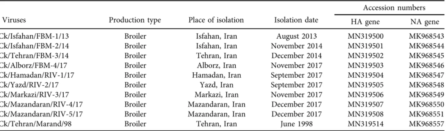
A total of ten H9N2 viruses were obtained from the Department of Avian Disease Research and Diagnostic, the Razi Vaccine and Serum Research Institute (Table 1).
The recent H9N2s were recovered from the respiratory system (trachea and lung) of broilers with severe respira- tory signs and high mortality. For virus propagation, all isolates were inoculated into the allantoic cavity of 9-day- old specific-pathogen-free (SPF) embryonated chicken eggs (Venkey’s, Maharashtra, India). After 48 h of incubation, the amnio-allantoicfluids were harvested and tested for the presence of H9N2 virus by haemagglutinin activity (OIE, 2019).
Extraction of viral genomic RNA and reverse transcription (RT)-PCR
The allantoic fluid from infected embryonated eggs was extracted and purified using the High Pure Viral RNA kit (Roche Life Science, Mannheim, Germany), in accordance with the manufacturer’s recommendations. The purity of the extracted RNA was assessed by calculating the ratio of readings at 260 and 280 nm.
Next, cDNAs of the studied viruses were synthesised with uni12 primer (50-AGCAAAAGCAGG-30) using the RevertAid cDNA synthesis kit (Thermo Fisher Scientific, Massachusetts, US), following the user manual. The full length of HA and NA genes of the above-mentioned H9N2s were amplified using influenza virus universal primers described previously (Hoffmann et al., 2001). The PCR mix was performed in a 50-
m
l reaction mix containing 33m
l ofnuclease-free water, 5
m
L of 103 Ampliqon Ammonium Buffer, 1m
L of 25 mM MgCl2, 2m
L 10 mM dNTP mix, 2m
Lof each primer (10
m
M), 1m
L of 2.5 units/m
L AmpliqonAccuPOLÔ DNA Polymerase (AMPLIQON, Odense M, Denmark) and 4
m
L of cDNA. The PCR thermal profile was set up at 958C/2 min, and 35 cycles at 958C/30 s, 508C/30 s and 72 8C/4 min followed by a final extension of 72 8C/5 min.Table 1.Information related to H9N2 viruses used in this study
Viruses Production type Place of isolation Isolation date
Accession numbers
HA gene NA gene
Ck/Isfahan/FBM-1/13 Broiler Isfahan, Iran August 2013 MN319500 MK968543
Ck/Isfahan/FBM-2/14 Broiler Isfahan, Iran November 2014 MN319501 MK968544
Ck/Tehran/FBM-3/14 Broiler Tehran, Iran December 2014 MN319502 MK968545
Ck/Alborz/FBM-4/17 Broiler Alborz, Iran November 2017 MN319503 MK968546
Ck/Hamadan/RIV-1/17 Broiler Hamadan, Iran September 2017 MN319504 MK968547
Ck/Yazd/RIV-2/17 Broiler Yazd, Iran September 2017 MN319505 MK968548
Ck/Markazi/RIV-3/17 Broiler Markazi, Iran November 2017 MN319506 MK968549
Ck/Mazandaran/RIV-4/17 Broiler Mazandaran, Iran December 2017 MN319507 MK968550
Ck/Mazandaran/RIV-5/17 Broiler Mazandaran, Iran December 2017 MN319508 MK968551
Ck/Tehran/Marand/98 Broiler Tehran, Iran June 1998 MN319514 MK968557
Cloning and transformation
After running on 1% agarose gel, each amplicon was excised and purified by the GeneJET Gel extraction Kit (Thermo Fisher Scientific, Massachusetts, US). The purified PCR frag- ments were ligated into pJET1.2/blunt according to CloneJET PCR Cloning Kit instructions (Thermo Fisher Scientific, Massachusetts, US) and then transformed into competent Escherichia coliTOP10 cells by exposing to heat shock at 428C for 1 min. The transformants were spread on Luria-Bertani (LB) agar supplemented with 50
m
g/mL ampicillin. Analysis of recombinant clones containing the expected insert was per- formed by either colony PCR amplification using the same primers for full-gene amplification or restriction endonuclease BglII. The positive plasmids were extracted from an overnight culture of bacteria using the High Pure Plasmid Isolation Kit (Roche Life Science, Mannheim, Germany).Nucleotide sequencing and phylogenetic analysis
Recombinant plasmids were submitted to a commercial sequencing service (Faza Pajooh Company, Tehran, Iran) for sequencing, using the pJET1.2 universal forward and reverse primer (HA and NA genes), as well as two internal primers for the HA gene (primer sequences will be provided upon request). All sequence data were assembled and analysed using the BioEdit software version 7.0.5 (Hall, 1999).
Nucleotide and deduced amino acid sequences were aligned with the Clustal W method in the MEGA software version 7.0.26 (Kumar et al., 2016). BLAST analysis (https://blast.
ncbi.nlm.nih.gov/Blast.cgi) was performed to retrieve iden- tical sequences related to our queries from the GenBank database. Phylogenetic analyses were carried out using the neighbour-joining method with the maximum composite likelihood model by calculating 1,000 replicates for boot- strap value in the MEGA 7.0.26 software (Kumar et al., 2016). Potential N-linked glycosylation sites in HA and NA protein were predicted using the NetNGlyc 1.0 server (http://www.cbs.dtu.dk/services/NetNGlyc).
Evolutionary analysis
To identify the natural selection on the coding sequences of HA and NA genes, the ratio of non-synonymous to syn- onymous substitutions (u 5 dN/dS) was measured using single likelihood ancestor counting (SLAC) in the web-based Datamonkey suite (Weaver et al., 2018). The dataset is composed of 59 Iranian HA and 65 NA complete coding sequences retrieved from GenBank for the period of 1998–
2017. The ratio ofufor each amino acid site in the HA and NA proteins was used to scan for evidence of positive or negative selection withu> 1,u51 andu< 1 indicating a positive selection, neutral evolution and negative selection, respectively. TheP-value threshold of 0.05 was set for evi- dence of positive and negative selection.
The dataset of Iranian HA and NA sequences was sub- jected to recombination analysis with recombination detec- tion program (RDP) version beta 4.56 (Martin et al., 2015).
Seven algorithms including RDP (Martin and Rybicki,
2000), GENECONV (Padidam et al., 1999), BootScan (Sal- minen et al., 1995), MaxChi (Smith, 1992), Chimaera (Posada and Crandall, 2001), SiScan (Gibbs et al., 2000) and 3Seq (Boni et al., 2007) were used for finding evidence of recombinant sequences. If a recombination was detected with at least three algorithms in any analysed sequence, it was regarded as a valid recombination and removed from the dataset (HA 5 58 and NA 5 61) used for Bayesian skyline plot reconstruction available in the BEAST software, version 1.10.4 (Suchard et al., 2018). Because of the higher performance of the codon-based SRD06 nucleotide substi- tution model (Fusaro et al., 2011), we employed this model with an uncorrelated lognormal relaxed clock and gamma distributed rate under a skyline coalescent tree prior. The Markov chain Monte Carlo (MCMC) for both genes was run by setting 50 million steps with sampling every 10,000 generations. To estimate polydynamic history genetic changes within the Iranian HA and NA sequences of H9N2 viruses, two Bayesian skyline plots were reconstructed using the tracer software, version 1.7.1 (Rambaut et al., 2018).
Nucleotide sequence accession numbers
The twenty sequences of H9N2 HA (MN319500–MN319508, MN319514) and NA (MK968543–MK968551, MK968557) genes obtained in this study have been deposited in GenBank.
RESULTS
Study of genetic identities and phylogenetic analysis
According to BLAST searches, the studied vaccine strain (Marand) was more similar to viruses previously isolated from the Japanese parakeet in 1997. However, Pakistani strains were the closest relative to other studied sequences.
Concerning HA and NA genes of FBM-1, a higher per cent of identity was shown with Iranian H9N2 viruses isolated in 2010 and 2011, which are closely related to H9N2 strains isolated from Pakistan between 2005 and 2008 (Bashashati et al., 2013).
Pairwise comparison of nucleotide sequences of the Ira- nian HA and NA genes revealed that identities among viruses ranged from 88.2 to 99.8% and 93.1 to 100%, respectively (data not shown). The deduced amino acid sequences of HA and NA shared 91–99.8% and 88.6–100% homology, respectively (data not shown). At the nucleotide level, all studied viruses except FBM-1 had about 12% divergence with the current vaccine strain, which showed quite a low ho- mology with significant genetic distances.
All studied isolates and published Iranian sequences available in GenBank were subjected to phylogenetic analysis (Fig. 1). The Iranian HA sequences were clustered into 4 distinct genetic groups in G1/like sub-lineage, which con- tained viruses isolated from different periods of times: (a) 1998–2004, (b) 2005–2009, (c) 2010–2013 and (d) 2014–
2017 (Fig. 1a). In the NA phylogenetic tree, there were five distinct clusters that are composed of H9N2s in (i) 1998– 2004, (ii) 2006–2008, (iii) 2009–2012, (iv) 2010–2013 and
(v) 2014–2017 (Fig. 1b). As can be seen, the topology of both HA and NA phylogenetic trees is very similar.
Evolutionary features
Positive and negative selection pressures of the coding sequence of HA and NA were estimated using the Data- monkey software as described above. Purifying selection was observed in Iranian HA and NA genes with mean dN/dS values of 0.177 and 0.231, respectively. The only positively selected amino acid substitution found among both studied genes of Iranian H9N2 viruses was located at position 190 (henceforth, H3 numbering was denoted throughout the
manuscript) in HA protein by aP-value threshold of <0.05.
In this regard, 95 and 72 amino acid residues in the HA and NA genes of Iranian H9N2s were found to be under negative selection, respectively.
The genetic diversity of Iranian HA and NA genes showed a slight increase over a period of 20 years (Fig. 2). After the first record of the H9N2 AIV in 1998, the viral population size remained relatively stable until 2001. Fluctuations in viral di- versity were observed in HA Bayesian skyline plot as three phases. Regarding Iranian HA genes, effective population size had experienced a period of instability since 2005 that lasted for about a decade (Fig 2a). Bayesian skyline plot analysis for the NA genes of the Iranian H9N2 viruses did not show any Fig. 1.Phylogenetic tree for HA (a) and NA (b) genes of the studied H9N2 viruses along with other Iranian and representative viruses
retrieved from GenBank. Both trees were generated in MEGA software version 7.0.26 using the neighbour-joining method with the maximum composite likelihood model. The studied isolates are marked with black circles. The black triangle indicates the vaccine strain.
Bootstrap values≥70 are shown next to the branches
major changes in 1998–2017. There was only a gradual rise in the NA population from 2005 to 2017 (Fig. 2b).
Genetic analysis of the HA and NA genes
Deduced amino acid sequences of the HA and NA genes were aligned and compared with each other and other Iranian representatives retrieved from the GenBank (Supplementary Tables 1–2). All coding sequences of the analysed H9N2s consisted of 1,683 nucleotides (560 amino acids) except for one virus. A 12-nucleotide deletion resulting from the deletion of four amino acids at residues from 220 to 223 was observed in the FBM-1 virus. Although a 24-nucleotide deletion in HA1 was found in H7N2 viruses isolated from live bird markets in the United States (Suarez et al., 1999), this rare deletion had not been observed in H9N2 viruses before. Virulence molec- ular determinants of HA are mainly described by specific amino acid residues in the cleavage site and receptor binding site (RBS). In addition, the presence or absence of glycosylation sites around RBS plays a role in the pathogenicity of AI.
Although the vaccine strain (Marand) had an RSSR motif at the cleavage site, all other H9N2 viruses isolated after 2013 shared a different motif (KSSR) at this site of the HA protein.
Lack of multiple basic amino acids at the connecting peptide is
regarded as a signature of LPAI (Steinhauer, 1999). By the analysis of RBSs, six substitutions were found in the studied isolates. Of these substitutions, Q226L in RBS, which are associated with binding preference to human-likea2,6-linked sialic acid receptors, were observed in nine recently isolated viruses. Thus, it can be stated that these viruses have zoonotic potential (Wan et al., 2008). The analysis of potential glyco- sylation sites with an NXS/T motif (where X represents any amino acid except proline) revealed that there werefive (21, 94, 128, 285/289 and 292/296) and one (154) conserved sites in the HA1 and HA2 parts of the protein, respectively. In comparison with the vaccine strain, all nine H9N2s lost two potential glycosylation sites at amino acid residues 158 of HA1 and 219 of HA2. Furthermore, RIV-1 gained another glycan at position 133 of the HA1 portion and lost one at amino acid residue 213 of the HA2 part. The comparison of the antigenic sites of 10 Iranian H9N2s with reference to the previous studies (Kaverin et al., 2004; Peacock et al., 2016) revealed that recent H9N2s have seven alterations in these epitopes in comparison to the vaccine strain (Table 2). A deletion of amino acid residue 222 in the H9-A site was found in FBM-1 due to the deletion of four amino acid residues at positions 220–223. Moreover, mutation R172Q at the H9-B antigenic site was only observed in RIV-1 virus among the viruses analysed.
The complete NA genes of 10 H9N2s were determined and the coding sequence of all viruses encoded a protein of 469 amino acids. No stalk deletions at amino acids 38–39 and 63–65 were observed in the NA protein of H9N2 viruses.
Pathogenicity indicators of NA are composed of enzyme active site, stalk length, haemadsorbing (HB) site and glycosylation sites. Analysis of active sites revealed that no mutation was found among the analysed viruses in known amino acid res- idues related to neuraminidase resistance. Analysis of three surface loops of the HB site in the NA protein revealed five substitutions (amino acid residues 366, 367, 372, 401 and 403) in two loops (370 and 400) of this site in the analysed viruses.
A different pattern of glycosylation sites was observed; seven Iranian H9N2 viruses showed a similar pattern of glycosyla- tion site, while three of them (Marand, FBM-3 and FBM-4) contained one more glycosylation in the NA protein. More- over, recent isolates have lost a potential glycosylation site in position 70 compared to the vaccine strain.
DISCUSSION
Although vaccination against the H9N2 virus has been routinely adopted for two decades in Iran since the late 1990s, outbreaks of the disease occur with each passing year.
H9N2s have a significant economic impact on poultry production throughout the Iranian industrial poultry sector.
The currently used vaccine strains against H9N2 were originally derived from viruses back in 1998, when the first H9N2 virus was isolated in Iran (Bashashati et al., 2013).
The aim of the present study was to perform sequencing and phylogenetic analysis of 10 viruses to determine the mo- lecular evolution and genetic variation of the HA and NA genes of H9N2 viruses.
Fig. 2.Bayesian skyline plots of Iranian HA (a) and NA (b) sequences of H9N2 viruses since itsfirst report. The dark solid
line and the blue shaded region indicate effective population size and 95% highest posterior density interval, respectively. The vertical axis shows the effective population size and the horizontal
axis indicates the isolation year of the samples
The topologies of HA and NA phylogenetic trees were very similar to each other and all the Iranian viruses fell into G1 sub-lineage in agreement with previous studies (Bashashati et al., 2013; Norouzian et al., 2014; Bahari et al., 2015). On the basis of phylogenetic analysis, the Iranian HAs are classified into four branches whereas NA genes fall intofive categories.
Recently studied H9N2 viruses were grouped into a cluster labelled‘2014–2017 group’that shows the alteration of clusters over time. Moreover, the recent viruses show a distant genetic relationship at the nucleotide and amino acid levels (about 90%) with a common vaccine strain (Marand). BLAST anal- ysis revealed that all tested strains were closely related to vi- ruses (95–99%) isolated from Pakistan, which shares a border with Iran. Except for the FBM-1 virus, all other studied strains had a 1,683 nucleotides long coding sequence. Four amino acid deletions at positions 220–223 close to the RBS were found in the FBM-1 virus. This distinguishing characteristic wasfirst reported in H7N2 viruses from live poultry markets in the northeast of the United States. This viral genotype showed eight amino acid deletions at positions 221–228 within the RBSs. Furthermore, the deletion of stalk in NA was detected in this genotype as it may compensate for the HA stalk deletion (Suarez et al., 1999). However, the studied iso- lates did not show any deletion in the stalk region of NA. In this regard, further investigations need to be conducted on the impact of this deletion on HA binding of the FBM-1 virus.
The demographic history of Iranian H9N2 viruses revealed that the diversification of H9N2 is increasing gradually over time. For the first three years after 1998, the effective population size of H9N2 viruses had a stable trend, perhaps because of the implementation of vaccina- tion against H9N2. Although Iranian H9N2s have evolved under purifying selection, one amino acid residue at posi- tion 190 in the HA protein is under positive selection. An extensive study on selection pressures for all segments of Central Asian and Middle Eastern H9N2 viruses revealed that most coding positions were under purifying selection.
Amino acid positions 160, 190 and 226 in the HA protein were detected to be under positive selection, located at the RBS, which may have an effect on receptor recognition (Fusaro et al., 2011).
Mutation of the amino acid residue at 226 in the RBS facilitates the transmission of AIV to mammalian species.
HAs of avian viruses show a preference for binding to sialic acid a2,3-linked to galactose, while human viruses tend to use a2,6-linked sialic acid as abundant receptors in the upper respiratory tract (Matrosovich et al., 2009). Among the 10 analysed Iranian H9N2 isolates, 9 recently isolated viruses showed that the Q226L mutation, compared with the older strain (Marand), shows a preference to binding to a2,6-linked sialic acid (Obadan et al., 2019). This mutation plays a significant role in overcoming the host barrier Table 2.Antigenic sites of HA genes of the Iranian H9N2 strains
Viruses
Antigenic site according to the study ofPeacock et al. (2016)
Antigenic site according to the study ofKaverin et al. (2004)
H9-A H9-B Site I Overlapping site Site II
155 193 222 227 244 122 120a 149 172 135 157 162 133 189 198 145 193 226
Marand (98) T S L Q R Q T R R T K P T T T G S Q
EU477247 (99) –b N – – – – – – – – – – – – – D N L
EU477249 (00) – – – – – – – – – – – – – – – – – –
EU477246 (02) – – – – – – – – – – – – – – – – – Q
EF063726 (03) – – – – – – – – – – – – – – – – – –
EF063730 (04) – – – – – – – – M – R – – – – – – L
EF063733 (05) – – – I – – – – R – K – – – – – – –
EU477245 (07) – – – – – – – – – – – – – – – – – –
GU071968 (08) – – – – – – – – – – – – – – – – – –
JX456178 (09) – – – – – – – – – – – – – – – – – –
JN646748 (10) – – – – – – – – – – – – – – – – – –
JQ970436 (11) – – – – – – – – – – – – – – – – – –
JX294920 (12) – – – – – – – – Q – – – – – – – – –
FBM-1 (13) – – –c – – – – – R – – – – – – – – –
FBM-2 (14) – – – – – – A – – – – – D – – N – –
FBM-3 (14) – – – – – – – – – – – – – – – – – –
FBM-4 (17) – – – – – – – – – – – – – – – – – –
RIV-1 (17) – – – – – – – – Q – – – N – – – – –
RIV-2 (17) – – – – – – – – R – – – D – – – – –
RIV-3 (17) – – – – – – – – – – – – – – – – – –
RIV-4 (17) – – – – – – – – – – – – – – – – – –
RIV-5 (17) – – – – – – – – – – – – – – – – – –
aThis position was numbered according to H9 due to absence of this site in subtype H3.
bDash indicates that the amino acid residues are the same as above.
cThis site was deleted in FBM-1.
between avian and human, resulting in human infections with H9N2 viruses (Guo et al., 1999; Saito et al., 2001).
Five conserved amino acids (S367, S370, S372, N400 and W403) in the HB site were determined among all nine sub- types of NA with some exceptions (Varghese et al., 1997;
Uhlendorff et al., 2009). Offive mutations in the 370 and 400 loops of the HB site, the analysed viruses possessed three substitutions in amino acid residues at positions 367, 372 and 403 in the conserved positions. Any mutation in the HB site of H9N2 viruses suggested the improvement offitness in land- based poultry from aquatic birds as their natural reservoir (Matrosovich et al., 2001). Since the HB site plays a role in the replication of the virus in avian hosts, it is not known whether these substitutions can alter the pathogenicity of H9N2.
A comparison of recent H9N2s at antigenic sites with the vaccine strain exhibited a remarkable amino acid divergence.
According to previous studies, of 17 determined antigenic sites by escape mutants (position 193 is common in two studies), seven substitutions occurred in recent H9N2 viruses (Kaverin et al., 2004; Peacock et al., 2016). Mutation in antigenic sites of the HA protein of H9N2s may produce antigenic variants and enable the virus to escape vaccine-induced neutralising anti- bodies, leading to vaccination failure (Peacock et al., 2016).
The results of the current study indicate that substitution in pivotal genetic markers of circulating field Iranian H9N2s continues to occur. Therefore, further studies are needed to evaluate the biological propensities of these mutations and assess the pathogenicity of circulating viruses. Although there have been reports on protection against clinical disease via vaccination, the H9N2 vaccine fails to protect from infection and virus shedding when antigenic variants are circulating (Lee et al., 2016; Xia et al., 2017). Accordingly, antigenic characterisation and systematic monitoring of prevailing H9N2s will be necessary to identify antigenically well-matched strains for a timely updating of the vaccine strain.
ACKNOWLEDGEMENTS
We would like to express our thanks to Mohsen Mah- moudzadeh for help in virus isolation. This study was sup- ported by the Razi Vaccine and Serum Research Institute under grant no. 13-18-1851-066-97020-971045.
SUPPLEMENTARY MATERIAL
The online version of this article offers supplementary material:https://doi.org/10.1556/004.2020.00048.
REFERENCES
Alexander, D. J. (2007): An overview of the epidemiology of avian influenza. Vaccine25, 5637–5644.
Bahari, P., Pourbakhsh, S. A., Shoushtari, H. and Bahmaninejad, M.
A. (2015): Molecular characterization of H9N2 avian influenza viruses isolated from vaccinated broiler chickens in northeast Iran. Trop. Anim. Health Prod.47, 1195–1201.
Bashashati, M., VasfiMarandi, M. and Sabouri. F. (2013): Genetic diversity of early (1998) and recent (2010) avian influenza H9N2 virus strains isolated from poultry in Iran. Arch. Virol.
158, 2089–2100.
Boni, M. F., Posada, D. and Feldman, M. W. (2007): An exact nonparametric method for inferring mosaic structure in sequence triplets. Genetics176, 1035–1047.
Butt, A. M., Siddique, S., Idrees, M. and Tong, Y. (2010): Avian influenza A (H9N2): computational molecular analysis and phylogenetic characterization of viral surface proteins isolated be- tween 1997 and 2009 from the human population. Virol. J.7, 319.
Fusaro, A., Monne, I., Salviato, A., Valastro, V., Schivo, A., Amarin, N. M., Gonzalez, C., Ismail, M. M., Al-Ankari, A. R., Al-Blowi, M. H., Khan, O. A., Maken Ali, A. S., Hedayati, A., Garcia Garcia, J., Ziay, G. M., Shoushtari, A., Al Qahtani, K. N., Capua, I., Holmes, E. C. and Cattoli, G. (2011): Phylogeography and evolutionary history of reassortant H9N2 viruses with potential human health implications. J. Virol.85, 8413–8421.
Gibbs, M. J., Armstrong, J. S. and Gibbs, A. J. (2000): Sister- scanning: a Monte Carlo procedure for assessing signals in recombinant sequences. Bioinformatics16, 573–582.
Guan, Y., Shortridge, K. F., Krauss, S. and Webster, R. G. (1999):
Molecular characterization of H9N2 influenza viruses: were they the donors of the‘internal’genes of H5N1 viruses in Hong Kong? Proc. Natl. Acad. Sci. U.S.A.96, 9363–9367.
Guo, Y., Li, J. and Cheng, X. (1999): Discovery of men infected by avian influenza A (H9N2) virus [in Chinese]. Zhonghua Shi Yan He Lin Chuang Bing Du Xue Za Zhi13, 105–108.
Hall, T. A. (1999): BioEdit: a user-friendly biological sequence alignment editor and analysis program for Windows 95/98/NT.
Nucleic Acids Symp. Ser.41, 95–98.
Hoffmann, E., Stech, J., Guan, Y., Webster, R. G. and Perez, D. R (2001): Universal primer set for the full-length amplification of all influenza A viruses. Arch. Virol.146, 2275–2289.
Homme, P. J. and Easterday, B. C. (1970): Avian influenza virus infections. I. Characteristics of influenza A-turkey-Wisconsin- 1966 virus. Avian Dis.14, 66–74.
Kaverin, N. V., Rudneva, I. A., Ilyushina, N. A., Lipatov, A. S., Krauss, S. and Webster, R. G. (2004): Structural differences among hemagglutinins of influenza A virus subtypes are re- flected in their antigenic architecture: analysis of H9 escape mutants. J. Virol.78, 240–249.
Kumar, S., Stecher, G. and Tamura, K. (2016): MEGA7: Molecular evolutionary genetics analysis version 7.0 for bigger datasets.
Mol. Biol. Evol.33, 1870–1874.
Lee, D. H., Fusaro, A., Song, C. S., Suarez, D. L. and Swayne, D. E.
(2016): Poultry vaccination directed evolution of H9N2 low pathogenicity avian influenza viruses in Korea. Virology 488, 225–231.
Martin, D. and Rybicki, E. (2000): RDP: detection of recombination amongst aligned sequences. Bioinformatics16, 562–563.
Martin, D. P., Murrell, B., Golden, M., Khoosal, A. and Muhire, B.
(2015): RDP4: detection and analysis of recombination patterns in virus genomes. Virus Evol.1, vev003.
Matrosovich, M. N., Krauss, S. and Webster, R. G. (2001): H9N2 influenza A viruses from poultry in Asia have human virus-like receptor specificity. Virology281, 156–162.
Matrosovich, M., Stech, J. and Klenk, H. D. (2009): Influenza receptors, polymerase and host range. Rev. Sci. Tech.28, 203–217.
Norouzian, H., Bashashati, M. and Vasfimarandi, M. (2014):
Phylogenetic analysis of neuraminidase gene of H9N2 avian influenza viruses isolated from chicken in Iran during 2010–
2011. Iran. J. Microbiol.6, 91–97.
Obadan, A. O., Santos, J., Ferreri, L., Thompson, A. J., Carnaccini, S., Geiger, G., Gonzalez Reiche, A. S., Raj~ao, D. S., Paulson, J. C.
and Perez, D. R. (2019): Flexibilityin vitroof amino acid 226 in the receptor-binding site of an H9 subtype influenza A virus and its effectin vivoon virus replication, tropism, and trans- mission. J. Virol.93, pii: e02011-18.
OIE (2019): Avian influenza (infection with avian influenza viruses).
In: Manual of diagnostic tests and vaccines for terrestrial animals.
Office International des Epizooties, Paris, France. pp. 821–843.
Padidam, M., Sawyer, S. and Fauquet, C. M. (1999): Possible emergence of new geminiviruses by frequent recombination.
Virology265, 218–225.
Park, K. J., Kwon, H. I., Song, M. S., Pascua, P. N., Baek, Y. H., Lee, J. H., Jang, H. L., Lim, J. Y., Mo, I. P., Moon, H. J., Kim, C. J.
and Choi, Y. K. (2011): Rapid evolution of low-pathogenic H9N2 avian influenza viruses following poultry vaccination programmes. J. Gen. Virol.92, 36–50.
Peacock, T., Reddy, K., James, J., Adamiak, B., Barclay, W., Shelton, H. and Iqbal, M. (2016): Antigenic mapping of an H9N2 avian influenza virus reveals two discrete antigenic sites and a novel mechanism of immune escape. Sci. Rep.6, 18745.
Posada, D. and Crandall, K. A. (2001): Evaluation of methods for detecting recombination from DNA sequences: computer simulations. Proc. Natl. Acad. Sci. U.S.A.98, 13757–13762.
Pu, J., Wang, S., Yin, Y., Zhang, G., Carter, R. A., Wang, J., Xu, G., Sun, H., Wang, M., Wen, C., Wei, Y., Wang, D., Zhu, B., Lemmon, G., Jiao, Y., Duan, S., Wang, Q., Du, Q., Sun, M., Bao, J., Sun, Y., Zhao, J., Zhang, H., Wu, G., Liu, J. and Webster, R.
G. (2015): Evolution of the H9N2 influenza genotype that facilitated the genesis of the novel H7N9 virus. Proc. Natl.
Acad. Sci. U.S.A.112, 548–553.
Qi, W., Zhou, X., Shi, W., Huang, L., Xia, W., Liu, D., Li, H., Chen, S., Lei, F., Cao, L., Wu, J., He, F., Song, W., Li, Q., Li, H., Liao, M. and Liu, M. (2014): Genesis of the novel human-infecting influenza A(H10N8) virus and potential genetic diversity of the virus in poultry, China. Euro. Surveill.19, 20841.
Rambaut, A., Drummond, A. J., Xie, D., Baele, G. and Suchard, M.
A. (2018): Posterior summarization in Bayesian phylogenetics using Tracer 1.7. Syst. Biol.67, 901–904.
Saito, T., Lim, W., Suzuki, T., Suzuki, Y., Kida, H., Nishimura, S. I.
and Tashiro, M. (2001): Characterization of a human H9N2 influenza virus isolated in Hong Kong. Vaccine20,125–133.
Salminen, M. O., Carr, J. K., Burke, D. S. and McCutchan, F. E.
(1995): Identification of breakpoints in intergenotypic recombi- nants of HIV type 1 by bootscanning. AIDS Res. Hum. Retrovir.
11, 1423–1425.
SJCEIRS Working Group (2013): Assessing thefitness of distinct clades of influenza A (H9N2) viruses. Emerg. Microb. Infect.2, e75.
Smith, J. M. (1992): Analyzing the mosaic structure of genes. J. Mol.
Evol.34, 126–129.
Song, X. F., Han, P. and Chen, Y. P. (2011): Genetic variation of the hemagglutinin of avian influenza virus H9N2. J. Med. Virol.83, 838–846.
Steinhauer, D. A. (1999): Role of hemagglutinin cleavage for the pathogenicity of influenza virus. Virology258, 1–20.
Suarez, D. L., Garcia, M., Latimer, J., Senne, D. and Perdue, M.
(1999): Phylogenetic analysis of H7 avian influenza viruses isolated from the live bird markets of the Northeast United States. J. Virol.73, 3567–3573.
Suchard, M. A., Lemey, P., Baele, G., Ayres, D. L., Drummond, A. J.
and Rambaut, A. (2018): Bayesian phylogenetic and phylody- namic data integration using BEAST 1.10. Virus Evol. 4, vey016.
Uhlendorff, J., Matrosovich, T., Klenk, H. D. and Matrosovich, M.
(2009): Functional significance of the hemadsorption activity of influenza virus neuraminidase and its alteration in pandemic viruses. Arch. Virol.154, 945–957.
Varghese, J. N., Colman, P. M., van Donkelaar, A., Blick, T. J., Sahasrabudhe, A., McKimm-Breschkin, J. L. (1997): Structural evidence for a second sialic acid binding site in avian influenza virus neuraminidases. Proc. Natl Acad. Sci. U.S.A.94, 11808–
11812.
Wan, H., Sorrell, E. M., Song, H., Hossain, M. J., Ramirez-Nieto, G., Monne, I., Stevens, J., Cattoli, G., Capua, I., Chen, L. M., Donis, R. O., Busch, J., Paulson, J. C., Brockwell, C., Webby, R., Blanco, J., Al-Natour, M. Q. and Perez, D. R. (2008): Replication and transmission of H9N2 influenza viruses in ferrets: evaluation of pandemic potential. PLoS One3, e2923.
Weaver, S., Shank, S. D., Spielman, S. J., Li, M., Muse, S. V. and Kosakovsky Pond, S. L. (2018): Datamonkey 2.0: a modern web application for characterizing selective and other evolutionary processes. Mol. Biol. Evol.35, 773–777.
Xia, J., Cui, J., He, X., Liu, Y. Y., Yao, K. C., Cao, S. J., Han, X. F. and Huang, Y. (2017): Genetic and antigenic evolution of H9N2 subtype avian influenza virus in domestic chickens in south- western China, 2013–2016. PLoS One12, e0171564.