C
hronic venous disease (CVD) is one of the most com- mon cardiovascular disorders in Northern and Western Europe; however, the causative factors predisposing individu- als to the disease are still under investigation. Age, sex, obe- sity, and family history were found to be major risk factors for CVD.1–3 Altered venous biomechanics may also contribute to the pathogenesis of venous disease.4,5Twin studies have been extensively used to characterize the interaction of genetic and environmental factors on car- diovascular phenotypes.6,7 A CVD twin study reported 84%
phenotypic concordance between monozygotic (MZ) twins, compared with only 39% between dizygotic (DZ) twins.8 Fiebig et al9 evaluated the relative genetic and environmental impact upon CVD risk by estimating the heritability of the disease in 4033 nuclear families, comprising 16 434 individu- als from all over Germany. The heritability of CVD proved to be high, thereby suggesting a notable genetic component in the pathogenesis of the disease.9 Altered venous biomechanics may be a predisposing phenotypic factor for the development of this disease. However, the level of genetic determination of venous biomechanics is less known. Brinsuk et al,10 using
the impedance plethysmography technique in a twin study of 46 twin pairs, found that both venous compliance and venous capacity are substantially influenced by genetic factors.
No previous attempts, however, to reveal the heritability of venous elasticity have been done, when using more accurate ultrasonographic techniques and making the venous elasticity measurements in different postures and under controlled pres- sure changes.11–13
The present study was done to differentiate between inher- ited and environmental factors in venous biomechanical phe- notype by comparison of MZ and DZ twins. The aim of our present work was to evaluate the heritability of in situ static and dynamic venous biomechanics.
Materials and Methods
SubjectsA total of 102 twin pairs were enrolled: 78 MZ twin pairs (112 women and 44 men, aged 42.4±16.8 years, mean age±SD) and 24 same-sex DZ twin pairs (36 women and 12 men, aged 50.5±16.1 years, mean age±SD). Subjects were recruited as part of the International Twin Study 2009 project. A sample of healthy twins aged >18 years and living in Hungary was measured at 2 large hospitals in Budapest in
Received on: July 5, 2011; final version accepted on: October 8, 2012.
From the Research Group for Inflammation Biology and Immunogenomics of Hungarian Academy of Sciences and Semmelweis University, Budapest, Hungary (A.A.M., Z.K., I.P., R.G.K.); Military Hospital, Department of Cardiology, Budapest, Hungary (A.A.M., Z.K., I.P., R.G.K.); Department of Radiology and Oncotherapy (A.D.T., D.L.T., V.B.), Faculty of Pharmacy (Á.L.), and Experimental Research Department and Department of Human Physiology (E.M., G.L.N.), Semmelweis University, Budapest, Hungary; Central European University, Budapest, Hungary (L.L.); and The Methodist Hospital DeBakey Heart and Vascular Center, Houston, TX (Z.G.).
Correspondence to Andrea Ágnes Molnár, Military Hospital, Department of Cardiology, Róbert Károly krt. 44, Budapest 1134, Hungary. E-mail molnandi@gmail.com
© 2012 American Heart Association, Inc.
Arterioscler Thromb Vasc Biol is available at http://atvb.ahajournals.org DOI: 10.1161/ATVBAHA.112.300062
Objective—Altered venous biomechanics may contribute to the pathogenesis of venous diseases, and their heritability is less known.
Methods and Results—Seventy-eight monozygotic twin pairs (aged 42.4±16.8 years) and 24 same-sex dizygotic twin pairs (aged 50.5±16.1 years) were examined. Anteroposterior and mediolateral diameters of the common femoral vein were measured by ultrasonography. Measurements were made both in supine and in standing body positions, with or without controlled forced expiration (Valsalva test). High correlation of diameter, capacity, and distensibility values was found between twin pairs. The univariate heritability (A), shared (C), and unshared (E) environmental effects model has shown 39.3% genetic component of the variance of low pressure, 37.9% of high-pressure venous capacity, and 36.4%
of maximal capacity changes, even after elimination of sex, age, and body weight effects. Bivariate Cholesky analysis revealed substantial covariance of inherited body weight and venous capacity components (57.0%–81.4%).
Conclusion—Femoral vein capacity and elasticity depend ≈30% to 40% on genetic factors, and this value in the standing body position can reach 50%. A relatively high genetic covariance was found between weight and femoral vein capacity and elasticity. Our work might yield some new insights into the inheritance of venous diseases that are associated with altered venous biomechanics and help elucidate the involved genes. (Arterioscler Thromb Vasc Biol.
2013;33:152-157.)
Key Words: biomechanics ◼ heritability ◼ ultrasound ◼ Valsalva ◼ vein ◼ venous
Andrea Ágnes Molnár, Ádám Domonkos Tárnoki, Dávid László Tárnoki, Zoltán Kulcsár, Levente Littvay, Zsolt Garami, István Préda, Róbert Gábor Kiss, Viktor Bérczi, Ágnes Lannert,
Emil Monos, György László Nádasy
00
2009 and 2010. We used a multiple self-reported question approach to assess zygosity suggested by Heath et al.14 The most likely zygosity was assigned based on the 7 self-reported responses.14 First-born (twin A) and second-born (twin B) members of the twin pairs were identified. Exclusion criteria were pregnancy, medical conditions possibly interfering with compliance during examination (eg, unable to carry out Valsalva test, unable to keep the supine or erect body position), and acute infection within 3 weeks of measurement. All subjects were asked to refrain from smoking 3 hours, eating 1 hour, and drinking alcohol and coffee 10 hours before their visit.
The subjects’ written informed consent was obtained before study entry, and the program was approved by the ethical commissions of the cooperating institutes.
Study Protocol
Physical examination included blood pressure measurement, heart rate, and observation of superficial leg veins and potential symp- toms of CVD. Anthropometric parameters, such as height and body weight, were also recorded.
Anteroposterior and mediolateral diameters of the common femo- ral vein at both sides were measured by ultrasonography (Toshiba Power Vision and Esaote My Lab; 5–10 MHz linear array probes).
The image resolution (0.1 mm) provided by the high-frequency probes is sufficient to track diameter changes with >1% accuracy.
Identification process of the veins consisted of a compression test (light pressure exerted with the transducer on the femoral vessels) and venous flow measurement, including distal compression test.
Compression of the veins with the transducer was avoided during diameter measurements. The diameters of common femoral veins were measured 1 cm above the junction of the great saphenous vein.
Measurements were made both in the supine and in the standing body positions, with or without controlled forced expiration (Valsalva test).
The Valsalva test was performed with the nose clipped; the patients were asked to produce a forced expiration while holding a tube in their mouth connected to an electromanometer until the requested pressure was reached and maintained. Valsalva pressures of 0 and 60 mm Hg were applied. Stable venous diameters were reached in ≈3 to 4 seconds. The monitor image was frozen in the fourth to sixth sec- onds of Valsalva, and in each case, both anteroposterior and mediolat- eral diameters were measured. We considered it important to measure both anteroposterior and mediolateral diameters at all pressures and body positions because the shape of veins may change with increase in pressure. It was also important to make the measurements both in the reclined and in the erect body positions. Minimum venous pres- sure is at no Valsalva in the reclined position. From the pathological point of view, however, the erect body position is more relevant, and maximum possible venous pressures can be reached with Valsalva and in the erect position. Venous pressure in large veins equilibrates quickly with the applied Valsalva pressure. When computing the dis- tensibility, it was supposed that pressure in these large veins, located not too far from the affected body cavities, is approximately identi- cal (plus gravitation effect in the erect body position) with controlled Valsalva pressure. This approach seems to be justified because some direct clinical measurements demonstrated an effective pressure equilibration among distant large veins.15–17
Capacity per unit length was computed in such a way that the cross section was considered elliptic and the equation Q = rarbπ was used to compute cross-sectional area (Q), where ra and rb are the 2 radii of the ellipse. Distensibility was defined as D=ΔV/V0ΔP, where ΔV is the change in capacity of vessel, V0 is the initial capacity, and ΔP is the pressure change.
Statistics
Heritability estimates were determined based on the consideration that greater levels of MZ than DZ within-pair similarity indicate a genetic influence on a phenotype, whereas similarity of co-twin cor- relations suggests that the variance is a result of shared environmental sources. Structural equation modeling was performed by using the Mplus Version 6.18 Empirical CIs were calculated with a Bollen–Stine
Bootstrap.19 Univariate quantitative genetic modeling was performed to decompose the phenotypic variance of the considered parameters into heritability (A), shared (C), and unshared (E) environmental effects (ACE analysis).20,21 The additive genetic component (A) mea- sures the effects because of genes at multiple loci or multiple alleles at 1 locus. The shared environmental component estimates contribu- tion of a common family environment for both twins (eg, familiar socialization, shared womb, air pollution, childhood diet), whereas the unshared environmental component estimates the effects that sep- arately apply to each individual twin (eg, smoking, physical activity) and also accounts for measurement errors. All data were adjusted for age, sex, and body weight. Opposite-sex twin pairs were excluded as their inclusion could bias the heritability estimates upward if sex-spe- cific or X-chromosome effects are present. For age and body weight adjustments, linear normalizations were used. For some selected pairs of phenotypic parameters, a bivariate Cholesky decomposition was carried out to derive the magnitude of covariation between the investigated phenotypes of interest and to estimate what proportion of this correlation is attributable to common underlying genetic and environmental factors. To estimate the amount of overlap between genes or environment that influence the 2 parameters, genetic and environmental correlations between those phenotypes were calcu- lated. P<0.05 was accepted as the significance level of the correlation coefficient. Values of the inherited (A) component >0.3 were consid- ered as evidence of substantial heritability.
Results
Systolic and diastolic pressures and body mass indexes were not different for the MZ and DZ groups (systolic, 128.5±16.8 and 128.2±17.2 mm Hg; diastolic, 75.7±12.2 and 74.8±14.0 mm Hg; body mass index, 25.5±4.9 and 26.1±5.0 kg/m2, for MZ and DZ, respectively). Current smoker rate was 16.0%
and 17.6% in MZ and DZ twins, respectively.
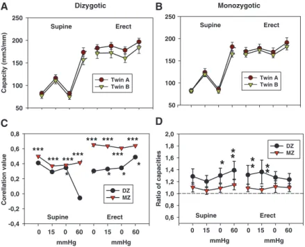
Anteroposterior and mediolateral diameters and capacity characteristically changed during the study protocol; they dilated as the pressure increased as an effect of altered posture and increased Valsalva pressure (Figure 1).
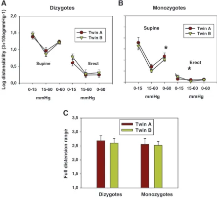
Femoral vein diameters, capacity, and distensibility showed very high level of parallelism in twin pairs (Figures 1–3). Values for first- and second-born twins are very close to each other, both in DZ and MZ twins (compare A and B blocks of Figures 1–3), but MZ twins are closer than DZs.
Corresponding to this, correlation values of twin pairs are shown in Figures 1C and 2C. Correlations between twin pairs are much higher for MZ twins than for DZs. We computed the ratio of the 2 co-twins (Figures 1D and 2D), which was closer to the unit in MZs and was significantly different from those of DZs at all pressure levels.
The correlations between DZ twin pairs were evaluated with and without opposite-sex DZ twin pairs. When opposite-sex DZ twins were omitted, intrapair correlation significantly decreased in case of femoral vein full-distension range. This suggests that some sex chromosome or sex- specific environmental effects on venous biomechanics may be present, but we did not have the statistical power to rigorously test this. Also, for this reason, opposite-sex DZ pairs were omitted. As heritability of body weight is high (82.7% in our sample; Table 1, first row), age, sex, and weight adjustments were performed while assessing the heritability of venous biomechanics. Parameters characterizing the level of inheritance and environmental effects in forming the geometric and elastic properties of the veins after age, sex, and weight adjustment are shown in the further part of
Table 1 (without the data of opposite-sex DZ twins). Data are given as heritability (A), shared (C), and unshared environmental (E) components of the intrapair variance values using the best-fitting univariate ACE model (with their 95% CIs). Results indicate a 39.3% genetic component of the variance of low-pressure venous capacity (reclined body position without Valsalva maneuver). The highest heritability of capacity has been proven at erect body position without the Valsalva maneuver (51.7%), when intraluminal venous pressures are high. However, further elevation of intraluminal pressure (erect body position using 60 mm Hg Valsalva pressure) heritability of venous capacity was 37.9%. The genetic component of maximum relative rise of capacity (full-distension range) has been shown to be somewhat similar (36.4%; Table 1, last row).
Because weight and the investigated venous parameters showed both sizable and significant phenotypic correlation and heritability, a bivariate Cholesky decomposition model was conducted to investigate a common genetic background of these traits. Table 2 shows standardized genetic, common, and unique environmental components of the covariance in venous capacity at different pressures with body weight that was estimated using the best bivariate Cholesky model. There was a sufficient correlation between femoral vein capacity and body weight without Valsalva both in the reclined and in the erect body positions. The high A values (0.60 and 0.81 for the erect and reclined body positions, respectively) show that there is a substantial inherited component in this correlation. Capacities after Valsalva correlated somewhat less with body position, and any statement on inheritance is rendered uncertain by the
Monozygotic
Twin A Twin B Supine
Erect
0,8 DZ 1,4
B
D
Dizygotic
Mediolateral diameter (mm)
8 10 12 14 16 18
Twin A Twin B Supine
Erect
C A
0,6 0,8 1,0 1,2 1,4
DZ A/B ratio MZ A/B ratio 0 15 0 60 0 15 0 60
Supine Erect
** * ** ** ** ** * **
Correllation value
-0,4 -0,2 0,0 0,2 0,4 0,6
DZ MZ
0 15 0 60 0 15 0 60
mmHg mmHg
mmHg mmHg
Supine Erect
*** ***
*** *
*** *** *** ***
**
Figure 1. Mediolateral diameter of the common femoral vein at different body positions and at different pressures. A, Dizygotic twins. B, Mono- zygotic twins. C, Correlation between twin pairs.
Significance levels of the correlation coefficient are shown: ***P<0.001, **P<0.01, and *P<0.05.
D, Ratio of the first co-twin to the second val- ues: ***P<0.001, **P<0.01, *P<0.05, significantly higher scatter of dizygotic twin than monozy- gotic twin ratios. Twins A and B are first-born and second-born members of the twin pairs, respectively.
Dizygotic
Capacity (mm3/mm)
50 100 150 200 250
Twin A Twin B
Supine Erect
Monozygotic
50 100 150 200 250
Twin A Twin B
Supine Erect
A B
Ratio of capacities
0,6 0,8 1,0 1,2 1,4 1,6 1,8 2,0
DZ MZ
0 15 0 60 0 15 0 60
* * *
* ** *
Corellation value
-0,4 -0,2 0,0 0,2 0,4 0,6 0,8
DZ MZ
0 15 0 60 0 15 0 60
mmHg mmHg
mmHg mmHg
Supine Erect
Supine Erect
*** ******
*** ***
***
***
***
* * *
* C D
Figure 2. Capacity of the common femoral vein at different body positions and at different pressures.
A, Dizygotic twins. B, Monozygotic twins. C, Cor- relation between twin pairs. Significance levels of the correlation coefficient are shown: ***P<0.001,
**P<0.01, and *P<0.05. D, Ratio of the first co- twin to the second values: ***P<0.001, **P<0.01,
*P<0.05, significantly higher scatter of dizygotic twin than monozygotic twin ratios. Twins A and B are first-born and second-born members of the twin pairs, respectively.
high scatter of the estimated 95% CI of the A and E values (data not shown). However, our data make another statement possible on pressurized capacity. There remains a high phenotypic correlation between pressurized and unpressurized femoral vein capacities (0.90 and 0.62, erect and reclined) with a substantial inherited (A=0.57 and 0.44, erect and reclined, respectively) covariance, even after body weight adjustment.
Insignificant genetic covariance was found between weight and pressurized femoral vein capacities at 60 mm Hg in reclined position (not shown). However, genetic covariance decomposition is a technique that traditionally calls for larger samples that could produce even more precise results.
Discussion
The specific aim of the present study was to determine the heritability of static and dynamic venous biomechanical prop- erties of the common femoral vein. Twin studies were per- formed using a combined ultrasonographic technique with controlled Valsalva maneuver. This technique yields direct but noninvasive in vivo geometric and distensibility data of
extremity veins in humans in altering body positions.11–13,16 The only previous publication regarding heritability of venous mechanical properties was by Brinsuk et al,10 who indirectly measured leg vein capacity and compliance using impedance plethysmography on 46 twin pairs.
The major finding of our investigation was that genetic factors are responsible for at least 30% of the biomechani- cal properties of the common femoral vein at lower venous pressures even after sex, age, and body weight adjustments.
The genetic determination of femoral vein capacity was even higher in the erect body position (51.7%). Although these data showed that there existed a weight-independent inherited component of venous capacity, the correlation between 0 mm Hg Valsalva capacity and body weight was substantial, and a bivariate Cholesky analysis (Table 2) also showed that there is a substantial inherited component in the covariance between capacity and body weight. The range of capacity changes (full-distension range) also indicated an age-, sex-, and body weight–independent 36.4% inheritance (Table 1). Capacity values measured with 60 mm Hg Valsalva have shown a very high correlation with values measured without Valsalva even
Monozygotes
Twin A Twin B Supine
Erect
*
*
Dizygotes B
Log distensibility (3+10logmmHg-1)
0,0 0,5 1,0 1,5 2,0
Twin A Twin B
Supine Erect
0-15 15-60 0-60 0-15 15-60 0-60 0-15 15-60 0-60 0-15 15-60 0-60
A
mmHg mmHg
mmHg mmHg
Full distension range
1,0 1,5 2,0 2,5 3,0 3,5
Twin A Twin B
Dizygotes Monozygotes
C
Figure 3. Distensibility of the common femoral vein at different body positions and at different pressures. A, Dizygotic twins. B, Monozygotic twins. Significance levels of the correlation coefficient are shown: ***P<0.001, **P<0.01, and *P<0.05. C, Full-distension range (ratio of capacities, 60 mm Hg Valsalva erect/0 mm Hg Valsalva reclined). Twins A and B are first-born and second-born members of the twin pairs, respectively.
Table 1. ACE Decomposition of Intra–Twin-Pair Correlations of Some In Vivo Biomechanical Parameters of the Femoral Vein to Show the Contribution of Inherited (A), Shared (C), and Unshared (E) Environmental Components
Parameter A C E
Body weight 0.827 (0.519–0.931) 0.00 (0.00–0.357) 0.173 (0.080–0.268)
Capacity reclined at 0 mm Hg 0.393 (0.193–0.601) 0.00 (0.00–0.319) 0.607 (0.414–0.783)
Capacity reclined at 60 mm Hg 0.369 (0.161–0.608) 0.00 (0.00–0.405) 0.631 (0.400–0.813)
Capacity erect at 0 mm Hg 0.5170 (0.280–0.720) 0.050 (0.00–0.351) 0.433 (0.296–0.628)
Capacity erect at 60 mm Hg 0.379 (0.092–0.655) 0.161 (0.00–0.526) 0.460 (0.308–0.656)
Full-distension range of capacity (erect 60 mm Hg/reclined 0 mm Hg) 0.364 (0.117–0.580) 0.00 (0.00–0.00) 0.636 (0.419–0.880) With age and weight adjustment, opposite-sex dizygotic twins were excluded. The 95% confidence intervals are in parentheses.
after age, sex, and body weight adjustment, and this correla- tion had a genetic (A) component showing a substantial (57%
and 44%) genetic influence. In addition, a probable sex influ- ence was found in the heritability of femoral vein full-disten- sion range.
The highest level of sex-, age-, and body weight–indepen- dent genetic component (A) for any biomechanical parameter was found for femoral vein capacity measured in the erect body position (51.7% [95% CI, 28.0%–72.0%]). This is inter- esting because this parameter is determined by a complexity of several other parameters, such as the morphological lumen of the femoral vein, its initial distensibility, and also the effec- tivity of sympathetic orthostatic control. Further Valsalva pressure load (erect, 60 mm Hg), however, reduced inherited component of capacity back to 37.9%, very close to reclined parameters. Another important observation of ours was that the full range of capacity changes (from 0 mm Hg reclined to 60 mm Hg Valsalva erect) has also shown a substantial (36.4%
[95% CI, 11.7%–58.0%]) inherited component. This can be a factor in the well-described inheritance of CVDs. We can only theorize what individual components can be responsible for it:
intrinsic factors, such as minor genetic variances of connec- tive tissue components of venous wall, and extrinsic (outside the venous wall) factors, such as minor genetic alterations of the renin–angiotensin–aldosterone system, peripheral or cen- tral neural mechanisms determining venous tone and affecting the blood volume and body fluid balance, differences in sur- rounding fascial and musculoskeletal tissues, and so on. The clinically observed thickening of the vessel wall of varicose veins seems to be associated with an increase in thick and dis- organized collagen bundles resulting from the imbalance in the synthesis of collagen type I and collagen type III.22 The responsiveness of veins to adrenergic stimulation is also influ- enced by genetic factors.23,24 The genetic variabilities of the angiotensinogen, angiotensin convertase enzyme, and angio- tensin receptor genes have been elaborately analyzed in con- nection with their potential contribution to the hypertension disease and arterial wall structural changes.25–27 Their impact on the geometric and elastic properties of the venous wall and CVD is yet to be analyzed.
In an earlier report, Brinsuk et al10 determined the heri- tability of venous function in 46 twin pairs. After a resting phase in the supine position, venous function was determined in both legs by impedance plethysmography. Heritability was estimated using a path modeling approach. Unadjusted
heritability was 0.6 (P≤0.05) for venous capacity and 0.9 (P≤0.05) for venous compliance. The heritability estimate for venous capacity decreased to 0.3 after adjustment for age, body mass index, and body fat. The heritability estimate for venous compliance remained essentially unchanged after adjustment for sex and age.10 Our results confirm her findings on a much larger twin population more diversified in age by using a more direct measurement technique to study in vivo human venous mechanics. In addition, our technique gave a possibility for a separate analysis of geometric and elastic properties and those at different levels of intraluminal pressure. We measured both anteroposterior and mediolateral diameters of common femo- ral vein at all pressures and body positions to minimize poten- tial error in the computation of venous capacity. However, measuring diameter in only 2 directions can limit the accuracy of capacity computation.
Aging decreases venous distensibility, and similar altera- tions were found in postthrombotic and thrombophilic patients without clinically proven deep vein thrombosis.13,16,17,28,29
Asymptomatic, altered venous biomechanics may be a pre- disposing factor to symptomatic CVD. Although CVD is relatively common, causal factors predisposing individuals to the pathological dilation, elongation, and tortuosity of the saphenous vein and its tributaries are still poorly understood.
A CVD twin study reported 84% phenotypic concordance between MZ twins, compared with only 39% between DZ twins.8 Fiebiget al9 determined the heritability of CVD and assessed the relative impact of genetic (familial background, sex) and nongenetic factors (age, body mass index) on CVD risk based on family analyses. Data from a large number of individual index patients and the disease status, age, and sex of their first-degree relatives led them to conclude that the additive genetic component of CVD is ≈17%. They concluded that the heritability of CVD was high, suggesting a notable genetic component in the pathogenesis of the disease.9 Our present observations give a more detailed information about the role of inheritance in the formation of some venous pheno- typic biomechanical components on the base of which CVD can develop.
In conclusion, we demonstrated in a larger sample twin study that venous biomechanical behavior is substantially influenced by genetic factors. Unshared environmental effects account for a moderate-to-large portion of the vari- ance. A moderate genetic covariance exists among certain venous diameter capacities and between weight and certain Table 2. Phenotypic Correlations of Certain In Vivo Biomechanical Parameters of the Common Femoral Vein With Body Weight and With Each Other and Results of ACE Decomposition of the Covariance as Determined by the Best Bivariate Cholesky Model
Parameter Phenotypic Correlation With
Correlation Value and
Significance A C E
Capacity reclined at 0 mm Hg
Body weight 0.220 (P<0.005) 0.814 (0.194–1.205) 0.000 (0.000–0.000) 0.186 (–0.206 to 0.803) Capacity erect at 0 mm Hg Body weight 0.327 (P<0.005) 0.599 (–0.028–0.969) 0.000 (0.000–1.031) 0.401 (0.101–0.771) Capacity reclined at 0
mm Hg
Capacity reclined at 60 mm Hg 0.623 (P<0.001) 0.439 (0.181–0.677) 0.000 (0.000–0.000) 0.561 (0.320–0.818) Capacity erect at 0 mm Hg Capacity erect at 60 mm Hg 0.904 (P<0.001) 0.570 (0.375–0.742) 0.000 (0.000–0.000) 0.430 (0.255–0.621)
Dizygotes of opposite sex were excluded. With the exception of the first 2 lines, body weight adjustment was applied. The 95% confidence intervals are in parentheses. A indicates additive genetic factors; C, shared environmental factors; and E, unique environmental factors.
venous diameter capacities. Maximum inherited component was found in the capacity in the erect body position. Altered venous capacity may be a predisposing phenotypic factor for the development of CVD. Venous capacity may be altered before the onset of the symptoms of CVD. Higher intralumi- nal venous pressure may lead to some degree of venous stasis, which initiates inflammatory process on the venous wall, thus altering the tissue composition and biomechanical properties of the venous wall even at the subclinical stage of different venous diseases. Such can later develop in tortuosity, buck- ling, varicosis, and chronic venous valvular insufficiency.30–32 The substantial genetic influence of venous capacity found in our results may highlight the importance of early screening, detection, and prevention of related venous diseases in high- risk patients. Elucidation of the genes that influence venous biomechanics at different pressures and in different body posi- tions may provide in the future important new insights into the pathophysiology of venous diseases that are associated with altered venous biomechanics. Finally, the results highlight the importance of early screening, detection, and prevention of related venous diseases in high-risk patients.
Acknowledgments
We thank Dora Melicher and Andrea Kovács for help with the data management (questionnaires).
Sources of Funding
The measurements were made using the human resources and equipment of the Department of Cardiology, Military Hospital, Budapest, and of the Department of Radiology and Oncotherapy and Experimental Research Department and Department of Human Physiology, Semmelweis University, Budapest.
Disclosures
None.
References
1. Brand FN, Dannenberg AL, Abbott RD, Kannel WB. The epidemi- ology of varicose veins: the Framingham Study. Am J Prev Med.
1988;4:96–101.
2. De Backer G. Epidemiology of chronic venous insufficiency. Angiology.
1997;48:569–576.
3. Evans CJ, Fowkes FG, Ruckley CV, Lee AJ. Prevalence of varicose veins and chronic venous insufficiency in men and women in the gen- eral population: Edinburgh Vein Study. J Epidemiol Community Health.
1999;53:149–153.
4. Lee AJ, Evans CJ, Allan PL, Ruckley CV, Fowkes FG. Lifestyle factors and the risk of varicose veins: Edinburgh Vein Study. J Clin Epidemiol.
2003;56:171–179.
5. Freeman R, Lirofonis V, Farquhar WB, Risk M. Limb venous compli- ance in patients with idiopathic orthostatic intolerance and postural tachycardia. J Appl Physiol. 2002;93:636–644.
6. Friedrich CL. Twins in cardiovascular genetic research. Hypertension.
2001;37:350–356.
7. Tarnoki AD, Tarnoki DL, Stazi MA, et al. Heritability of cen- tral blood pressure and arterial stiffness: a twin study. J Hypertens.
2012;30:1564–1571.
8. Niermann H. A study of heritability of 89 skin diseases based on examina- tion of 370 twin pairs. In: Niermann H, eds. Dermatology of Twins. Berlin, Göttingen, Heidelberg, New York: Springer; 1964:1–108 (in German).
9. Fiebig A, Krusche P, Wolf A, Krawczak M, Timm B, Nikolaus S, Frings N, Schreiber S. Heritability of chronic venous disease. Hum Genet.
2010;127:669–674.
10. Brinsuk M, Tank J, Luft FC, Busjahn A, Jordan J. Heritability of venous function in humans. Arterioscler Thromb Vasc Biol.
2004;24:207–211.
11. Lobato EB, Florete OG Jr, Paige GB, Morey TE. Cross-sectional area and intravascular pressure of the right internal jugular vein during anes- thesia: effects of Trendelenburg position, positive intrathoracic pressure, and hepatic compression. J Clin Anesth. 1998;10:1–5.
12. Armstrong PJ, Sutherland R, Scott DH. The effect of position and differ- ent manoeuvres on internal jugular vein diameter size. Acta Anaesthesiol Scand. 1994;38:229–231.
13. Molnár AA, Apor A, Kiss RG, Préda I, Monos E, Bérczi V, Nádasy GL.
New results in the research of the biomechanics of the venous system.
Orv Hetil. 2008;149:1801–1809.
14. Heath AC, Nyholt DR, Neuman R, Madden PA, Bucholz KK, Todd RD, Nelson EC, Montgomery GW, Martin NG. Zygosity diagnosis in the absence of genotypic data: an approach using latent class analysis. Twin Res. 2003;6:22–26.
15. Arbeille P, Fomina G, Roumy J, Alferova I, Tobal N, Herault S.
Adaptation of the left heart, cerebral and femoral arteries, and jugular and femoral veins during short- and long-term head-down tilt and space- flights. Eur J Appl Physiol. 2001;86:157–168.
16. Bérczi V, Molnár AA, Apor A, Kovács V, Ruzics C, Várallyay C, Hüttl K, Monos E, Nádasy GL. Non-invasive assessment of human large vein diameter, capacity, distensibility and ellipticity in situ: dependence on anatomical location, age, body position and pressure. Eur J Appl Physiol. 2005;95:283–289.
17. Molnár AA, Apor A, Kristóf V, Nádasy GL, Préda I, Hüttl K, Acsády G, Monos E, Bérczi V. Generalized changes in venous distensibility in post- thrombotic patients. Thromb Res. 2006;117:639–645.
18. Muthén LK, Muthén BO. Mplus. Statistical Analysis with Latent Variables. User’s Guide (1998–2010). Los Angeles: Muthén & Muthén;
2010;5:84–85.
19. Bollen KA, Stine RA. Bootstrapping goodness-of-fit measures in struc- tural equation models. Sociol Meth Res. 1992;21:205–229.
20. Jinks JL, Fulker DW. Comparison of the biometrical genetical, MAVA, and classical approaches to the analysis of human behavior. Psychol Bull. 1970;73:311–349.
21. Neale MC, Cardon LR. Methodology for Genetic Studies of Twins and Families. Dordrecht, the Netherlands: Kluwer Academic Publishers;
1992;1–12:1–250.
22. Sansilvestri-Morel P, Rupin A, Badier-Commander C, Kern P, Fabiani JN, Verbeuren TJ, Vanhoutte PM. Imbalance in the synthesis of collagen type I and collagen type III in smooth muscle cells derived from human varicose veins. J Vasc Res. 2001;38:560–568.
23. Gupta A, Carruthers SG. Familial studies of heritability of alpha1-adren- ergic receptor responsiveness in superficial veins. Clin Pharmacol Ther.
1997;62:322–326.
24. Dishy V, Sofowora GG, Xie HG, Kim RB, Byrne DW, Stein CM, Wood AJ. The effect of common polymorphisms of the beta2-adrenergic receptor on agonist-mediated vascular desensitization. N Engl J Med.
2001;345:1030–1035.
25. Rudnicki M, Mayer G. Significance of genetic polymorphisms of the renin-angiotensin-aldosterone system in cardiovascular and renal dis- ease. Pharmacogenomics. 2009;10:463–476.
26. Hegele RA, Brunt JH, Connelly PW. A polymorphism of the angioten- sinogen gene associated with variation in blood pressure in a genetic isolate. Circulation. 1994;90:2207–2212.
27. Bonithon-Kopp C, Ducimetière P, Touboul PJ, Fève JM, Billaud E, Courbon D, Héraud V. Plasma angiotensin-converting enzyme activity and carotid wall thickening. Circulation. 1994;89:952–954.
28. Monahan KD, Dinenno FA, Seals DR, Halliwill JR. Smaller age-associ- ated reductions in leg venous compliance in endurance exercise-trained men. Am J Physiol Heart Circ Physiol. 2001;281:H1267–H1273.
29. Molnár AA, Apor A, Kristóf V, Nádasy GL, Szeberin Z, Monos E, Acsády G, Préda I, Bérczi V. Generalized alterations in the biomechani- cal properties of large veins in non-thrombotic thrombophilic young patients. Int Angiol. 2008;27:247–252.
30. Raffetto JD, Qiao X, Koledova VV, Khalil RA. Prolonged increases in vein wall tension increase matrix metalloproteinases and decrease constric- tion in rat vena cava: Potential implications in varicose veins. J Vasc Surg.
2008;48:447–456.
31. Martinez R, Fierro CA, Shireman PK, Han HC. Mechanical buckling of veins under internal pressure. Ann Biomed Eng. 2010;38:1345–1353.
32. Atta HM. Varicose veins: role of mechanotransduction of venous hyper- tension. Int J Vasc Med. 2012;2012:538627.
Emil Monos and György László Nádasy
Levente Littvay, Zsolt Garami, István Préda, Róbert Gábor Kiss, Viktor Bérczi, Àgnes Lannert,
Print ISSN: 1079-5642. Online ISSN: 1524-4636
Copyright © 2012 American Heart Association, Inc. All rights reserved.
Greenville Avenue, Dallas, TX 75231
is published by the American Heart Association, 7272 Arteriosclerosis, Thrombosis, and Vascular Biology
doi: 10.1161/ATVBAHA.112.300062 2012;
2013;33:152-157; originally published online November 1, Arterioscler Thromb Vasc Biol.
http://atvb.ahajournals.org/content/33/1/152
World Wide Web at:
The online version of this article, along with updated information and services, is located on the
http://atvb.ahajournals.org//subscriptions/
at:
is online Arteriosclerosis, Thrombosis, and Vascular Biology
Information about subscribing to Subscriptions:
http://www.lww.com/reprints
Information about reprints can be found online at:
Reprints:
document.
Question and Answer
Permissions and Rights page under Services. Further information about this process is available in the
which permission is being requested is located, click Request Permissions in the middle column of the Web Copyright Clearance Center, not the Editorial Office. Once the online version of the published article for
can be obtained via RightsLink, a service of the Arteriosclerosis, Thrombosis, and Vascular Biology
in
Requests for permissions to reproduce figures, tables, or portions of articles originally published Permissions: