Herzschrittmachertherapie + Elektrophysiologie
Case Reports
Herzschr Elektrophys 2020 · 31:228–231 https://doi.org/10.1007/s00399-020-00682-y Received: 19 March 2020
Accepted: 12 April 2020 Published online: 2 May 2020
© Springer Medizin Verlag GmbH, ein Teil von Springer Nature 2020
David Pilecky1· Robert Fischer1· Tanja Wiesinger1· Michael Gröbner1· Mate Vamos2· Dietmar Elsner1
1Department of Internal Medicine III, Klinikum Passau, Passau, Germany
22nd Department of Medicine and Cardiology Center, University of Szeged, Szeged, Hungary
Anterior wall ST-elevation myocardial infarction in biventricular paced rhythm
Electronic supplementary material
The online version of this article (https://doi.
org/10.1007/s00399-020-00682-y) contains supplementary material, which is available to authorized users.
Medical history
The case of a 78-year-old male patient with known ischemic cardiomyopathy who presented with acute retrosternal chest pain and hypotension is reported.
The patient had undergone cardiac resyn- chronization therapy with defibrillator (CRT-D) upgrade with implantation of an epicardial left ventricular (LV) lead in a basolateral position 2 years previ- ously (.Fig.1). Subsequent atrioventric-
Fig. 19Chest X-ray after implan- tation of a cardiac resynchronization therapy with de- fibrillator system with epicardial left ventricular lead ular node ablation was performed due to atrial fibrillation refractory to pharmaco- logical treatment and symptomatic heart failure.
Observations
The patient’s electrocardiogram (ECG) on admission (.Fig.2b) showed atrial fibrillation and permanent biventricular pacing at 70 bpm, as well as loss of R-wave in V3 and new 0.2–0.4 mm discordant ST-elevations in V3–V5 without recip- rocal ST-depression. Since these ST-ab- normalities were not seen on the previ- ous routine ECG recording (.Fig.2a), STEMI was suspected. Urgent bed-side echocardiography strengthened the di- agnosis by revealing akinesia of the api-
228
Herzschrittmachertherapie + Elektrophysiologie 2 · 2020Fig. 28Previous routine follow-up electrocardiogram (ECG) (a) and ECG at presentation (b)
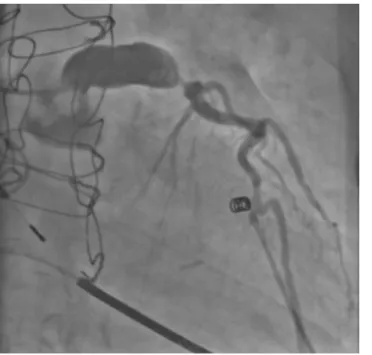
Fig. 39Coronary angiogram showing a left main coro- nary aneurysm and subtotal occlusion of the left ante- rior descending artery (right ante- rior oblique 14°, caudal 15°)
Herzschrittmachertherapie + Elektrophysiologie 2 · 2020
229
cal, anterior, and anteroseptal segments (Video 1).
Therapy and its course
After administration of aspirin, heparin, and morphine, symptoms were allevi- ated. Emergent cardiac catheterization demonstrated an aneurysmal left main and subtotal occlusion in the proximal part of the left anterior descending artery (LAD) with delayed flow (.Fig.3) as the culprit lesion. Percutaneous coro- nary intervention (PCI) with placement of a drug-eluting stent in the proximal LAD was successfully performed. ECG changes regressed completely after PCI.
Laboratory tests showed a marked in- crease in levels of cardiac enzymes (peak troponin I > 125 ng/ml) and the patient developed cardiogenic shock. He was treated accordingly and was discharged from hospital 13 days later.
Discussion
The diagnosis of STEMI can be chal- lenging in the presence of ventricular pacing due to the modified depolariza- tion and repolarization pattern of the my- ocardium. In the setting of right ventric- ular pacing, ST-elevation >1 mm con- cordant with QRS complex and discor- dant ST-segment elevations >5 mm have been shown to be the most specific ECG changes, albeit with low to moderate sensitivity[1]. For this reason, modified Sgarbossa criteria have been developed for the diagnosis of STEMI in left bun- dle branch block [2], which relate the extent of discordant ST elevation to the size of the S wave and may also apply for right ventricular pacing. Due to a lack of systematic studies and varying QRS mor- phology depending on lead position and stimulation timing, the diagnosis of my- ocardial ischemia can be more difficult in the case of biventricular pacing. In cer- tain cases, temporary inhibition of pacing in non-pacemaker-dependent patients or switching to right ventricular stimulation can be attempted to promote early diag- nosis. There are only three published case reports of STEMI in biventricular paced rhythm [3–5]. These all describe cases of anterior/anterolateral STEMI, where
Abstract · Zusammenfassung
Herzschr Elektrophys 2020 · 31:228–231 https://doi.org/10.1007/s00399-020-00682-y
© Springer Medizin Verlag GmbH, ein Teil von Springer Nature 2020
D. Pilecky · R. Fischer · T. Wiesinger · M. Gröbner · M. Vamos · D. Elsner
Anterior wall ST-elevation myocardial infarction in biventricular paced rhythm
Abstract
There is a lack of evidence on electrocardio- graphic criteria for ST-elevation myocardial infarction (STEMI) in patients with biventricu- lar paced rhythm. In all previous case reports of STEMI in biventricular paced rhythm, concordant ST-elevations and/or discordant ST-elevations >5 mm were present. This report describes the case of a patient with anterior STEMI and discordant ST-elevations of less than 5 mm during biventricular
stimulation with epicardial left ventricular lead and highlights the importance of comparing the electrocardiogram to previous recordings when STEMI is suspected.
Keywords
Cardiac resynchronisation therapy · Biven- tricular stimulation · Myocardial ischemia · Electrocardiography · Sgarbossa criteria · Epicardial lead
ST-Hebungs-Myokardinfarkt der Vorderwand bei biventrikulär stimuliertem Rhythmus
Zusammenfassung
Bezüglich der elektrokardiographischen Diagnosekriterien eines ST-Hebungs-Myo- kardinfarkts (STEMI) während biventrikulärer Schrittmacherstimulation liegt keine klare Evidenz vor. In allen vorherigen Fallberichten über STEMI bei biventrikulärer Schritt- macherstimulation wurden konkordante ST-Hebungen und/oder diskordante ST- Hebungen >5 mm beschrieben. Unsere Kasuistik schildert den Fall eines Patienten mit STEMI der Vorderwand und diskordanten ST-
Hebungen <5 mm während biventrikulärer Stimulation mit epikardialer linksventrikulärer Sonde und betont, wie wichtig es bei Verdacht auf STEMI ist, das Elektrokardiogramm mit früheren Aufzeichnungen zu vergleichen.
Schlüsselwörter
Kardiale Resynchronisationstherapie · Biven- trikuläre Stimulation · Myokardischämie · Elektrokardiographie · Sgarbossa Kriterien · Epikardiale Sonde
concordant ST-elevations with recipro- cal ST-depressions and/or discordant ST- elevations in leads with negative QRS complexes were present. To the authors’
knowledge, this is the first report of ECG abnormalities associated with myocar- dial infarction in a patient with a CRT system and epicardial LV lead. In the cur- rent case, only discordant ST-elevations of less than 5 mm but more than 25% of the S wave were seen. Typical symptoms and comparison of the admission ECG with a previous follow-up ECG facilitated the correct diagnosis. Although no to- tal coronary occlusion was present at the time of cardiac catheterization, the clin- ical presentation, echocardiography, and cardiac enzyme kinetics were together indicative of transmural ischemia. Fur- ther research is warranted to determine ECG criteria for STEMI in biventricular and/or epicardial paced rhythm.
Corresponding address
Dr. med. David Pilecky Department of Internal Medicine III, Klinikum Passau Innstraße 76, 94032 Passau, Germany
pileckyd@gmail.com
Compliance with ethical guidelines
Conflict of interest.D. Pilecky, R. Fischer, T. Wiesinger, M. Gröbner, M. Vamos and D. Elsner declare that they have no competing interests.
For this article no studies with human participants or animals were performed by any of the authors. For images or other information within the manuscript which identify patients, consent was obtained from them and/or their legal guardians.
230
Herzschrittmachertherapie + Elektrophysiologie 2 · 2020References
1. Sgarbossa EB, Pinski SL, Gates KB, Wagner GS (1996) Early electrocardiographic diagnosis of acute myocardial infarction in the presence of ventricular paced rhythm. GUSTO-I investigators.
Am J Cardiol 77:423–424
2. Smith SW, Dodd KW, Henry TD, Dvorak DM, Pearce LA (2012) Diagnosis of ST-elevation myocardial infarction in the presence of left bundle branch block with the ST-elevation to S-wave ratio in a modified Sgarbossa rule. Ann Emerg Med 60:766–776
3. Ukena C, Mahfoud F, Buob A, Bohm M, Neu- berger H-R (2012) ST-elevation during biventricu- lar pacing. Europace 14:609–611
4. Karumbaiah K, Omar B (2013) ST-elevation my- ocardial infarction in the presence of biventricular paced rhythm. J Emerg Med 45:e35–40 5. Pandit A, Hakim F, Chandrasekaran K, Srivathsan K
(2014) ST segment elevation myocardial infarction in biventricular paced rhythm. Heart Lung Circ 23:e184–187
Herzschr Elektrophys 2020 · 31:238 https://doi.org/10.1007/s00399-016-0438-2 Online publiziert: 30. Mai 2016
© Springer-Verlag Berlin Heidelberg 2020
Marcus Wiemer
Klinik für Kardiologie und Internistische Intensivmedizin, Johannes Wesling Klinikum, Minden, Deutschland
RETRACTED ARTICLE:
Differenzierung von
„Misunderstanding“ und
„Mismanagement“
This article has been retracted by the Publisher as it was published accidentally without consent of the Editor in Chief.
Korrespondenzadresse
PD Dr. med. Marcus Wiemer Klinik für Kardiologie und Internistische Intensivmedizin, Johannes Wesling Klinikum Hans-Nolte-Str. 1, 32429 Minden, Deutschland marcus.wiemer@muehlenkreiskliniken.de
Herzschrittmachertherapie + Elektrophysiologie 2 · 2020