1 2 3 4 5 6 7 8 9 10 11 12 13 14 15 16 17 18 19 20 21 22 23 24 25 26 27 28 29 30 31 32 33 34 35 36 37 38 39 40 41 42 43 44 45 46 47 48 49 50 51 52 53 54 55 56 57
Enhancement of Ion Pairing of Sr(II) and Ba(II) Salts by a Tritopic Ion-Pair Receptor in Solution
Bence Kutus,
[a]Jun Zhu,
[b]Jian Luo,
[b]Qi-Qiang Wang,
[b]Alexandru Lupan,
[c]Amr A. A. Attia,
[c]De-Xian Wang,*
[b]and Johannes Hunger*
[a]Tritopic ion-pair receptors can bind bivalent salts in solution;
yet, these salts have a tendency to form ion-pairs even in the absence of receptors. The extent to which such receptors can enhance ion pairing has however remained elusive. Here, we study ion pairing of M2+ (Ba2+, Sr2+) and X (I , ClO4 ) in acetonitrile with and without a dichlorooxacalix[2]arene[2]
triazine-related receptor containing a pentaethylene-glycol moiety. We find marked ion association already in receptor-free solutions. When present, most of the MX+ ion-pairs are bound
to the receptor and the overall degree of ion association is enhanced due to coordinative, hydrogen-bonding, and anion-π interactions. The receptor shows higher selectivity for iodides but also stabilizes perchlorates, despite the latter are often considered as weakly coordinating anions. Our results show that ion-pair binding is strongly correlated to ion pairing in these solutions, thereby highlighting the importance of taking ion association in organic solvents into account.
1. Introduction
Ion receptors have reached by now an elaborate design,[1–6]yet when coordinating a single ion, the corresponding counter-ion affects both binding strength and selectivity. To also control the binding of the counter-ion, ion-pair (IP) receptors, which have cation and anion recognition moieties in the same molecular scaffold, have become the focal point of recent ion sensing studies.[7–11] Since these receptors benefit from synergistic effects between the co-bound ions, such as electrostatic and allosteric interactions, they exhibit enhanced binding affinities.
Additionally, the modification of the recognition sites allows for fine-tuning the selectivity, thus, a plethora of receptors has been designed for efficient binding of alkali metal (MX) and tetraalkylammonium salts (R4NX).[7–39]As such, IP receptors have emerged as potential candidates for numerous applications, such as salt extraction,[12–17]transmembrane transport,[18–23]and catalysis.[24,25]
The ability to tailor cationic and anionic binding sites also enables the design of multitopic receptors. In contrast to MX
receptors, only a few structures that bind the cation and both anions of bivalent (MX2) salts, have been reported to date.[39–42]
Such MX2 receptors could improve the extraction of alkaline metal earth cations, like Sr2+ and Ba2+,[43–46] from aqueous solutions or the selective extraction of the hazardous90Sr from calcium-containing radioactive wastes.[47 49]
The binding of MX2salts to such receptors has been mostly derived from titration experiments. In such titrations typically the receptor is probed (NMR chemical shift or optical absorption/fluorescence), and thus interaction of individual ions with the receptor can be quantified given that ion binding results in salient variations of the receptor’s chemical environ- ment. In turn, it is challenging to detect weak interactions of the anion or cation of an IP with the receptor, and it is impossible to account for the formation of bare IPs that are not directly bound to the organic molecule. The latter is in particular relevant to bivalent MX2 salts, as they have a high tendency to form IPs in solution – i. e. cations and anions form long-lived aggregates in solution – even in the absence of a guiding molecular scaffold.[50]Thus, one fundamental question about the function of IP receptors has remained elusive: can these receptors efficiently bind pairs of ions and thereby enhance the overall degree of ion association? That is, does the formation of receptor-IP complexes increase the overall concen- tration of associated ions (i.e. bare ion-pairs and receptor-bound ion-pairs)? Quantifying this receptor-induced enhancement of ion association can thus provide essential information about the function of such receptors in solution.
To address this question we focus in the present study on a tritopic IP receptor, for which cation binding can be rationally designed using appropriately sized pentaethylene glycol chains,[1–3,7–11] while anion coordination can be achieved by interaction of anions with electron-deficient aromatic triazine rings.[51–63] The bridging oxygen atoms conjugate with the triazines such that the aromatic trimeric fragment tends to form a pre-organized V-shaped pocket in which two triazines serve [a] Dr. B. Kutus, Dr. J. Hunger
Department of Molecular Spectroscopy, Max Planck Institute for Polymer Research, 55128 Mainz, Germany
E-mail: hunger@mpip-mainz.mpg.de
[b] J. Zhu, Dr. J. Luo, Prof. Dr. Q.-Q. Wang, Prof. Dr. D.-X. Wang
Beijing National Laboratory for Molecular Sciences, CAS Key Laboratory of Molecular Recognition and Function, Institute of Chemistry, University of Chinese Academy of Sciences, Chinese Academy of Sciences, Beijing, 100190, China
E-mail: dxwang@iccas.ac.cn [c] Dr. A. Lupan, Dr. A. A. A. Attia
Faculty of Chemistry and Chemical Engineering, Babeş-Bolyai University, 400028 Cluj-Napoca, Romania
Supporting information for this article is available on the WWW under https://doi.org/10.1002/cphc.202000507
© 2020 The Authors. Published by Wiley-VCH GmbH. This is an open access article under the terms of the Creative Commons Attribution Li- cense, which permits use, distribution and reproduction in any medium, provided the original work is properly cited.
1 2 3 4 5 6 7 8 9 10 11 12 13 14 15 16 17 18 19 20 21 22 23 24 25 26 27 28 29 30 31 32 33 34 35 36 37 38 39 40 41 42 43 44 45 46 47 48 49 50 51 52 53 54 55 56 57
as homoditopic binding sites for anions.[59,61] Receptor 1[42]
(Scheme 1) is based on a triazine-containing aromatic trimer fragment for anion recognition and a pentaethylene glycol chain for cation chelation.1 has been reported to form stable complexes with Ca2+salts but shows low affinity to Mg2+.[42]
Here we report on ion pairing of Sr(ClO4)2, Ba(ClO4)2, and SrI2
dissolved in acetonitrile in the presence and absence of receptor1. To explore to what extent1can induce ion pairing and the corresponding structures of the IPs formed, we use a combination of experiments. To quantify ion association in solution, we use dielectric relaxation spectroscopy (DRS), which is sensitive to the rotation of dipolar species and as such can detect both IPs bound by the molecular scaffold of the receptor and bare IPs. We compare these results to those obtained from
1H nuclear magnetic resonance spectroscopy (1H NMR) titra- tions. To obtain information on the composition and structure of the formed complexes, we use electrospray ionization mass spectrometry (ESI-MS), single crystal X-ray diffraction (XRD), and density functional theory (DFT) calculations. We find that all salts have a marked tendency to form IPs in acetonitrile in the absence of 1. The addition of 1, which in solution adopts an open form favorable for ion binding, stabilizes IPs and thus results in the enhancement of ion association.
Experimental Section
Sample Preparation
Samples were prepared using HPLC grade acetonitrile (Fisher Scientific), deuterated acetonitrile, or HPLC-MS grade acetonitrile (VWR Chemicals) as solvents. Salts for DRS and ESI-MS experiments SrI2(Alfa Aesar, 99.99 %), Sr(ClO4)2· 3H2O (Alfa Aesar, 98 %), BaI2(Alfa Aesar, 99.99 %) and Ba(ClO4)2 (Acros Organics, 99 %) were used
without further purification. To avoid the contribution of water to the dielectric spectra, Sr(ClO4)2· 3H2O was dried in vacuo at 160–
170°C until constant weight was reached. For1H nuclear magnetic resonance spectroscopic (NMR) titrations, all metal (Sr(ClO4)2· 6H2O, Ba(ClO4)2· 3H2O) and tetrabutylammonium salts (Bu4NCl, Bu4NBr and Bu4NI) were used as received. Receptor1was synthesized according to the procedure reported in Ref. [42].
Dielectric Relaxation Spectroscopy (DRS)
DRS probes the frequency-dependent macroscopic polarization of a sample in an external electric field[64 66] with field frequency ν, which is generally expressed in terms of the complex permittivity, be nð Þ:
b
e nð Þ ¼e0ð Þn e00ð Þn ik
2pne0 (1)
with ɛ’(ν) and ɛ”(ν) being the frequency-dependent dielectric permittivity and loss, respectively; andɛ0is the permittivity of free space. For conducting samples, the translational motion of mobile ions gives rise to Ohmic loss (last term of Eq. 1), which scales with the conductivity, k, of the sample. We assume k to be real and independent ofν(i. e. the dc conductivity).
At microwave frequencies, polarization stems predominantly from rotation of species with an electrical dipole moment. Thus, besides its sensitivity to dipolar molecules, DRS is particularly sensitive to the formation of IPs in solution, as the oppositely charged ions of an IP are separated by a well-defined separation distance, yielding an intrinsically high dipole moment. As the dipole moment increases with increasing distance between cation and anion, DRS can distinguish between different IP species, like contact or solvent- separated ion-pairs.[65,66]For any dipolar relaxation (e. g. solvent or IPs), a dispersion in the real part,ɛ’(ν), and a peak in the imaginary part,ɛ”(ν), are observed.
Thebe(ν) spectra were recorded at room temperature ((23�2)°C), using an Anritsu Vector Network Analyzer (model MS4647 A). The frequency range at 0.2�ν/GHz�50 was covered using a fre- quency-domain reflectometer, equipped with a coaxial open-ended probe based on 1.85 mm connectors. Spectra at 60�ν/GHz�125 were recorded using an open-ended probe connected with 1 mm connectors to an external frequency converter module (Anritsu 3744 A mmW).[67]To calibrate the setup, air, conductive silver paint, and acetonitrile[68]were used as calibration standards.
To study solutions of the salts in acetonitrile using DRS, samples withcsaltup to 0.14–0.16 M were prepared. To study the binding of salts to1in acetonitrile, two series of solutions were prepared. First, c1was varied from 0 to 0.11 M at a constantcsalt(0.10 M), except for BaI2, which is not sufficiently soluble in acetonitrile. Second,csaltwas increased from 0 to 0.14 M at constant c1 (0.05 M). All solution compositions are listed in Tables S1 and S2 in the Supporting Information (SI).
1H Nuclear Magnetic Resonance (NMR) Spectroscopy
To supplement the quantitative findings from the DR spectroscopic measurements, we performed1H NMR titrations. The data evalua- tion was performed with the PSEQUAD[69] and Bindfit[70] software packages. More experimental details are given in the SI.
Scheme 1.Structure of the dichlorooxacalix[2]arene[2]triazine-related ion- pair receptor1.[42]
1 2 3 4 5 6 7 8 9 10 11 12 13 14 15 16 17 18 19 20 21 22 23 24 25 26 27 28 29 30 31 32 33 34 35 36 37 38 39 40 41 42 43 44 45 46 47 48 49 50 51 52 53 54 55 56 57
Single-Crystal X-ray Diffraction
Mixtures of1and M(ClO4)2(M=Ba, Sr) were dissolved in CH3OH/
CHCl3, and ethyl ether was allowed to slowly diffuse into the solution at 273 K to produce single crystals for X-ray analysis. Single crystal X-ray diffraction data were collected on a MM007HF Saturn724+ diffractometer using MoK/αradiation (λ=0.71073 Å) at a temperature of 173 K. The intensity data were collected by the omega scans techniques, scaled, and reduced with theCrystalClear software.[71]X-rays were provided by a fine-focus sealed X-ray tube operated at 50 kV and 24 mA. Integrated reflection intensities were produced and the correction of the collected intensities for absorption was done usingCrystalClear.The structures were solved by direct methods using SHELXT[72] and refined using full-matrix least-squares methods implemented in theSHELXL[73]program. All non-hydrogen atoms were refined anisotropically, and hydrogen atoms attached to carbon atoms were fixed at their ideal positions.
Electrospray Ionization Mass Spectrometry
To assess the composition of ionic/molecular complexes in solution, electrospray ionization (ESI) mass spectra were recorded using an Advion Expression-L Compact Mass Spectrometer equipped with a single quadrupole separator, providing an average resolution of 0.5m/zunits. Here, the range of 100�m/z�1200 was scanned in the positive-ion mode. Experiments were performed using samples with csalt=c1=0.01 M (SrI2, Sr(ClO4)2, BaI2, Ba(ClO4)2) or csalt=c1= 0.02 M (SrI2).
Density Functional Theory (DFT) Calculations
The geometries of 1 and the ion-pair complexes at various configurations were optimized with the Gaussian 09[74] software using the B3LYP hybrid DFT functional[75,76]and the def2-TZVP or its def2-TZVPD variant including diffuse functions[77] to take non- covalent interactions into account. For all calculations, Grimme’s D3 empirical dispersion correction[78] was employed. Implicit solvent effects were taken into account applying the conductor-like polar- izable continuum model (CPCM)[79]with acetonitrile as solvent.
2. Results and Discussion
2.1. Ion Pairing in the Absence of the Receptor
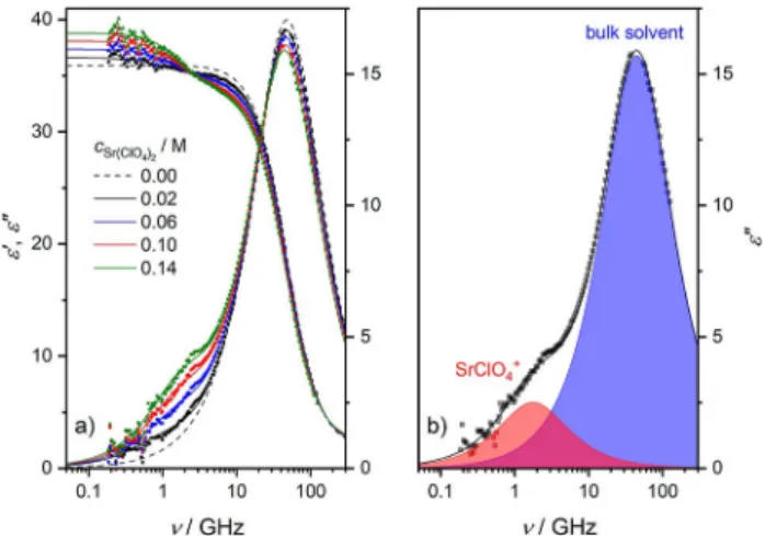
To explore ion pairing of the studied salts in the absence of the receptor molecules, we study the dielectric relaxation of solutions of Sr(ClO4)2(Figure 1a), SrI2, and Ba(ClO4)2(Figures S1a and S2a, SI) in acetonitrile. For all samples we observe a relaxation at ~ 50 GHz, evidenced by a peak in the dielectric loss and a dispersion in the dielectric permittivity spectra (Figure 1), due to the solvent acetonitrile.[68]
As can be seen for Sr(ClO4)2in Figure 1a (also for SrI2and Ba (ClO4)2, see Figures S1a and S2a in the SI), upon dissolution of salt a shoulder in the imaginary part at ~ 1 GHz emerges with increasing csalt, indicative of the formation of dipolar IPs.[65,66]
Due to this low-frequency relaxation, which also goes along with a dispersion in ɛ’(ν), the static permittivity (ɛs, the low- frequency plateau ofɛ’(ν)) exhibits an increase with increasing salt concentration. As such, the increase in sample polarization due to the formation of dipolar solute species, i. e. IPs, overcompensates its decrease due to the dilution of dipolar
solvent molecules. This suggests that the studied iodide and perchlorate salts do not fully dissociate in acetonitrile due to its lower solvent permittivity and weaker solvation,[50]giving rise to the formation of dipolar IPs (i. e., SrI+, SrClO4
+, and BaClO4 +).
To analyze the spectra quantitatively, we fit a relaxation model to the data. For the present samples we find that a combination of two Debye-type relaxations[64] accounting for the solvent (AN) and the ion-pair (MX+) relaxations, respectively, provides an excellent description of the experimental spectra with the least number of adjustable parameters:
be nð Þ ¼ SMXþ
1þi2pntMXþ
þ SAN
1þi2pntAN
þe1 ik
2pne0 (2)
where SMX+ and SAN are the MX+ and the solvent relaxation amplitudes, respectively, while τMX+ and τAN represent the corresponding relaxation times. The infinite-frequency permit- tivity,ɛ1, comprises all contributions at frequencies higher than covered in our experiment. Such decomposed dielectric loss spectra of the 0.14 M salt solutions are depicted in Figures 1b, S1b, and S2b (see SI).
To obtain quantitative information about the degree of ion pairing, we use the Cavell equation,[65,66,80]which relatesSMX+ to the equilibrium concentration ([MX+]) and effective dipole moment (μMX+) of the MX+ion-pairs:
SMXþ¼ es
esþAMXþð1 esÞ � NA
3kBTe0
�½MXþ� �mMXþ2 (3)
where AMX+ is the so-called cavity-field factor (determined by the geometry of the rotating particle), NA is the Avogadro number,kB is the Boltzmann constant, andTis the thermody- namic temperature.
Figure 1.(a) Relative permittivity (ɛ’, triangles, left axis) and dielectric loss (ɛ”, squares, right axis) spectra of 0–0.14 M Sr(ClO4)2solutions. Solid lines are the results of fitting Eq. 2 to the data; dashed line shows the spectrum of pure acetonitrile, taken from Ref. [68]. (b) Contribution of acetonitrile (blue- shaded area) and SrClO4+ion-pairs (red-shaded area) toɛ”for 0.14 M Sr(ClO4)2, as obtained from the fit. Symbols represent the experimental data, and the black solid line is the result of the fit. In both panels, the last term of Eq. 2 has been subtracted fromɛ”for visual clarity.
1 2 3 4 5 6 7 8 9 10 11 12 13 14 15 16 17 18 19 20 21 22 23 24 25 26 27 28 29 30 31 32 33 34 35 36 37 38 39 40 41 42 43 44 45 46 47 48 49 50 51 52 53 54 55 56 57
In order to calculate [MX+] from SMX+, the value of μMX+, which predominantly depends on the spatial separation between cation and anion, needs to be known. In salt solutions, both contact (CIP, direct contact between the cation and anion) and solvent-shared (SIP, cation and anion are separated by one solvent molecule) IPs are conceivable species.[50,65,66] Despite also the existence of triple IPs (consisting of one cation and two anions or one anion and two cations) has been inferred from infrared spectra for Mg(ClO4)2and Ca(ClO4)2in acetonitrile, only one dipolar relaxation mode has been detected in the DRS spectra for these salts,[68]in line with our present findings. Given the low salt concentrations of the present samples, at which triple ion aggregates are minor species also for Mg(ClO4)2and Ca(ClO4)2, we ascribe the low-frequency relaxation mode to CIPs or SIPs.
To determine which IP species prevails in solution, we consider the two limiting cases: exclusive formation of either
CIPs or SIPs. Based on the geometric model described in detail in Ref. [80], we calculate μCIP, μSIP, ACIP, and ASIP (Table S3, SI) using data for ionic radii, solvent radii, and polarizabilities from Refs. [81–83]. From these values, we obtain [MX+] using Eq. 3.
Assuming that [MX+]=[CIP] or [SIP], we calculate the IP formation constants,KMX+, for the CIP (KCIP) and SIP (KSIP) species via Eq. 4:
KMXþ¼½MXþ� �c� Mþ
½ � �½X �¼ ½MXþ� �c�
ðcsalt ½MXþ�Þ � ð2csalt ½MXþ�Þ (4) where [M+] and [X ] are the free cation and anion concen- trations and cø the standard molar concentration (1 M). As shown in Figure 2, the equilibrium constants for the two limiting cases (SIP and CIP) decrease with increasing salt concentration due to increased charge screening.[50] Yet the curves are offset as a result of the different absolute values of mMXþ for the CIP and SIP species. To elucidate which IP species predominates association equilibria, the formation constants have to be compared to literature data obtained from independent experimental techniques.
To exclude differences arising from different experimental sensitivities and ionic strengths, such comparison should be based on the standard thermodynamic association constant, K0MX+ (i. e. the limiting value of KMX+ at infinite dilution). To obtain K0MX+, we extrapolate the values of KMX+ to zero ionic strength using a Guggenheim-type equation:[65,68]
logKMXþ¼logK0MXþ
2ADHjzþz j ffiffi
pI
1þBDHd ffiffi
p þI CIþDI1:5 (5)
where ADH and BDH are the Debye-Hückel constants at T= 23°C,[68]dis the distance of charge separation, andCandDare adjustable parameters. We calculate the ionic strength, I, from csalt by correcting for the IPs formed (that is,I=3csalt–2[MX+]) and fit Eq. 5 to the data in Figure 3. Error bars (�σ) for logKMX+
and I were estimated assuming σ(SMX+)=0.3. The fitted parameters of Eq. 5 for each IP are listed in Table S4, SI.
Figure 2 demonstrates that for SrClO4
+ (and also for SrI+ and BaClO4
+, see Figures S3 and S4 in the SI), Eq. 5 describes the ionic strength dependence for both constants,KCIPandKSIP, Figure 2.Experimental ion-pair formation constants, logKSrClO4+, assuming
the formation of contact (CIP, red symbols) or solvent-separated (SIP, black symbols) SrClO4+ion-pairs as a function of ionic strength (I). Solid lines show fits using Eq. 5; error bars were calculated assumingσ(SMX+)=�0.3. Dashed lines indicate the thermodynamic formation constants (logK0SrClO4+) at infinite dilution, obtained in this work or reported in Ref. [84].
Figure 3.Structures of the open (a) and twisted (b) conformers of receptor1, optimized at the B3LYP-D3/def2-TZVP level. Implicit solvent effects were taken into account applying the CPCM approach. The calculated effective dipole moments are 8.3 D (a) and 1.6 D (b).
1 2 3 4 5 6 7 8 9 10 11 12 13 14 15 16 17 18 19 20 21 22 23 24 25 26 27 28 29 30 31 32 33 34 35 36 37 38 39 40 41 42 43 44 45 46 47 48 49 50 51 52 53 54 55 56 57
very well. This comparison shows that the extrapolated values (marked as dashed lines in Figure 2) agree well with those derived from previous conductometric experiments for the same salts,[84]if we assume the exclusive formation of CIPs (see also Table 1). Hence, this agreement provides evidence for CIP being the dominant ion-pair species for the studied salts in acetonitrile.
We note that this notion partially contrasts earlier findings for solutions of Mg(ClO4)2and Ca(ClO4)2in acetonitrile, where – despite CIPs also dominate – the formation of both CIP and SIP atcsalt<0.2 M has been suggested.[68]This apparent discrepancy can be rationalized on the basis of different ionic radii: smaller ionic radii give rise to higher ionic surface charge density and thus to stronger solvation. As such, the interaction of acetonitrile with the smaller Mg2+/Ca2+cations is stronger than with the larger ions Sr2+ and Ba2+. In turn, weaker solvation of Sr2+ and Ba2+ relative to Mg2+ and Ca2+, likely makes SIPs less significant for the salts studied in this work.
Overall, our findings for these solutions imply that despite perchlorate is often considered as weakly coordinating anion, we find that both Sr(ClO4)2 and Ba(ClO4)2 tend to form IPs in acetonitrile, in line with previous studies.[68,84] The fraction of ions that form IPs (% [MX+]/csaltatcsalt=0.1 M, Table 1) exceeds 40 % for the perchlorates, while for SrI2our results suggest that more than 50 % of all ions are bound in CIPs. In turn, only a fraction of ions is present in solution as free ions. Upon addition of IP receptors, which will be discussed below, binding of both, free ions and ion-pairs to the receptor can occur.
2.2. The Structure of Receptor 1 in Acetonitrile
Before discussion of binding of salts to the receptor, we first investigate the structure of receptor 1 in solution. Crystallo- graphic experiments have shown that1exists in two markedly different conformations in the solid state,[42]here referred to as
‘open’ and ‘twisted’ conformer (Figure 3). Given the different symmetries of both conformers, studying the dielectric relaxa- tion of 1 can provide information on the most stable conformation in acetonitrile.
For solutions of 0.05 M of 1, we detect a small-amplitude relaxation at ca. 1 GHz in the DR spectrum (amplitudeS1�0.3, relaxation timeτ1�130 ps, see the decomposed loss spectrum
in Figure S5, SI). Based on Eq. 3, this relaxation amplitude corresponds to a dipolar species with a dipole moment ofμ1= (8.3�0.8) D. We compare this value with the structures of the open and twisted conformers (Figure 3), obtained from geome- try optimizations at the B3LYP-D3/def2-TZVP level of theory, taking implicit solvent effects into account. These calculations suggest that the calculated dipole moment of the open form is μcalc=8.3 D, which is in excellent agreement with the exper- imental value. Conversely, due to its high symmetry, the twisted conformer has a much lower dipole moment (μcalc=1.6 D).
Thus, our results indicate that in solution the open conformer prevails. In this geometry the ion binding sites are pre- organized such that both cations and anions can readily access the binding pockets, in contrast to the twisted form.
2.3. Ion Pairing in the Presence of the Receptor
2.3.1. Qualitative Findings for the Binding of Salts Both in the Solid and Solution Phases
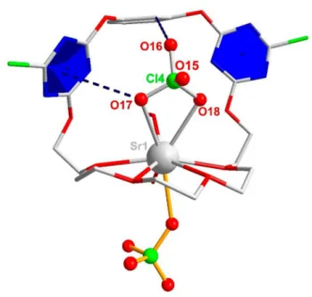
Having established the relaxation dynamics of the binary solutions, we now turn to ternary samples where both receptor 1and salt are present. Through the diffusion of ethyl ether to a mixture of receptor and alkali earth metal salts in CH3OH/CHCl3, we obtained single crystals of the [1· Sr(ClO4)2] · H2O · CH3OH (Figure 4 and Table S5, SI) and [1· Ba(ClO4)2] · 2H2O (Figure S6 and Table S6, SI) complexes. From the crystal structures, we find the cation is coordinated equatorially by the oxygens of the glycol chain, and are axially coordinated by the anion. The respective coordination number (CN) is 9 for Sr2+ and 10 for
Table 1. Formation constants (logK0MX+�σ at (23�2) °C) of MX+ ion- pairs, assuming contact (CIP) or solvent-separated ion-pairs (SIP) as obtained from the dielectric relaxation amplitudes. Also listed are the values from Ref. [84] (25°C) determined using conductometry. The last column lists the degree of ion pairing as obtained in the present study at csalt=0.1 M.
Reaction SIP CIP Ref. [84] % [MX+]/
csalt
Sr2++IÐ SrI+ 1.95�0.03 3.39�0.13 53
Sr2++ClO4
Ð SrClO4+
1.76�0.03 2.87�0.04 2.58�0.01 46
Ba2++ClO4 Ð BaClO4+
1.72�0.04 2.67�0.04 2.69�0.02 43
Figure 4.Crystal structure of the [1· Sr(ClO4)2] · H2O · CH3OH ion-pair complex.
The anion-πdistance is indicated by a dashed line between the O17 atom and the blue triazine plane (dO17–plane=3.393 Å). Also shown is the intra- molecular H bond between the Hfaryl proton and O16 (dO16···Hf=2.486 Å).
1 2 3 4 5 6 7 8 9 10 11 12 13 14 15 16 17 18 19 20 21 22 23 24 25 26 27 28 29 30 31 32 33 34 35 36 37 38 39 40 41 42 43 44 45 46 47 48 49 50 51 52 53 54 55 56 57
Ba2+; the higher CN of the latter reflects its higher ionic radius, providing room for more ligands.
In the structure of1· Sr(ClO4)2(Figure 4), one ClO4 is located outside the receptor, interacting electrostatically with the cation. The second anion resides in the aromatic binding pocket, being stabilized by a triazine moiety through anion-π interaction, as indicated by the short distance between the aromatic ring and the O17 atom of the anion (3.393 Å).
Moreover, an additional hydrogen bond between the O16 atom of ClO4 and the Hf proton of the central aromatic ring is formed. Based on the distance (dO16···Hf=2.486 Å), this hydrogen bond can be considered weak.[85,86]
The crystal structure of the 1· Ba(ClO4)2 IP complex shows similar binding motifs, with each Ba2+ coordinated by ten oxygens, four from the glycol chain, one from a water molecule and five from three ClO4 anions (Figure S6, SI). The perchlo- rates are additionally coordinated by triazine rings, evidenced by the short distances between the O15, O16 atoms and the triazine planes (dO -plane=2.944 and 3.063 Å). The perchlorates also act as bridge to link two IP complexes to form a dimer.
To obtain information on the binding of salts to 1 in the solution phase, we carried out ESI-MS measurements in solutions with csalt=c1=0.01 or 0.02 M. In the positive-ion mode, we observe peaks due to1· SrI+,1· SrClO4
+,1· BaI+ and 1· BaClO4
+ as well as to the bare and solvated 1· Sr2+ and 1· Ba2+complexes (Figures S7–S11, SI). This suggests that both, CIPs and cations bound to1, coexist in solution. Based on the relative peak intensities, the 1· MX+ complexes prevail at 1 : 1 and higher c1:csalt ratios (except for BaI2, see Figure S10, SI).
Conversely, peaks corresponding to the free IPs are absent for all salts. Despite one cannot fully exclude their formation based solely on the mass spectra, their absence suggests that dissociation of 1· MX+ is energetically more demanding than dissociation of MX+. Thus,1· MX+ complexes are the dominant ion-pair species in the presence of the receptor.
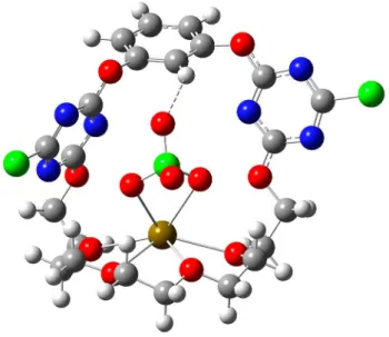
To gain further insights into the structure of the receptor- bound IPs, we optimized the geometry of the 1· SrClO4
+, 1· BaClO4
+, 1· SrI+ and 1· BaI+ species at the B3LYP-D3/def2- TZVPD level of theory (Figure 5 as well as Figures S12–S14, SI), as these species dominate DRS relaxations atc1:csalt�1 : 1 ratio (see below). In the perchlorate species the anion forms a hydrogen bond to the aryl proton (Hf). The bond lengths are 2.520 Å (1· BaClO4
+) and 2.419 Å (1· SrClO4
+), with the latter agreeing well with the one found in the 1· Sr(ClO4)2 crystal.
Together with the C H···O bond angles (139.0° and 149.7°, respectively), these characteristics are common for weak C H···O hydrogen bonds.[85,86] As for the iodide complexes, we find strong interaction between I and the same aryl proton.
The calculated distances (1· SrI+: 3.382 Å, 1· BaI+: 3.163 Å) indicate the formation of a strong C H···I bond.[87]From these findings we conclude that in addition to anion-πinteraction, an intramolecular C H···X hydrogen bond also contributes to the stabilization of the receptor-bound anion.
2.3.2. Quantifying Ion Pairing in the Presence of 1 by DRS
To study the formation of IPs in the presence of1, we recorded dielectric spectra of the ternary samples. Upon addition of1to solutions of 0.10 M Sr(ClO4)2, SrI2and Ba(ClO4)2(Figure 6a as well as Figures S15a–17a, SI), we find a shift of the IP relaxation, i. e.
the dispersion inɛ’and the shoulder inɛ”at ~ 1 GHz, to lower frequencies. The relaxation amplitude increases slightly upon addition of1(see also discussion below). The small variation of the IP amplitude directly implies that the spatial separation of the underlying dipolar species is only little affected by the presence of1, given that the overall concentration of IPs does Figure 5.Structure of the SrClO4+ion-pair bound to receptor1, optimized at the
Figure 6.(a) Relative permittivity (ɛ’, triangles, left axis) and dielectric loss (ɛ”, squares, right axis) spectra for a 0.10 M Sr(ClO4)2solution with (red symbols) and without (black symbols) receptor1. Solid lines are the results of fitting Eq. 2 to the data; dashed line shows the spectrum of pure acetonitrile, taken from Ref. [68]. The last term of Eq. 2 has been subtracted fromɛ”for visual clarity. (b) Ion-pair relaxation time in 0.10 M Sr(ClO4)2as a function of receptor concentration.
1 2 3 4 5 6 7 8 9 10 11 12 13 14 15 16 17 18 19 20 21 22 23 24 25 26 27 28 29 30 31 32 33 34 35 36 37 38 39 40 41 42 43 44 45 46 47 48 49 50 51 52 53 54 55 56 57
not decrease in the presence of1 (see Eq. 3). The decrease of the solvent relaxation amplitude is due to the reduced concentration of the solvent, but its peak position (~ 50 GHz= (2πτAN) 1) in the dielectric loss spectrum is hardly affected by the presence of the receptor.
Also for the ternary samples Eq. 2 describes the experimen- tal spectra well; the contributions of both relaxations to the overall loss spectra for solutionscsalt=c1=0.10 M are plotted in Figures 15b–17b in the SI. The presence of only two discernible relaxations (solvent and ion-pairs) implies that we cannot resolve separate relaxations due to receptor-bound salts and due to bare IPs. Yet, the variation of the peak position of the solute mode – in contrast to the solvent peak – indicates the binding of MX2salts (or IPs) to1: we find the extracted values of the IP relaxation time, τMX+, to increase with increasing concentration of1 (Figure 6b and S18, SI). For diffusive rotation, τMX+ is proportional to the hydrodynamic volume of the rotating species and to the viscosity of the sample.[88]The low concentration of solutes and the insensitivity of τAN to the addition of1 renders increasing viscosity unlikely. Rather, the large increase of τMX+ provides evidence for the formation of receptor-bound complexes: the 1· MX+ species have a larger volume than the bare MX+ IPs. As such, the DRS relaxation times indicate that for all studied salts,1· MX+ is the major IP species at high concentrations of 1, in line with the ESI-MS spectra. This notion is further supported by 1H NMR titration experiments, which indicate that 1· MX2 complexes are only significant for an excess of iodide (see discussion in the SI together with Figures S19–S24 and Table S7).
To relate the relaxation amplitudes, SMX+, to IP concen- trations, the contribution of both MX+ and1· MX+ species to SMX+ has to be taken into account. For quantitative analysis of ion pairing using Eq. 3, the dipole moment of1· MX+,μ1· MX+, is required. Based on the direct contact between the cation and the anion in the crystal structure (Figures 4 and S6, SI), one might expect μ1· MX+�μMX+, which is confirmed by DFT calculations that show the dipole moments of the receptor- bound and bare IPs to agree within less than 3 D (see Table S3, SI). Experiments on solutions of BaI2+1 further support the similar dipole moments of1· MX+and MX+(see the discussion together with Figures S25–S27 in the SI).
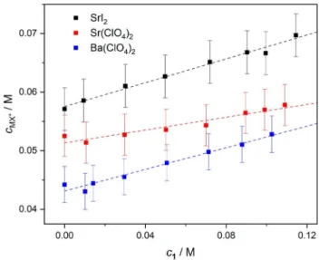
Thus, assuming μ1· MX+ � μMX+, the total IP concentration, cMX+ (�[MX+]+[1· MX+]), in the presence of the receptor can be extracted from SMX+ using Eq. 3. The thus obtained values for cMX+ (Figure 7) show an increase in [MX+]+[1· MX+] by
~ 10–25 %, upon addition of ~ 0.1 M 1 to the 0.1 M salt solutions. Consequently, ion association is enhanced in the presence of1. This increase in ion pairing is consistent with the 15–25 % decrease of the conductivities (Figure S28, SI) of the samples as the 1· MX+ complexes hardly contribute to the overall conductivity due to their reduced mobility or charge/
volume ratio.[89] We note that similar conclusions can be obtained from the experiments where csaltgradually increases (0.02–0.14 M) at c1=0.05 M: addition of 1 results in higher relaxation times, higher IP concentrations as well as lower conductivities (Figures S29–S34, SI).
Our results thus show that the presence of 1 enhances IP formation for all studied salts. For the overall degree of ion association (([MX+]+[1· MX+])/csalt), we find 67 % for SrI2, 57 % for Sr(ClO4)2and 50 % for Ba(ClO4)2in the presence of 0.10 M 1 (see Table 1). Overall, the trend in the ion association strength is the same (SrI2>Sr(ClO4)2>Ba(ClO4)2) with and without 1.
Assuming that the formation of 1· MX+ prevails at c1/csalt�1 ratios (that is, c1· MX++cMX+�c1· MX+), we can estimate the cumulative stability constants of the 1· MX+ IP complexes for samples containing 0.10 M1:
K1�MXþ¼½1�MXþ� � ðc;Þ2
½M2þ� � ½I � � ½1�
¼ ½1�MXþ� � ðc;Þ2
ðcsalt ½1�MXþ�Þ � ð2csalt ½1�MXþ�Þ � ðc1 ½1�MXþ�Þ (6)
The thus calculated constants are listed in Table 2. Accord- ingly, we find stronger association for Sr2+salts as compared to Ba2+ showing that Sr2+ matches better the size of the polyethylene-glycol binding cavity. Also, this trend is consistent Figure 7.Concentrations of bare/receptor-bound MX+ion-pairs for 0.10 M SrI2, Sr(ClO4)2and Ba(ClO4)2solutions as a function of concentration of1. The values ofcMX+and their error bars were calculated via Eq. 3, assuming σ(SMX+)=�0.3. The dashed lines are guide to the eye.
Table 2. Stability constants (logK�σ, at (23�2)°C) corresponding to the reactions in the first column, obtained in this work via DR spectroscopic measurements atI�0.17–0.19 M.
Reaction logK
1+Sr2++IÐ 1· SrI+ 2.66�0.04
1+SrI+Ð 1· SrI+ 1.67�0.04
1+Sr2++ClO4Ð 1· SrClO4+ 2.35�0.03 1+SrClO4+Ð 1· SrClO4+ 1.52�0.03 1+Ba2++ClO4Ð 1· BaClO4+ 2.19�0.03 1+BaClO4+Ð 1· BaClO4+ 1.44�0.03
1 2 3 4 5 6 7 8 9 10 11 12 13 14 15 16 17 18 19 20 21 22 23 24 25 26 27 28 29 30 31 32 33 34 35 36 37 38 39 40 41 42 43 44 45 46 47 48 49 50 51 52 53 54 55 56 57
with that of the cation-binding constants derived from1H NMR titrations using perchlorate salts (i. e.K1· Sr2+>K1· Ba2+, see Table S7 in the SI). These constants are however consistently higher than K1· SrClO4+ andK1· BaClO4+,obtained from DRS (Table 2). This discrepancy can be rationalized by the notion that DRS is sensitive only to the formation of dipolar 1· MX+ complexes, while NMR detects all species that contain a cation (i. e.1· M2+
+1· MX+):1H NMR chemical shifts are primarily sensitive to the coordination of cations, while the binding of ClO4 does not alter the protons’ chemical environment. Consequently, NMR yields higher equilibrium concentrations and thus higher formation constants (for discussion, see the SI).
Overall, the ion association equilibria in the ternary systems studied in the present work consists of ion pairing and the binding of free ions as well as IPs by the receptor. Although the formation of 1· SrI+ species refers to thermodynamic equili- brium and therefore we cannot derive if such complexes are formed via the binding of free ions or IPs (or both), it is possible to compare the formation constants of these processes: we estimate the equilibrium constant for binding of IPs to 1 (K’1· MX+) using the values ofK1· MX+ (referring to the binding of free ions, Eq. 6) andKMX+(referring to ion pairing, Eq. 4):
K1�MX0 þ¼½1�MXþ� �c;
½1� � ½MXþ� ¼K1�MXþ
KMXþ (7)
For this estimation we calculate the values of log KMX+ at the same ionic strength at which we determined the logK’1· MX+
constants (I�0.16–0.19 M) using Eq. 5. The results (Table 2) suggest that1is also somewhat more efficient in binding SrI+ IPs as compared to SrClO4
+and BaClO4
+. This difference can be explained by the stronger binding of Sr2+ as compared to Ba2+ as well as by the formation of strong hydrogen bonds to iodide as compared to perchlorate, as inferred from1H NMR titrations and DFT geometries, respectively.
3. Conclusions
In the absence of 1, SrI2, Sr(ClO4)2 as well as Ba(ClO4)2tend to form 1 : 1 contact ion-pairs (CIPs) in acetonitrile to a significant extent. The degree of ion pairing can be as high as ~ 50 % for the I and ~ 40 % for the ClO4 salts, already at low salt concentrations (<0.15 M). The neat receptor 1 exists predom- inantly in an open form in solution, to which a cation can bind without major structural reorganization.
In the presence of 1 we find simultaneous binding of cations and anions to the receptor. The ESI-MS results indicate that the receptor-bound CIP species, i. e.1· SrI+,1· SrClO4
+ and 1· BaClO4
+ prevail in solution over SrI+, SrClO4
+ and BaClO4 +
IPs. The formation of these receptor-bound IP complexes is confirmed by the increasing DRS relaxation times in the presence of 1. Quantitative analysis of the DRS results shows the overall degree of ion association to increase by approx- imately 10–25 % in the presence of1, relative to the receptor- free solutions. Despite the overall enhancement of ion pairing in solution by the receptor is only moderate, a large fraction of
IPs is complexed by 1 in solution. Structural analysis of the complexes reveals that their higher stability (as compared to the bare IPs) can be traced to the formation of coordinative, anion-πas well as hydrogen bonding interactions.
We find that the overall degree of salt binding by1is higher for I salts than for ClO4 salts. The extent of ion association follows the order of 1· SrI2>1· Sr(ClO4)2 >1· Ba(ClO4)2, which also resembles the ion association trends of the bare salt solutions. These paralleling trends can be explained by that the cation-anion distance, i. e. the electrostatic interaction between the co-bound ions, remaining essentially unaffected upon binding to1. This holds also for perchlorate salts, even though perchlorate is often considered as weakly coordinating anion.
Generally, our study shows that ion-pair recognition is intimately related to ion pairing in solution, thereby high- lighting the importance of taking the latter equilibrium into account when studying salt binding in organic solvents.
Acknowledgements
The authors are grateful to Stephan Türk for performing the ESI- MS measurements. B.K. gratefully acknowledges financial support from the Alexander von Humboldt Foundation. A.A.A.A. expresses his gratitude for funding from the Romanian Ministry of Education and Research (Grant PN-III-P1-1.1-PD-2016-1585). JH acknowl- edges funding from the European Research Council (ERC) under the European Union’s Horizon 2020 research and innovation program (grant agreement n°714691. This work was funded by the Deutsche Forschungsgemeinschaft (DFG) HU 1860/8 and the National Science Foundation of China (NSFC) 21761132024.Open access funding enabled and organized by Projekt DEAL.
Conflict of Interest
The authors declare no conflict of interest.
Keywords: cooperative effects · dielectric relaxation spectroscopy · host-guest systems · ion pairing · ion-pair receptor
[1] J.-M. Lehn,Supramolecular Chemistry: Concepts and Perspectives, VCH, Weinheim,1995.
[2]Supramolecular Chemistry, Vol 1: Molecular Recognition, Receptors for Cationic Guests(Ed.: G. W. Gokel), Pergamon, Oxford,1996.
[3] A. Arduini, A. Casnati, A. Pochini, R. Ungaro,Curr. Opin. Chem. Biol.1997, 1, 467–474.
[4] P. D. Beer, P. A. Gale,Angew. Chem. Int. Ed.2001,40, 486–516;Angew.
Chem.2001,113, 502–532.
[5] M. J. Langton, C. J. Serpell, P. D. Beer,Angew. Chem. Int. Ed.2016,55, 1974–1987;Angew. Chem.2016,128, 2012–2026.
[6] M. G. M. Antonisse, D. N. Reinhoudt,Chem. Commun.1998, 443–448.
[7] G. J. Kirkovits, J. A. Shriver, P. A. Gale, J. L. Sessler,J. Inclusion Phenom.
Macrocyclic Chem.2001,41, 69–75.
[8] B. D. Smith inIon-Pair Recognition by Ditopic Macrocyclic Receptors In Macrocyclic Chemistry(Ed.: K. Gloe), Springer, Dordrecht,2005, pp. 137–
151.
[9] S. K. Kim, J. L. Sessler,Chem. Soc. Rev.2010,39, 3784–3809.