EREDETI KÖZLEMÉNY
FUNCTIONAL NEUROTOXICITY AND TISSUE METAL LEVELS IN RATS EXPOSED SUBACUTELY TO TITANIUM
DIOXIDE NANOPARTICLES VIA THE AIRWAYS
Tamara HORVÁTH1, Tünde VEZÉR1, Gábor KOZMA2, András PAPP1
1Department of Public Health, University of Szeged Faculty of Medicine, Szeged
2Department of Applied and Environmental Chemistry, University of Szeged Faculty of Science and Informatics, Szeged
Introduction and aims– Nanoparticles of titanium dioxide are suspected neurotoxic agents and have numerous appli- cations possibly resulting in human exposure by several ways including inhalation. In the present work, rats were exposed to spherical TiO
2nanoparticles of two different sizes by the intratracheal route. It was investigated how the neuro-functional alterations, detected by electrophysiologi- cal and behavioral methods, were related to the concentra- tion of Ti in the tissue samples and what the influence of the size of the NPs was.
Materials and methods– Rats (young adult Wistar males, 10/group) were exposed to TiO
2nanoparticles of ca. 10 and 100 nm diameter (suspension medium: neutral PBS with 1% hydroxyethyl cellulose) by intratracheal instillation in 5 and 18 mg/kg b.w. dose; 5 days per week for 6 weeks.
Controls were instilled with saline, and vehicle controls, with the suspension medium. To see general toxicity, body weight was checked daily, and organ weights were measured at the end of experiment. Grip strength test, to assess motor function damage, was done before and after the 6-week treatment. Finally, the rats were anesthetized with urethane, spontaneous cortical activity and sensory evoked potentials were recorded, then the rats were dissected and tissue sam- ples were taken for Ti level measurement.
Results– Body weight gain indicated no general toxicity, and no significant change in the relative organ weights, except that of the lungs, was seen. However, change of time-to-fall in the grip strength test, and latency of cortical evoked po - tentials, were altered in the treated groups, indicating func- tional damage. Correlation of these alterations with the corti- cal Ti level was dissimilar for the two sizes of nanoparticles.
FUNKCIONÁLIS NEUROTOXICITÁS ÉS SZÖVETI FÉMSZINTEK PATKÁNYBAN TITÁN-DIOXID- NANORÉSZECSKÉK SZUBAKUT LÉGÚTI ADAGOLÁSÁT KÖVETÔEN
Horváth T, MD; Vezér T, MD; Kozma G, MD; Papp A, MD Ideggyogy Sz 2018;71(1–2):000–000.
Bevezetés és célkitûzés– A feltételezetten neurotoxikus hatású titán-dioxid-nanorészecskék kiterjedt alkalmazása emberi expozícióval járhat, ami többféle módon, többek közt belégzés útján mehet végbe. A jelen munkában kétféle méretû gömbszerû TiO
2-nanorészecskével kezeltünk patkányokat, intratrachealis adagolással. Megvizsgáltuk, hogyan függ össze az elektrofiziológiai és magatartási mód- szerekkel kimutatott idegrendszeri funkcióváltozás a szöveti Ti-szintekkel és hogyan hat erre a részecskék mérete.
Anyagok és módszerek– Fiatal felnôtt hím Wistar- patkányokat (hat csoport, 10-10 állat) kezeltünk heti öt napon, hat héten át, 10 és 100 nm körüli átmérôjû TiO
2- nanorészecskékkel, 1% hidroxietil-cellulózt tartalmazó fosz- fátpufferelt fiziológiás oldatban szuszpendálva, 5 és 18 mg/ttkg dózisban. A vivôanyagos kontrollcsoportot a szuszpendáló közeggel, míg a kezeletlen kontrollokat fiziológiás sóoldattal instilláltuk. Az általános toxicitást a testtömeg naponta történt mérésével, illetve a kísérlet végén a szervtömegek meghatározásával mutattuk ki. A motoros funkciók károsodását a hathetes kezelés elôtt és annak végén végzett kapaszkodási próbával vizsgáltuk. Végül ure- tános altatásban kérgi alapaktivitást és szenzoros kiváltott potenciálokat vezettünk el, majd az állatokat felboncoltuk és szövetmintákat vettünk fémszint-meghatározásra.
Eredmények– A testtömeg-gyarapodás nem utalt általános toxicitásra, és relatív szervtömegre tett hatás csak a tüdônél látszott. A kezelt állatokban azonban funkcionális károsodásra utaló változás volt a kapaszkodási próbánál a leesési idôben, és a kérgi kiváltott potenciálok laten- ciájában. Ezen változások és a kérgi Ti-szint korrelációja a kétféle méretû nanorészecske esetében eltérô volt.
Correspondent: Dr. András PAPP, Department of Public Health, University of Szeged Faculty of Medicine;
6720 Szeged, Dóm tér 10. E-mail: papp.andras@med.u-szeged.hu Érkezett: 2017. november 17. Elfogadva: 2017. december 5.
| English| http://dx.doi.org/10.18071/isz.71.0001 | www.elitmed.hu
R
ecent advances in nanosciences and nanotech- nology gave rise to numerous industrial pro - cesses and products based on nanoparticles (NPs) that is, particles with <100 nm typical diameter. By now, NPs have been used among others in health care, energy production, agriculture and environ- mental protection1, which results in their release into the environment. In nanoparticulate state, sub- stances show different physical and chemical prop- erties than those seen in more conventional physical states, leading to biological, and hence, toxicologi- cal, interactions not seen with traditional materials, which also means novel health risks. When inhaled by humans, NPs are either deposited in the nasopharynx of or get down to the alveoli, and reach distant body parts by migration along fibres of the olfactory and other nerves, or by crossing the alveolar and capillary wall and entering systemic circulation2. Generation of reactive oxygen species (ROS) and other surface reactions known to be cru- cial in the biological effects of NPs, are promoted by their high number and large specific surface area3. Beyond that, NPs are known to migrate to mitochondria within the cells and directly interfere with oxidative phosphorylation, also resulting in ROS and finally in cell and tissue damage2.TiO2 in NP form is a frequently applied engi- neered nanomaterial, present in coatings, and in skin care products like sunscreens, as radiation blocking agent4. Its anatase crystal form has photo- catalytic properties in UV light which is utilized in antibacterial and anti-fouling applications, and in coatings and building materials that break down air pollutants5. This broad and growing range of appli- cation has raised questions about possible health risks6, including nervous system damage7.
Chemical safety is a primary requirement today8, toxicological evaluation of novel materials should therefore take place not later than their introduction to everyday use. In occupational exposure to NPs, lung damage is the number one concern9, but – regarding the known oxidative stress generating potency of TiO2 NPs (directly and via mitochond - rial damage10) and the especial sensitivity of neu-
rons to oxidative stress (due to highly active mito- chondrial energy production because of the high energy demand, to abundance of unsaturated struc- tural lipids, and to low antioxidant defence capaci- ty11) – nervous system effects are also expected. In humans exposed to airborne titanium-based pig- ment particles at the workplace, neurological symp- toms were indeed found12, but most results suggest- ing human nervous system affection came for in vitro investigations on cells13, 14.
Rat-based models of nervous system effects of TiO2 NPs are scarce in the literature4. Inhalation exposure of rats to 10 mg/m3 nano-TiO2 aerosol (daily 6 hours, 5 days a week for 4 weeks) resulted in increased level of inflammation markers and decreased expression of synaptophysin in the brain without relevant deposition of Ti15. In most other rat studies using airway application, pulmonary toxici- ty was investigated16and in some, the access of the NPs beyond the lungs was questioned17. On indirect application of TiO2NPs (young rats exposed during lactation via dam’s milk who were treated orally) impaired synaptic plasticity in the dentate gyrus and significant Ti deposition in the hippocampus were found18. When adult rats received oral administra- tion of TiO2 NPs, decreased brain acetylcholines - terase activity, together with increased GFAP reac- tivity and interleukin-6 level, was observed19. In a less realistic model – using intraperitoneal (ip.) injection – TiO2NPs given for 20 days resulted in increased anxiety in young Wistar rats, with elevat- ed Ti level in brain and other organs and signs of cellular damage20.
Considering the above mentioned importance of airborne exposure and nervous system effects, in the present work rats were exposed to spherical TiO2NPs of two different sizes via the airways by intratracheal (it.) instillation. Internal Ti exposure was determined, and functional neurotoxicity was investigated by electrophysiological and behavioral methods. It was also investigated how the neuro- functional alterations induced were related to the level of Ti in tissues and how these were influenced by the size and dose of the NPs.
Conclusion– The results provided further support to the functional neurotoxicity of TiO
2nanoparticles. The exact role of particle size, and the mechanisms involved, remain to be elucidated.
Keywords: nanoparticles, titanium dioxide, neurotoxicity, tissue metal levels
Következtetés– Az eredmények újabb érvet szolgáltatnak a TiO
2-nanorészecskék funkcionális neurotoxicitása mellett.
A részecskeméret szerepe és a toxikus mechanizmus továb- bi vizsgálatokat igényel.
Kulcsszavak: nanorészecskék, titán-dioxid, neurotoxicitás, szöveti fémszintek
Methods
ANIMALS
Young adult SPF Wistar rats were used, altogether 60, obtained from Toxi-Coop Ltd. (Budapest, Hungary). The animals (with 170±20 g body weight at start) were kept in polypropylene cages (3-4 rats/cage) under GLP-equivalent conditions.
(12-12 hours light/dark cycle with light on at 06:00;
temperature 22-24 °C; relative humidity 30-60%).
After one week of acclimation, the rats were ran- domized to 6 treatment groups of 10 rats each, on the basis of their body weight. The groups and cor- responding treatments are shown in Table 1.
PRODUCTION OF THE NANOPARTICLES
All chemicals for the synthesis of NPs were ob - tained from Sigma-Aldrich.
Spherical TiO2 NPs of two sizes were synthe- sized, at the Department of Applied and Envi - ronmental Chemistry, Faculty of Science and Informatics, the following way:
To obtain NPs of ca. 10 nm diameter, titanium isopropoxide (TTIP, 7.32 g) was added to 50 mL ethanol (absolute) and stirred for ten minutes.
Another 20 mL ethanol was mixed in 165.5 mL dis- tilled water and stirred simultaneously for the same duration. The ethanol-distilled water mix was then added by slow dropping (one drop in 5 seconds) to the TTIP solution being continuously stirred at high speed (1200 rpm). After adding all the TTIP, stir- ring went on for half an hour. The TiO2 NPs were collected from suspension by centrifuging and dried for 36 hours at 80 °C in air; their final diameter was 9.67±1.66 nm.
Larger NPs were prepared in a similar manner but other volumes and sequence was applied. 300 mL ethanol was added to 68.4 mL distilled water and stirred for 10 minutes. In this case the TTIP was given to the ethanol-water mix dropwise, at the same rate as mentioned above. These NPs had a diameter of 110±21.5 nm.
The medium for suspending the dried NPs was phosphate-buffered saline (PBS, pH 7.4) containing 1% hydroxyethyl cellulose (HEC), both obtained from the pharmacy of the Faculty of Medicine of the University of Szeged.
DOSES AND TREATMENT
Rats in the control group (C,see Table 1) received saline (NaCl 0.9%) and vehicle control (VC) rats received the medium described above (1% HEC in PBS) by it. instillation. Treated rats received TiO2 NPs suspended in the medium; the two sizes of TiO2NPs were administered in two doses as shown in Table 1. Treatment was done every workday (i.e., 5 days per week) over a 6 weeks period, between 8:00 and 10:00 a.m. It. instillation was per- formed in light volatile anesthesia as described in21. Our Ti doses were ca. one order of magnitude high- er than that applied in a similar dosing regime in15 which – 10 mg/m3for 6 hours – corresponds to ca.
1.25 mg/kg on the basis of ventilation volume data published in16, and is itself higher than the 0.3 mg/m3occupational limit9.
INVESTIGATIONS
General toxicity
General toxicity of the instilled TiO2NPs was char- acterized by the rats’ body weight gain. Body weight was measured before every treatment to cal- culate the exact doses. From these data, individual weight gain for every rat over the whole treatment period was obtained as the difference between body weight on the last and first treatment day.
After the electrophysiological recording describ - ed below, each rat was sacrificed by the twofold of the anesthetic dose of urethane. From the group VC and the metal-treated groups, 3 rats were randomly chosen for Ti level measurement. From these, a blood sample was taken from the left ventricle after opening the thorax, then the rat was transcardially perfused with 300 ml saline of 4 °C temperature to Table 1. Treatment groups, group coding, and treatments in the control and treated groups
Treatment Groups and Codes Vehicle and volume NP dose*
Control (C) saline, 1 ml/kg b.w. –
Vehicle Control (VC) HEC-PBS, 1 ml/kg b.w. –
Small spherical NPs, low dose (S-LD) HEC-PBS, 1 ml/kg b.w. 5 mg/kg b.w.
Small spherical NPs, high dose (S-HD) HEC-PBS, 1 ml/kg b.w. 18 mg/kg b.w.
Large spherical NPs, low dose (L-LD) HEC-PBS, 1 ml/kg b.w. 5 mg/kg b.w.
Large spherical NPs, high dose (L-HD) HEC-PBS, 1 ml/kg b.w. 18 mg/kg b.w.
*The doses were based on previous experience24; with increases to achieve more clear-cut effects.
remove blood from the organs. Samples of cerebral cortex and blood were stored at -20 °C. For mea - surement, the samples, after being dried to constant weight at 80 °C, were digested in 3 mL cc. HCl/g wet tissue for 90 min at 90 °C, then an equal vol- ume of cc. HNO3was added for a further 90 min of digestion in order to fully dissolve all TiO2 parti- cles. The liquid obtained was filtered on 0.45 mikrom hydrophilic membrane filter and diluted to 100 mL final volume. Ti level was determined by inductively coupled plasma mass spectrometry at the Department of Inorganic and Analytical Che - mistry, University of Szeged Faculty of Science and Informatics. All other rats were dissected, the organs brain, heart, kidneys, adrenals, liver, lungs, spleen and thymus were weighed, and relative organ weights (to 1/100 body weight) were calcu- lated.
Functional neurotoxicity
The rats’ motor function was assessed by the grip strength test22, 23. A wooden rod of ca. 8 mm dia - meter and rough surface was fixed horizontally in 60 cm height above a tray lined with a layer of wood chips litter. One by one, the rats were made grasp the rod with their front paws and let hang free. There were four trials in line for each rat and the length of time until falling was measured and averaged. Grip strength test was done before the treatment period (0thweek) and two days after the last treatment (6thweek). For each rat, the average time-to-fall on the 6thweek was divided by the cor- responding data of the 0thweek, and this ratio was used to quantify the effect of nano-TiO2.
Electrophysiological recording was done on the day following the 6th week grip strength test.
Preparation and recording was done in urethane anesthesia l (1000 mg/kg b.w. ip, the level of anes- thesia was checked by the hind leg withdrawal reflex). After mechanically fixing the skull, head skin was opened by a mid-sagittal cut, soft tissues were removed, and the left hemisphere was
exposed by removing the temporal bone along the inner circumference by means of a mini drill.
Wounds were sprayed with 10% lidocaine, the exposed cortex was protected with a thin layer of petroleum jelly, and the animal was put aside for at least 30 min for recovery. For recording, the rat was placed into the stereotaxic frame of the electrophys- iological apparatus. For sustaining normal body temperature, a thermostated (+36.5 °C) base plate was used to support the rat’s underside during the recording procedure. Silver electrodes were placed on the primary somatosensory (SS), visual (VIS) and auditory (AUD) areas. Spontaneous electrical activity was recorded from the sites for 6 min, and the relative spectral power of the frequency bands was determined by the software used for electro- physiological recording and analysis (Neurosys 1.1, Experimetria Ltd., Hungary). Then, sensory stimu- lation in trains of 50 was applied (SS: electric stim- uli to the contralateral whisker pad; 3-4 V, 0.05 ms, 1, 2 and 10 Hz; VIS: flashes of a high-luminance LED to the contralateral eye, 0.2 ms, 1 Hz; AUD:
clicks, 70 dB, 1 Hz to the contralateral ear through the hollow ear bar of the stereotaxic frame). The cortical evoked potentials (EPs) were recorded, averaged automatically, and onset latency was measured manually.
During the whole study, the principles of the Ethical Committee for the Protection of Animals in Research of the University of Szeged were strictly followed. The methods used in this work were licensed by the authority competent in animal wel- fare issues under No. XXI./151/2013.
Statistics
The sufficient number of animals in the groups was calculated by means of Power analysis (p≥0.8).
From the data, group means were calculated and checked for normality by the Kolmogorov-Smirnov test. Depending on the result of that, the main sta- tistical test used was parametric one-way ANOVA or non-parametric Kruskal-Wallis method. Post hoc
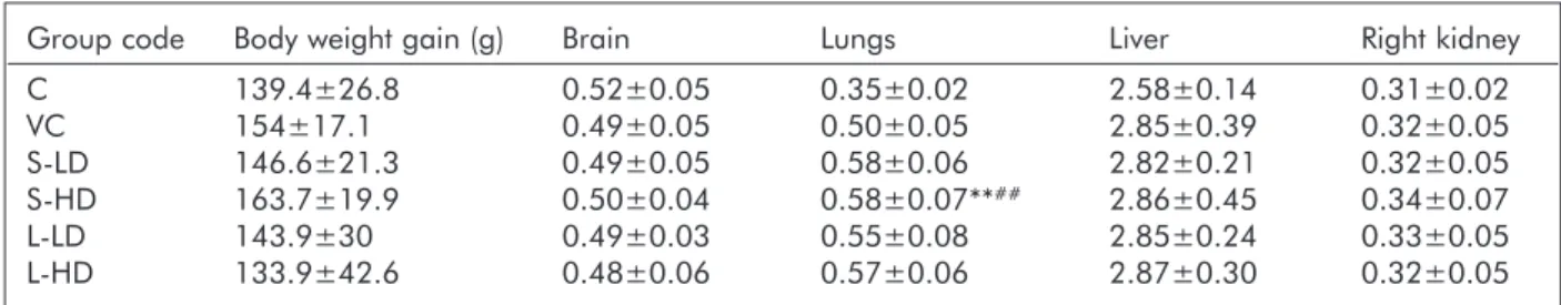
Table 2. Body weight gain and relative organ weights (to 1/100 body weight) in the control and treated groups
Group code Body weight gain (g) Brain Lungs Liver Right kidney
C 139.4±26.8 0.52±0.05 0.35±0.02 2.58±0.14 0.31±0.02
VC 154±17.1 0.49±0.05 0.50±0.05 2.85±0.39 0.32±0.05
S-LD 146.6±21.3 0.49±0.05 0.58±0.06 2.82±0.21 0.32±0.05
S-HD 163.7±19.9 0.50±0.04 0.58±0.07**## 2.86±0.45 0.34±0.07
L-LD 143.9±30 0.49±0.03 0.55±0.08 2.85±0.24 0.33±0.05
L-HD 133.9±42.6 0.48±0.06 0.57±0.06 2.87±0.30 0.32±0.05
Mean±SD, n=10. **: p<0.01 vs. C; ##: p<0.01 vs VC
analysis of group differ- ences was done by Tukey test and the Mann-Whit - ney U test. SPSS 24.0 (IBM Corporation, U.S.A.) was used.
Results
As judged from the group mean data of body weight gain, 6 weeks of TiO2 NP exposure exerted no gen- eral toxicity in the treated rats (Table 2). Also, among the organs, only the lungs appeared to be affected; their relative weight reflected the effect of the treatment procedure (group VC vs. C) and of the TiO2NP exposure.
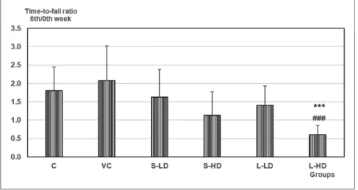
In the grip strength test, the decrease of time-to-fall from the 0thto the 6thweek (indicated by the 6thweek / 0th week ratio; Figure 1) was stronger in the treat- ed rats, with significant change in the L-HDgroup.
Among the electro- physiological phenomena recorded, spontaneous cortical activity was slightly shifted to higher frequencies in the treated rats (not shown). EP laten- cy, however, was substan- tially lengthened on expo- sure to TiO2 NPs, and in groups S-HD and L-HD the change was significant vs. Cand VC(Figure 2).
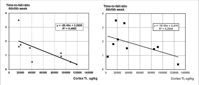
The causal relationship between TiO2 NP expo- sure and the mentioned functional alterations, sug- gested by the group-by- group dose dependence of the changes, was further tested by examining the correlation of cortical Ti levels, and corresponding parameters of EP latency Figure 1. Ratio of the grip strength test time-to-fall in the 6th and 0th week (see
Methods)
Mean+SD, n=10. *, **, ***: p<0.05, 0.01, 0.001 vs. C; #, ##, ###: p<0.05, 0.01, 0.001 vs. VC
Figure 2. Latency times of the averaged EPs in the somatosensory (A)and the visu- al and auditory (B)modalities in the control and treated rat groups
Mean+SD, n=10. *, **, ***: p<0.05, 0.01, 0.001 vs. C; #, ##, ###: p<0.05, 0.01, 0.001 vs. VC; °°, °°°: p<0.01, 0.001 vs. 1 Hz stimulation in the same group
(Figure 3) and grip strength test time-to-fall ratio (Figure 4). Although Ti measurement was done only on 3 rats’ samples per group, the diagrams show mostly fair correlations. The data show also that the effect of the smaller sized NPs was in most cases more clear-cut, resulting in relatively higher R2values.
Discussion
The neuro-functional alterations seen in the treated rats, and the correlation of these with the individual cortical Ti levels, pointed to the possible causative role of some TiO2 NP-dependent processes. Most probably, mitochondrial damage and oxidative stress were involved, both known to be present in cells and tissues in NP exposure2. In rats undergo-
ing artificial cerebral hy poxia-reperfusion, oxida- tive stress indicators in crea sed (including lipid per- oxidation, seen also in a previous work of us24with TiO2 NPs) and muscle force, examined by grip strength test, de creased25. This is in parallel with our findings in nano-TiO2 treated rats in the men- tioned and the present study.
Latency lengthening of the EPs could result both from mitochondrial lesion and oxidative damage.
The input-output relationship in18 indicated im - paired synaptic function in the nano-TiO2exposed rats. Decreased level of synaptophysin in rats after inhalation of nano-TiO2, as described in15also sug- gests the role of impaired transmission in EP laten- cy lengthening observed in our work. Beyond that, oxidative damage of membrane constituents, main- ly lipids11, impairs membrane-bound functions including synaptic transmission. In rats exposed the Figure 3. Correlation diagrams of the somatosensory evoked potential latency to cortical Ti level in rats treated with the smaller (left column) and larger (right column) TiO2NPs
same way with another metal oxide NPs, nano- MnO2, lengthening of EP latency in the treated rats was reversed by antioxidant treatment26supporting the above described mechanism of NP-induced neuro-functional damage.
Mitochondrial damage leads to energy shortage in neuronal and glial cells. Glutamate is co-trans- ported into the astrocytes with Na+ along its con- centration gradient which can be degraded if ion pumps do not operate properly due to energy short- age, ending up with excess perisynaptic glutamate and disturbed transmission27. Together with impaired axonal conduction this can result in depressed impulse propagation manifested in delayed cortical response to a peripheral stimulus.
The data in Figure 1and 2suggest that at equal mass dose the smaller NPs had a stronger effect on cortical EPs, and the larger ones, on grip strength.
One can suppose that 10 nm TiO2NPs had higher chance to reach the brain and 100 nm NPs stayed more in the peripheral circulation affecting nerves and muscles. With iridium NPs inhaled by rats, ca.
5 times less translocation of 75 nm size NPs than of 10 nm ones from the lungs to liver and kidneys was reported28, underlining the effect of NP size on crossing barriers. Likewise, breakdown of blood- brain barrier and neuronal damage caused by metal
NPs, given ip., was more severe with 20-30 nm than with 56-60 nm or >100 nm particles29. Blood-brain barrier damage was also observed in rats that inhaled nano-Ti15.
The present study left a few questions open. It remains to be clarified, by electron microscopy, whether and to what extent the chemically measured Ti content of the organ (first of all brain) samples indicates the presence of TiO2 NPs. Likewise, the actual role of the putative toxic mechanisms, espe- cially in the periphery, awaits further investigations.
Conclusion
The results provided further support to the function- al neurotoxicity of TiO2 nanoparticles, including central and peripheral actions. The exact role of particle size, and the mechanisms involved, remains to be elucidated.
ACKNOWLEDGMENTS
The authors are thankful to Prof. Gábor Galbács and co-workers (Department of Inorganic and Analytical Chemistry, University of Szeged Faculty of Science and Informatics) for Ti level determina- tion.
Figure 4. Correlation diagrams of the time-to-fall ratio in the grip strength test to cortical Ti level in rats treated with the smaller (left) and larger (right) TiO2NPs
REFERENCES
1.Buzea C, Pacheco Blandino II, Robbie K.Nanomaterials and nanoparticles: Sources and toxicity. Biointerphases 2007;2:MR17-MR172. https://doi.org/10.1116/1.2815690 2.Oberdörster G, Oberdörster E, Oberdörster J. Nano -
toxicology: An emerging discipline evolving from studies of ultrafine particles. Environ Health Perspect 2005;113:
823-39.
https://doi.org/10.1289/ehp.7339
3.Nel A, Yia T, Mädler L, Li N. Toxic potential of materials at the nanolevel. Science 2006;311:622-27.
https://doi.org/10.1126/science.1114397
4.Czajka M, Sawicki K, Sikorska K et al.Toxicity of titanium dioxide nanoparticles in central nervous system. Toxicol In Vitro 2015;29:1042-52.
https://doi.org/10.1016/j.tiv.2015.04.004
5.Chen J, Poon C.Photocatalytic construction and building materials: From fundamentals to applications. Building Environ 2009;44:1899-906.
https://doi.org/10.1016/j.buildenv.2009.01.002
6.Grande F, Tucci P. 2016. Titanium dioxide nanoparticles:
a risk for human health? Mini Rev Med Chem 2016;16:
762-9.
https://doi.org/10.2174/1389557516666160321114341 7.Migliore L, Uboldi C, Di Bucchianico S, Coppede F.Nano -
materials and neurodegeneration. Environ Mol Mutagen 2015;56:149-70.
https://doi.org/10.1002/em.21931
8.SCENIHR(Scientific Committee on Emerging and Newly Identified Health Risks). Risk assessment of products of nanotechnologies, 19 January 2009.
http://ec.europa.eu/health/ph_risk/committees/04_
scenihr/docs/scenihr_o_023.pdf (Letöltés ideje: 2017.
november 17.)
9.NIOSH. Occupational Exposure to Titanium Dioxide.
Current Intelligence Bulletin 63. Department of Health and Human Services (National Institute of Occupational Safety and Health) Publication No. 2011-160.
http://www.cdc.gov/niosh/docs/2011-160/ (Letöltés ideje:
2017. november 17.)
10.Nalika N, Parvez S. Mitochondrial dysfunction in titanium dioxide nanoparticle-induced neurotoxicity. Toxicol Mech Meth 2015;25:355-63.
https://doi.org/10.3109/15376516.2015.1020183
11.Guerra-Araiza C, Álvarez-Mejía AL, Sánchez-Torres S, Farfan-García E, Mondragón-Lozano R, Pinto-Almazán R, et al.Effect of natural exogenous antioxidants on aging and on neurodegenerative diseases. Free Rad Res 2013;47:451- 62.
https://doi.org/10.3109/10715762.2013.795649
12.Oleru UG.Respiratory and nonrespiratory morbidity in a titanium oxide paint factory in Nigeria. Am J Ind Med 1987;12:173-80.
https://doi.org/10.1002/ajim.4700120206
13.Coccini T, Grandi S, Lonati D, Locatelli C, De Simone U.
Comparative cellular toxicity of titanium dioxide nanopar- ticles on human astrocyte and neuronal cells after acute and prolonged exposure. Neuro Toxicology 2015;48:77-89.
https://doi.org/10.1016/j.neuro.2015.03.006
14.Valdiglesias V, Costa C, Sharma V, Kiliç G, Pásaro E, Teixeira JP, et al. Comparative study on effects of two dif- ferent types of titanium dioxide nanoparticles on human neuronal cells. Food Chem Toxicol 2013;57:352-61.
https://doi.org/10.1016/j.fct.2013.04.010
15. Disdier C, Chalansonnet M, Gagnaire F, Gaté L, Cosnier F, et al.Brain inflammation, blood brain barrier dysfunc- tion and neuronal synaptophysin decrease after inhalation exposure to titanium dioxide nano-aerosol in aging rats.
Scientific Report S 2017;7:12196.
https://doi.org/10.1038/s41598-017-12404-5
16.Yoshiura Y, Izumi H, Oyabu T, Hashiba M, Kambara T, Mizuguchi Y, et al.Pulmonary toxicity of well-dispersed titanium dioxide nanoparticles following intratracheal inst- illation. J Nanopart Res 2015;17:241.
https://doi.org/10.1007/s11051-015-3054-x
17.Baisch BL, Corson NM, Wade-Mercer P, Gelein R, Kennell AJ, Oberdörster G, et al.Equivalent titanium dioxide nano- particle deposition by intratracheal instillation and whole body inhalation: the effect of dose rate on acute respiratory tract inflammation. Part Fibre Toxicol 2014;11:5.
https://doi.org/10.1186/1743-8977-11-5
18.Gao X, Yin S, Tang M, Chen J, Yang Z, Zhang W et al.
Effects of developmental exposure to TiO2nanoparticles on synaptic plasticity in hippocampal dentate gyrus area:
an in vivo study in anesthetized rats. Biol Trace Elem Res 2011;143:1616-28.
https://doi.org/10.1007/s12011-011-8990-4
19.Grissa I, Guezguez S, Ezzi L, Chakroun S, Sallem A, Kerkeni E, et al.The effect of titanium dioxide nanopartic- les on neuroinflammation response in rat brain. Environ Sci Pollut Res Int 2016;20:20205-13.
https://doi.org/10.1007/s11356-016-7234-8
20.Ben-Younes NR, Amara S, Mrad I, Ben-Slama I, Jeljeli M, et al.Subacute toxicity of titanium dioxide (TiO2) nano- particles in male rats: emotional behavior and pathophysi- ological examination. Environ Sci Pollut Res 2015;22:
8728-37.
https://doi.org/10.1007/s11356-014-4002-5
21.Oszlánczi G, Horváth E, Szabó A, Horváth E, Sápi A, Kozma G, et al.Subacute exposure of rats by metal oxide nanoparticles through the airways: general toxicity and neuro-functional effects. Acta Biol Szeged 2010;54:165- 70.
22.Strohl KP, Thomas AJ, St.Jean P, Schlanker EH, Koletsky RJ, Schork NJ. Ventilation and metabolism among rat strains. J Appl Physiol 1997;82:317-23.
23.Feng Q, Ma Y, Mu S, Wu J, Chen S, Ou-Yang L, Lei W.
(2014) Specific reactions of different striatal neuron types in morphology induced by quinolinic acid in rats. PLoS ONE 2014;9:e91512.
https://doi.org/10.1371/journal.pone.0091512
24.Horváth T, Papp A, Kovács D, Kálomista I, Kozma G, Vezér T.Electrphysiological alterations and general toxic signs obtained by subacute administration of titanium diox- ide nanoparticles to the airways of rats. Ideggyogy Sz 2017;70:127-35.
https://doi.org/10.18071/isz.70.0127
25.Tabassum R, Vaibhav K, Shrivastava P, Khan A, Ahmed E, Javed H et al.Centella asiatica attenuates the neurobehavi- oral, neurochemical and histological changes in transient focal middle cerebral artery occlusion rats. Neurol Sci 2013;34:925-33.
https://doi.org/10.1007/s10072-012-1163-1
26.Sárközi K, Papp A, Horváth E, Máté Zs, Hermesz E, Kozma G et al. Protective effect of green tea against neuro-functi- onal alterations in rats treated withMnO2 nanoparticles. J Sci Food Agric 2017;97:1717-24.
https://doi.org/10.1002/jsfa.7919
27.Takahashi M, Billups B, Rossi D, Sarantis M, Hamann M, Attwell D.The role of glutamate transporters in glutamate homeostasis in the brain. J Exp Biol 1997;200:401-9.
28.Buckley A, Warren J, Hodgson A, Marczylo T, Ignatyev K, Guo C. Slow lung clearance and limited translocation of four sizes of inhaled iridium nanoparticles. Part Fibre Toxicol 2017;14:5.
https://doi.org/10.1186/s12989-017-0185-5
29.Sharma A, Muresanu DF, Patnaik R, Sharma HS.Size- and age-dependent neurotoxicity of engineered metal nanopar- ticles in rats. Mol Neurobiol 2013;48:386-96.
https://doi.org/10.1007/s12035-013-8500-0