Renal nitric oxide production in rat pregnancy: role of constitutive nitric oxide synthases
Cheryl A. Smith,1Beth Santymire,2Aaron Erdely,3Vasuki Venkat,4György Losonczy,5and Chris Baylis1,6
1Department of Physiology and Functional Genomics and6Department of Medicine, Division of Nephrology, University of Florida, Gainesville, Florida;2Department of Physiology, West Virginia University,3Toxicology and Molecular Biology Branch, National Institute of Occupational Safety and Health, Morgantown, West Virginia;4Department of Nephrology, University of California, San Diego, California; and5Department of Pulmonology, Semmelweis University, Budapest, Hungary
Submitted 26 May 2010; accepted in final form 10 July 2010
Smith CA, Santymire B, Erdely A, Venkat V, Losonczy G, Baylis C.Renal nitric oxide production in rat pregnancy: role of constitutive nitric oxide synthases.Am J Physiol Renal Physiol299: F830–F836, 2010. First published July 14, 2010; doi:10.1152/ajprenal.00300.2010.—Functional studies show that increased renal nitric oxide (NO) mediates the renal vasodilation and increased glomerular filtration rate that occur during normal pregnancy. We investigated whether changes in the constitu- tive NO synthases (NOS), endothelial (eNOS) and neuronal (nNOS), were associated with the increased renal NO production in normal midterm pregnancy in the rat. In kidneys from midterm pregnant (MP:
11–13 days gestation), late-term pregnant (LP: 18 –20 days gestation), and similarly aged virgin (V) rats, transcript and protein abundance for eNOS and the nNOS␣and nNOSsplice variants, as well as the rate of L-arginine-to-L-citrulline conversion, were determined as a measure of NOS activity. At MP, renal cortical abundance of the total eNOS protein and phosphorylated (Ser1177) eNOS was reduced, and
L-arginine-to-L-citrulline conversion in the cortical membrane fraction was decreased; these declines were also seen in LP. There were no changes in the eNOS transcript. In contrast,L-arginine-to-L-citrulline conversion in the soluble fraction of renal cortex increased at MP and then declined at LP. This MP increase was ablated byS-methylthio- citrulline, a nNOS inhibitor. Using Western blotting, we did not detect a change in the protein abundance or transcript of the 160-kDa nNOS␣, but protein abundance and transcript of the nNOS were increased at MP in cortex. Collectively, these studies suggest that the soluble nNOS is responsible for the increased renal cortical NO production during pregnancy.
endothelial nitric oxide synthase; neuronal nitric oxide synthase;
L-arginine-to-L-citrulline conversion; S-methylthiocitrulline; splice variants
A MARKED RENAL VASODILATION occurs during pregnancy and leads to increases in renal plasma flow (RPF) and glomerular filtration rate (GFR) (5, 11, 13). In rats, this increase in RPF and GFR is maximal by midterm and involves a parallel relaxation of afferent and efferent arterioles (5, 11). Functional studies suggest that nitric oxide (NO) mediates the renal vasodilation, since nonselective NO synthase (NOS) inhibitors prevent the gestational rise in GFR (6, 9, 12, 14, 25).
Although NO appears to modulate the renal hemodynamic response during pregnancy, attempts to define the exact origin of the increased NO remain inconclusive. The use of isoform-
“selective” NOS inhibitors has implicated neuronal and induc- ible NOS (nNOS and iNOS) (1, 3), while lack of endothelial
NOS (eNOS)-selective inhibitors has prevented direct investi- gation of eNOS. Alexander et al. (4) reported an increase in nNOS protein abundance in rats at midterm, which coincides with maximal renal vasodilation (5), while an unexpected decline in eNOS protein abundance was also observed. In contrast, Novak et al. (27) reported no change in renal eNOS and nNOS protein abundance with pregnancy. Apart from the unresolved question of NOS protein abundance, large changes in NOS activity are possible without alterations in protein abundance. For example, phosphorylation of eNOS at Ser1177 can lead to a 15- to 20-fold increase in NOS activity compared with unphosphorylated eNOS (18).
To better our understanding of the renal NO system in pregnancy, we have conducted a series of studies assessing renal constitutive NOS isoform mRNA abundance, protein abundance, and activity in kidneys of virgin and pregnant rats.
MATERIALS AND METHODS
Animals.Studies were performed on female Sprague-Dawley rats (n⫽51; Harlan Sprague Dawley, Indianapolis, IN) aged 4 –5 mo. The animal studies were reviewed and approved by the West Virginia University and the University of Florida Animal Care and Use Committees. All animals were housed separately, provided rat chow and water ad libitum, and maintained on a 12:12-h light-dark cycle.
Female rats were temporarily housed with males for 1–5 days. Mating was confirmed by the presence of sperm in the vagina (taken asday 1 of pregnancy) and a continual diestrus vaginal smear. Pregnancy was confirmed by an increased body weight and the presence of live fetuses “in utero” on the day of death.
Inseries 1, in vitro NOS activity was evaluated in tissues harvested from midterm-pregnant (MP: 11–13 days gestation, n ⫽ 7), late- pregnant (LP: 18 –20 days gestation,n⫽8), and similarly aged virgin (V,n⫽8) rats. Inseries 2, NOS mRNA and protein analysis was carried out in tissues harvested from MP (n⫽5), LP (n⫽5), and similarly aged V (n⫽5) rats. Inseries 3, NOS mRNA and protein analysis was carried out in tissues harvested from MP (n⫽ 4), LP (n⫽4), and V (n⫽5) rats.
Tissue harvest.Rats were anesthetized with pentobarbital sodium (0.7 mg/kg ip) or isoflurane. The kidneys were perfused blood-free with cold PBS, separated into cortex and medulla, flash frozen in liquid nitrogen, and stored at⫺80°C.
NOS activity.NOS activity was measured from the conversion of [3H]L-arginine to [3H]L-citrulline in kidney tissue, as described pre- viously (32). Briefly, tissues were homogenized and ultracentrifuged.
The supernatant (soluble) and membrane fractions were assayed. The soluble fraction contains most of the nNOS, while the membrane fraction contains mostly eNOS. Endogenous arginine was removed using Dowex, and the total protein content was determined using a Bradford assay (7). Samples were assayed in the presence of the Address for reprint requests and other correspondence: C. Baylis, 1600 SW
Archer Rd., Univ. of Florida, POB 100274, Gainesville, FL 32667 (e-mail:
baylisc@ufl.edu).
First published July 14, 2010; doi:10.1152/ajprenal.00300.2010.
presence of the nNOS-selective inhibitor S-methylthiocitrulline (SMTC, 1M).
Western blotting.Samples were homogenized, and proteins were separated by SDS-PAGE and transferred to a nitrocellulose mem- brane, as described previously (32). Ponceau S staining confirmed equivalent protein loads for cortex (200 g of total protein) and medulla (100g of total protein). Inseries 2, membranes were first probed with rabbit polyclonal phosphorylated (Ser1177) eNOS anti- body (Cell Signaling Technology, Danvers, MA) in a 1:250 dilution of 5% BSA in 0.1% Tris-buffered saline⫹Tween 20 (TBS-T) for 16 h at 4°C and then with anti-rabbit IgG horseradish peroxidase (HRP)- linked antibody (Cell Signaling Technology) in a 1:2,000 dilution of 5% nonfat milk in 0.1% TBS-T for 1 h at room temperature. Mem- branes were then stripped and reprobed for eNOS (mouse monoclonal antibody, Transduction Laboratories, San Jose, CA) at a 1:250 dilu- tion for 1 h. Next, goat anti-mouse IgG-HRP conjugate (Transduction Laboratories) at a 1:2,000 dilution was applied, and the membranes were incubated for 1 h. Duplicate membranes were run and probed for nNOS with a rabbit polyclonal antibody targeting the NH2terminus at a 1:5,000 dilution for 1 h (16) and then with goat anti-rabbit IgG-HRP (Bio-Rad, Hercules, CA) at a 1:3,000 dilution for 1 h at room temperature. Inseries 3, membranes were probed overnight with a 1:250 dilution of COOH-terminal rabbit polyclonal antibody (PA1–
033, Affinity BioReagents, Golden, CO) or for 1 h with a 1:5,000 dilution of the same NH2-terminal rabbit polyclonal antibody used in series 2(gift from Dr. Kim Lau) (22) and then for 1 h with a 1:3,000 dilution of secondary goat anti-rabbit IgG-HRP antibody (Bio-Rad).
Bands of interest were visualized using an ECL Western blot detec- tion kit (Amersham Biosciences, Piscataway, NJ). Densitometric analysis was performed using Optimas 6.2 imaging analysis software.
Protein abundance is reported as integrated optical density (OD) units.
The integrated OD was factored for Ponceau S staining (total protein loaded) and for an internal standard run on each membrane.
PCR.RNA was isolated from tissue using TRI Reagent (Sigma, St.
Louis, MO) and treated with DNase I (Ambion, Austin, TX), and 1–2g was reversed transcribed (SuperScript II RNase H⫺Reverse Transcrip- tase, Invitrogen, Bethesda, MD) with random primers (Invitrogen) in a total volume of 20 – 40l. For control RT reactions, the RT enzyme was omitted, and primers were designed to include intron-exon splice sites.
Primers were designed using GeneTool software (Biotools, Edmonton, AB, Canada) with annealing temperatures at 54 – 62°C. All PCR products were verified by restriction endonuclease digestion. Cyclophilin (CyP) was used as an internal reference, since CyP abundance remained con- sistent in V, MP, and LP rats.
For end-point PCR, preliminary experiments were conducted for each PCR product to ensure that the number of cycles represented the linear portion of the PCR OD curve. For each primer set [5= acgc- cgctgtctcttttc 3=(forward) and 5=tgccttctttcaccttc 3=(reverse) for CyP (M19533), 5=ccggctgccacctgatcctaacttg 3=(forward) and 5=tgcgcaat- gtgagtccgaaaatgtc 3= (reverse) for eNOS (XM342615), and 5=cggc- catcacgatattccctcag 3= (forward) and 5= cccgatctccagcagcatgttg 3=
(reverse) for “total” nNOS (NM052799); primer sets for nNOS␣and nNOS were previously published (23, 30)], the cDNA from all samples was amplified simultaneously using aliquots from the same PCR mixture. PCR was carried out using 1g of cDNA, 50 ng of each primer, 250 M deoxyribonucleotide triphosphates, 1⫻ PCR buffer, and 2 U of TaqDNA polymerase (Sigma) in a 50-l final volume. After amplification, 20l of each reaction were electropho-
Detection System (Bio-Rad). nNOS primers that would target all nNOS isoforms [5=aagcctatgccaagaccctgtgtgagatc 3=(forward) and 5=
ccagggcttcgtgctccaggtg 3= (reverse) for total nNOS (NM052799), 5=
cggcgtgctgcgggatca 3= (forward) and 5= tgcggatgcggctcgtcac 3= (re- verse) for eNOS (XM342615), and 5= gcccttgggtcgcgtctgct 3= (for- ward) and 5=caccctggcacatgaatcctgga 3=(reverse) for CyP (M19533)]
were designed. Real-time PCR, a more quantitative approach, was not used to detect nNOS␣and nNOSmRNA, since multiple bands may occur with nNOSproduct amplification (30). Samples were run in duplicate (2.5–5 l of cDNA per well), and the cycle times from duplicate wells were averaged. Preliminary experiments were con- ducted to optimize PCR conditions, ensure the absence of dimer formation, and affirm that a single PCR product was formed for each primer pair. For the relative quantification of gene expression, the comparative threshold cycle (Ct) method was employed (24). Valida- tion methods were conducted over a 10-fold range of cDNA and over a 2-fold range of primer concentrations from control and experimental kidneys to confirm equal efficiency of NOS and CyP. The averaged NOS Ctwas subtracted from the corresponding averaged CyP Ctfor each sample, resulting in ⌬Ct. The fold change was established by calculating 2⫺⌬⌬Ctfor experimental vs. control samples (16).
Statistical analysis.One-way ANOVA and Bonferroni’s multiple comparison test were used to compare mRNA and protein abundance differences of V, MP, and LP samples. Values are means ⫾ SE.
Significance was accepted atP⬍0.05.
RESULTS
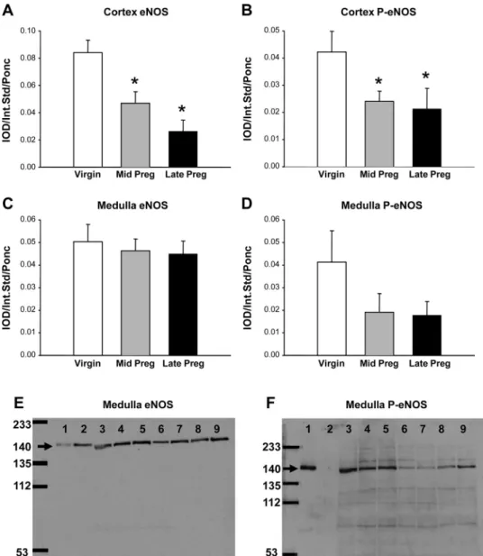
As shown in Fig. 1, the abundance of total eNOS (Fig. 1A) and phosphorylated eNOS (Fig. 1B) falls during pregnancy in renal cortex. In renal medulla, there were no significant changes in eNOS or phosphorylated eNOS abundance (Fig. 1, CandD). Representative blots for renal medulla in Fig. 1,E andF, show that the eNOS antibody recognized the eNOS- and phosphorylated eNOS-positive controls (lanes 1and2in Fig.
1E), while the phosphorylated (Ser1177) eNOS antibody recog- nized only the phosphorylated eNOS-positive control (lane 1in Fig. 1F). To measure eNOS transcript, we used CyP mRNA abundance as an internal control for end-point and real-time PCR, since CyP mRNA abundance did not change with preg- nancy (Table 1). Cortical and medullary eNOS mRNA abun- dance was unaffected by pregnancy, as shown by end-point and real-time PCR and two different primer pairs (Table 1).
We measured the total [3H]L-arginine-to-[3H]L-citrulline conversion, as a measure of NOS activity, in renal cortex and medulla membrane fractions (Fig. 2). [3H]L-arginine-to-[3H]L- citrulline conversion was significantly lower in LP than V rats in cortex. In medulla, membrane NOS activity fell in preg- nancy. There was, however, a significant increase in [3H]L- arginine-to-[3H]L-citrulline conversion in the soluble fraction of renal cortex of MP compared with V and LP rats (Fig. 3). In MP rats, the nNOS-selective NOS inhibitor SMTC reduced the cortical soluble [3H]L-arginine-to-[3H]L-citrulline conversion to V values. In medulla, there was no difference in total NOS activity in the soluble fraction during pregnancy, and SMTC
inhibited ⬎50% of [3H]L-arginine-to-[3H]L-citrulline conver- sion in V, MP, and LP rats.
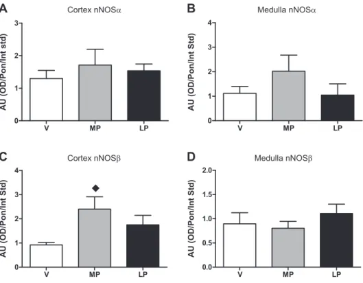
We also measured cortical and medullary nNOS␣ protein abundance. As shown in Fig. 4,AandB, there was no change in nNOS␣ protein abundance with pregnancy in animals in
series 3or in animals inseries 2(data not shown). In contrast, there was a marked rise in the cortical nNOSprotein abun- dance at MP (Fig. 4C) that occurred in parallel with the increase in NOS activity in the soluble fraction of cortex (Fig.
3). Representative blots for the nNOS␣ and nNOS proteins
Fig. 1. Relative abundance of kidney endo- thelial nitric oxide synthase (eNOS) and phosphorylated (Ser1177) eNOS (P-eNOS) in cortex and medulla of virgin, midterm preg- nant (Mid Preg), and late-term pregnant (Late Preg) rats.A–D: average densitometric values [integrated optical density (IOD)/in- ternal standard (Int Std)/Ponceau S (Ponc)]
for eNOS and P-eNOS in renal cortex and medulla. *Significantly different from virgin (P⬍0.05).EandF: representative Western blots for eNOS and P-eNOS in renal me- dulla. A molecular weight marker (in kDa) is shown atfar left.Lane 1, P-eNOS-positive control (lysate of VEGF-treated rat aortic endothelial cells); lane 2, eNOS-positive control (lysate of confluent unstimulated rat aortic endothelial cells);lanes 3and4, vir- gin;lanes 5and6, midterm pregnant;lanes 7–9, late-term pregnant.
Table 1. mRNA abundance: series 2 experiments
Renal Cortex Renal Medulla
V MP LP V MP LP
Real-time PCR
eNOS* 0.88 0.85 0.86 0.68
nNOS* 1.04 1.10 0.84 0.78
CyP† 19.28⫾0.15 19.38⫾0.38 17.59⫾0.45 17.92⫾0.06 17.68⫾0.06 17.72⫾0.06
End-point PCR‡
eNOS 0.37⫾0.04 0.43⫾0.07 0.36⫾0.04 0.87⫾0.19 0.87⫾0.15 0.73⫾0.04
nNOS 0.71⫾0.15 0.71⫾0.14 0.69⫾0.14 0.78⫾0.06 0.76⫾0.02 0.70⫾0.04
CyP 2.72⫾0.38 2.80⫾0.58 2.54⫾0.47 2.19⫾0.18 2.37⫾0.13 2.33⫾0.10
V, virgin; MP, midterm pregnant; LP, late-term pregnant; eNOS and nNOS, endothelial and neuronal nitric oxide synthase; CyP, cyclophilin. *eNOS and nNOS real-time PCR values are expressed as mean fold change in gene expression (2⫺⌬⌬Ct, where Ctis cycle time) compared with V. †CyP real-time PCR values (means⫾SE) are expressed in Ct. ‡End-point PCR values (means⫾SE) are expressed in arbitrary units; eNOS and nNOS were normalized to CyP.
(from animals inseries 3) are shown in Fig. 5. The total nNOS transcript was unchanged by real-time and end-point PCR (Table 1), and there was no change in nNOS␣mRNA in cortex or medulla during pregnancy, while nNOS transcript in- creased in cortex and medulla in MP rats (Fig. 6).
DISCUSSION
The main findings in the present study are that, in renal cortex of the MP rat, membrane NOS activity and total and phosphorylated (Ser1177) eNOS protein are not increased when the gestational renal vasodilation is at a maximum. In contrast, a marked rise in renal cortical NOS activity in the soluble fraction is evident, which can be ablated with SMTC, a nNOS-selective inhibitor (17, 26). Using an antibody to the NH2terminus of the 160-kDa nNOS␣, we saw no change in protein expression during pregnancy, and the nNOS␣ tran- script was also unchanged. Transcript and protein abundance of the functional splice variant nNOS was significantly ele- vated in renal cortex at MP, coincident with the gestational renal vasodilation.
Our findings confirm earlier observations that increased eNOS expression does not contribute to the NO-dependent renal vasodilation and hyperfiltration that occurs during preg- nancy. Previous studies indicate no change (27) or a decrease (4) in eNOS during pregnancy. Here, we report decreases in total cortical eNOS abundance at MP and LP, and we found no change in eNOS transcript abundance. Of course, protein abundance is not the only determinant of eNOS activity, since there are many possible posttranslational modifications that affect activity, including phosphorylation of eNOS at Ser1177,
to the eNOS.
In contrast to the membrane fraction, we observed a signif- icant increase in renal cortical soluble NOS activity at MP and a return in LP that parallels the time course of the gestational renal vasodilation in the rat (5, 11). iNOS and some nNOS reside in the soluble fraction. We used a relatively selective nNOS inhibitor (SMTC, Ki ⫽ 1.2 nM), which have been reported to exhibit ⬎100-fold selectivity for rat nNOS com- pared with iNOS,⬃10-fold selectivity for nNOS over eNOS in purified enzyme preparations, and 17-fold selectivity for nNOS over eNOS in tissues (17, 26). We found that SMTC com- pletely prevented the rise in soluble NOS activity in renal cortex in MP while having no impact on V or LP tissue homogenates. This suggests that the increased soluble NOS activity originated from a nNOS source. These findings are in agreement with studies by Abram and colleagues (1), who reported that acute infusion of another nNOS-selective inhib- itor, 7-nitroindazole, decreased RPF and GFR in conscious pregnant rats without affecting renal hemodynamics of virgins.
Although SMTC and 7-nitroindazole are chemically unrelated, both are used as selective nNOS inhibitors (17). As a caution- ary note, however, assumptions based on supposedly “isoform- selective” NOS inhibitors are fraught with problems. Alderton
Fig. 3. Rate of conversion of [3H]L-arginine to [3H]L-citrulline as a measure of NOS activity in soluble fraction of renal cortex and medulla. Solid columns, baseline state; hatched columns,S-methylthiocitrulline. *Significantly differ- ent from V and LP (P⬍0.05). #Significantly different from baseline (P⬍ 0.05).
Fig. 2. Rate of conversion of [3H]L-arginine to [3H]L-citrulline as a measure of NOS activity in membrane fraction of renal cortex and medulla of virgin (V), midterm pregnant (MP), and late-term pregnant (LP) rats. *Significantly different from V (P⬍0.05).
et al. (2) report that much of the confusion results from the differing criteria used to define selectivity. In fact, when selectivity is defined on the basis of potency under identical conditions and in the physiological range, few selective NOS inhibitors are highly selective. This may explain why nNOS- selective (1) and iNOS-selective (3) inhibitors have been shown to ablate the gestational rise in GFR. In view of this, we cannot rely on selective NOS inhibitors as a means of defining which NOS isoform(s) play(s) a role in renal hemodynamic changes that occur with pregnancy; therefore, we also looked at NOS protein and transcript abundance.
Novak et al. (27) reported no change in medullary nNOS protein abundance and could not detect the nNOS protein in renal cortex in V or MP; they observed no change in the cortical nNOS transcript in MP vs. V. In contrast, Alexander et
al. (4) reported a significant increase in whole kidney nNOS protein abundance that reached a peak at midterm (13 days gestation) and remained high throughout the rest of pregnancy.
These workers used a commercially available antibody to the COOH terminus of nNOS, which we now recognize will detect all the nNOS splice variants that are present. In the present study, using an antibody to nNOS␣(which recognizes only the unique NH2 terminus of nNOS␣), we found no change in protein abundance at MP, and this was confirmed at the transcript level.
We recently reported that there are nNOS splice variants in the normal rat kidney and that the proportions of these can change under pathological conditions (30). Of particular rele- vance to the present study, one of these splice variants, nNOS, is a functional enzyme that is entirely cytosolic in
Fig. 4. A and B: relative neuronal NOS (nNOS)-␣(nNOS␣) protein abundance inse- ries 3 rats (i.e., NH2-terminal antibody) in cortex and medulla in V, MP, and LP rats.C and D: relative nNOSabundance in cortex and medulla. nNOS-positive control is rat cer- ebellar lysate.⽧Significantly different from V (P⬍0.05).
Fig. 5.Top: representative whole membrane Western blots of renal cortex (300 g of total protein) and medulla (100g of total protein). An NH2-terminal antibody (22) was used for nNOS␣ (⬃160 kDa, arrow- head) detection (A), and a COOH-terminal antibody (PA1– 033, Affinity Bioreagents) was used for nNOS (⬃140 kDa, arrow) detection (B).Bottom: Ponceau S staining of corresponding lanes confirming similar pro- tein loads.
location (2). Lacking the unique PDZ-binding domain at the NH2 terminus, nNOS would be recognized by antibodies targeting the COOH, but not the NH2, terminus. We therefore conducted a preliminary study assessing the variable 5=-un- translated regions (5=-UTRs; exon 1a, 1b, and 1c) reported by Lee and colleagues (23) in the rat kidney. Using RT-PCR and exon 1a-exon 6 primer pairing, we detected two bands, indi- cating that nNOS␣ and nNOS transcripts had a common 5=-UTR (30), as previously reported by Huber et al. (19) in the rat small intestine. Although we report no change in nNOS␣ transcript abundance with exon 1a-exon 2 primer pairing and a significant increase in nNOStranscript abundance at MP with exon 1a-exon 6 primer pairing, we recognize that there may be additional transcripts for nNOS␣ and nNOS. Indeed, in rat kidney, Lee et al. (23) identified three nNOS transcripts with distinct 5=-UTR first exons (exon 1a, 1b, and 1c), all of which spliced to exon 2 (nNOS␣). Oberbaumer et al. (28) identified five nNOS mRNA variants: four encoded for nNOS␣(exon 2), and one encoded for nNOS(exon 3). Although these nNOS mRNA variants may be generated by alternative promoters encoding the same nNOS protein, it is possible that the various nNOS transcripts with different 5=-UTRs may have different functional properties that may influence mRNA translational efficiency, alter mRNA localization, or alter mRNA stability.
This may account for the differences in cortical and medullary nNOS␣ and nNOS transcript expression compared with nNOS␣ and nNOS protein abundance seen in the present study.
We used the previously characterized COOH-terminal nNOS antibody (30) to directly test whether nNOS might contribute to the gestational renal vasodilation and observed a marked increase in the protein abundance. Unlike the nNOS␣, nNOScontains a unique NH2terminus of 6 amino acids and lacks the first 236 amino acids of nNOS␣, which contains the PSD-95 disks large/ZO-1 homology (PDZ) and protein inhib- itor of NOS (PIN) domains. The PDZ domain of nNOS␣ mediates interaction with postsynaptic density protein 95
(PSD-95) of neurons and␣1-syntrophin (part of the dystrophin complex) of myocytes, which anchors nNOS␣ in the plasma membrane (8). Without a PDZ domain, nNOSwould occur in the cytosol, as shown by Huber et al. (19) in the rat intestine.
Furthermore, the PIN domain contains a binding site for the protein inhibitor of NOS (21), and without a PIN domain, nNOScannot be regulated by PIN. Therefore, nNOSmay be regulated by other mechanisms, such as alternate promoter usage and pre-mRNA splicing events (31). With no PDZ and PIN domains, the secondary and/or tertiary structure of nNOS may be similar to iNOS, and we speculate that iNOS-selective inhibitors may inhibit the activity of iNOS and nNOS. Also, with similar molecular weights, nNOS may comigrate with iNOS and/or be mistaken for iNOS in the absence of appro- priate positive controls.
Brenman and colleagues (8) reported that nNOS trans- fected in COS cells was catalytically active, with a Km for arginine similar to nNOS␣, and fully dependent on calcium/
calmodulin. Several in vivo studies using wild-type and nNOS␣⫺/⫺ mice also suggested a functional role for nNOS. For example, in wild-type mice, nNOSis a functional enzyme accounting for much of the citrulline formation in many brain areas (15). The nNOS␣⫺/⫺ and eNOS⫺/⫺ double-knockout mice exhibit normal penile erection mediated by nNOS(20).
In nNOS␣⫺/⫺mice, nNOSabundance increases, suggesting a compensatory upregulation of nNOS when nNOS␣-derived NO is deficient (15). In humans, nNOShas been localized in the spinal cord and is upregulated in astrocytes of the ventral horn and white matter in patients with amyotrophic lateral sclerosis (10). This suggests that nNOSmay be upregulated in disease states, as also reported by us in the rat kidney with chronic kidney disease (30). The present study suggests that renal cortex nNOSis upregulated in response to physiological stimuli in normal pregnancy, which we suggest may play an important role in the renal vasodilation at MP.
In summary, the increase in renal cortical NO generation during pregnancy cannot be attributed to increased abundance
dulla (B andD) assessed by end-point PCR.
There was no significant difference in cortical or medullary nNOS␣ mRNA abundance be- tween V, MP, and LP. However, nNOS mRNA abundance was significantly increased in MP rats compared with V and LP rats. AU, arbitrary units [i.e., optical density (OD) ⫻ area/cyclophilin (CyP)]. *Significantly differ- ent from V and LP (P⬍0.05).
or activity of eNOS. Rather, nNOSin the soluble fraction of renal cortex provides the likely source. Future studies should investigate the specific location and the signaling pathway responsible for activating the renal cortical nNOSin normal pregnancy.
GRANTS
This work was supported by National Institutes of Health Grants HL-31933 and DK-56843 to C. Baylis.
DISCLOSURES
No conflicts of interest, financial or otherwise, are declared by the authors.
REFERENCES
1. Abram SR, Alexander BT, Bennett WA, Granger JP.Role of neuronal nitric oxide synthase in mediating renal hemodynamic changes during pregnancy.Am J Physiol Regul Integr Comp Physiol281: R1390 –R1393, 2001.
2. Alderton WK, Cooper CE, Knowles RG.Nitric oxide synthases: struc- ture, function and inhibition.Biochem J357: 593–615, 2001.
3. Alexander BT, Cockrell K, Cline FD, Granger JP. Inducible nitric oxide synthase inhibition attenuates renal hemodynamics during preg- nancy.Hypertension39: 586 –590, 2002.
4. Alexander BT, Miller MT, Kassab S, Novak J, Reckelhoff JF, Kruck- eberg WC, Granger JP. Differential expression of renal nitric oxide synthase isoforms during pregnancy in rats.Hypertension33: 435–439, 1999.
5. Baylis C.Glomerular filtration and volume regulation in gravid animal models.Baillieres Clin Obstet Gynaecol8: 235–264, 1994.
6. Baylis C, Engels K.Adverse interactions between pregnancy and a new model of systemic hypertension produced by chronic blockade of endo- thelial derived relaxing factor (EDRF) in the rat.Clin Exp HypertensB11:
117–129, 1992.
7. Bradford MM. A rapid and sensitive method for the quantitation of microgram quantities of protein utilizing the principle of protein-dye binding.Anal Biochem72: 248 –254, 1976.
8. Brenman JE, Chao DS, Gee SH, McGee AW, Craven SE, Santillano DR, Wu Z, Huang F, Xia H, Peters MF, Froehner SC, Bredt DS.
Interaction of nitric oxide synthase with the postsynaptic density protein PSD-95 and␣1-syntrophin mediated by PDZ domains.Cell84: 757–767, 1996.
9. Cadnapaphornchai MA, Ohara M, Morris KG Jr, Knotek M, Ro- gachev B, Ladtkow T, Carter EP, Schrier RW.Chronic NOS inhibition reverses systemic vasodilation and glomerular hyperfiltration in preg- nancy.Am J Physiol Renal Physiol280: F592–F598, 2001.
10. Catania MV, Aronica E, Yankaya B, Troost D.Increased expression of neuronal nitric oxide synthase spliced variants in reactive astrocytes of amyotrophic lateral sclerosis human spinal cord.J Neurosci21: RC148, 2001.
11. Conrad KP.Renal hemodynamics during pregnancy in chronically cath- eterized, conscious rats.Kidney Int26: 24 –29, 1984.
12. Danielson LA, Conrad KP. Acute blockade of nitric oxide synthase inhibits renal vasodilation and hyperfiltration during pregnancy in chron- ically instrumented conscious rats.J Clin Invest96: 482–490, 1995.
13. Davison JM, Dunlop W. Renal hemodynamics and tubular function normal human pregnancy.Kidney Int18: 152–161, 1980.
14. Deng A, Engels K, Baylis C.Impact of nitric oxide deficiency on blood pressure and glomerular hemodynamic adaptations to pregnancy in the rat.
Kidney Int50: 1132–1138, 1996.
15. Eliasson MJ, Blackshaw S, Schell MJ, Snyder SH. Neuronal nitric oxide synthase alternatively spliced forms: prominent functional localiza- tions in the brain.Proc Natl Acad Sci USA94: 3396 –3401, 1997.
16. Erdely A, Freshour G, Smith C, Engels K, Olson JL, Baylis C.
Protection against puromycin aminonucleoside-induced chronic renal dis- ease in the Wistar-Furth rat.Am J Physiol Renal Physiol287: F81–F89, 2004.
17. Furfine ES, Harmon MF, Paith JE, Knowles RG, Salter M, Kiff RJ, Duffy C, Hazelwood R, Oplinger JA, Garvey EP.Potent and selective inhibition of human nitric oxide synthases. Selective inhibition of neuronal nitric oxide synthase byS-methyl-L-thiocitrulline andS-ethyl-L-thiocitrul- line.J Biol Chem269: 26677–26683, 1994.
18. Gallis B, Corthals GL, Goodlett DR, Ueba H, Kim F, Presnell SR, Figeys D, Harrison DG, Berk BC, Aebersold R, Corson MA.Identifi- cation of flow-dependent endothelial nitric-oxide synthase phosphoryla- tion sites by mass spectrometry and regulation of phosphorylation and nitric oxide production by the phosphatidylinositol 3-kinase inhibitor LY294002.J Biol Chem274: 30101–30108, 1999.
19. Huber A, Saur D, Kurjak M, Schusdziarra V, Allescher HD.Charac- terization and splice variants of neuronal nitric oxide synthase in rat small intestine.Am J Physiol Gastrointest Liver Physiol275: G1146 –G1156, 1998.
20. Hurt KJ, Sezen SF, Champion HC, Crone JK, Palese MA, Huang PL, Sawa A, Luo X, Musicki B, Snyder SH, Burnett AL. Alternatively spliced neuronal nitric oxide synthase mediates penile erection.Proc Natl Acad Sci USA103: 3440 –3443, 2006.
21. Jaffrey SR, Snyder SH.PIN: an associated protein inhibitor of neuronal nitric oxide synthase.Science274: 774 –777, 1996.
22. Lau KS, Grange RW, Isotani E, Sarelius IH, Kamm KE, Huang PL, Stull JT. nNOS and eNOS modulate cGMP formation and vascular response in contracting fast-twitch skeletal muscle.Physiol Genomics2:
21–27, 2000.
23. Lee MA, Cai L, Hubner N, Lee YA, Lindpaintner K. Tissue- and development-specific expression of multiple alternatively spliced tran- scripts of rat neuronal nitric oxide synthase.J Clin Invest100: 1507–1512, 1997.
24. Livak KJ, Schmittgen TD. Analysis of relative gene expression data using real-time quantitative PCR and the 2⫺⌬⌬CT method.Methods25:
402–408, 2001.
25. Molnar M, Hertelendy F.N-nitro-L-arginine, an inhibitor of nitric oxide synthesis, increases blood pressure in rats and reverses the pregnancy- induced refractoriness to vasopressor agents.Am J Obstet Gynecol166:
1560 –1567, 1992.
26. Narayanan K, Griffith OW.Synthesis ofL-thiocitrulline,L-homothioc- itrulline, andS-methyl-L-thiocitrulline: a new class of potent nitric oxide synthase inhibitors.J Med Chem37: 885–887, 1994.
27. Novak J, Rajakumar A, Miles TM, Conrad KP.Nitric oxide synthase isoforms in the rat kidney during pregnancy.J Soc Gynecol Investig11:
280 –288, 2004.
28. Oberbaumer I, Moser D, Bachmann S.Nitric oxide synthase 1 mRNA:
tissue-specific variants from rat with alternative first exons.Biol Chem 379: 913–919, 1998.
29. Singh R, Pervin S, Rogers NE, Ignarro LJ, Chaudhuri G.Evidence for the presence of an unusual nitric oxide- and citrulline-producing enzyme in rat kidney.Biochem Biophys Res Commun232: 672–677, 1997.
30. Smith C, Merchant M, Fekete A, Nyugen HL, Oh P, Tain YL, Klein JB, Baylis C.Splice variants of neuronal nitric oxide synthase are present in the rat kidney.Nephrol Dial Transplant24: 1422–1428, 2009.
31. Wang Y, Newton DC, Robb GB, Kau CL, Miller TL, Cheung AH, Hall AV, VanDamme S, Wilcox JN, Marsden PA.RNA diversity has profound effects on the translation of neuronal nitric oxide synthase.Proc Natl Acad Sci USA96: 12150 –12155, 1999.
32. Xiao S, Erdely A, Wagner L, Baylis C.Uremic levels of BUN do not cause nitric oxide deficiency in rats with normal renal function. Am J Physiol Renal Physiol280: F996 –F1000, 2001.
![Fig. 3. Rate of conversion of [ 3 H] L -arginine to [ 3 H] L -citrulline as a measure of NOS activity in soluble fraction of renal cortex and medulla](https://thumb-eu.123doks.com/thumbv2/9dokorg/1379030.113559/4.904.81.425.100.528/conversion-arginine-citrulline-measure-activity-soluble-fraction-medulla.webp)