Research Paper DOI: 10.1556/1326.2017.29.3.07
This is an open-access article distributed under the terms of the Creative Commons Attribution-NonCommercial 4.0 International License (https://creativecommons.org/licenses/by-nc/4.0/), which permits unrestricted use, distribution, and reproduction in any medium for non-commercial purposes, provided the original author and source are credited, a link to the CC License is provided, and changes - if any - are indicated.
First published online: October 24, 2016 ISSN 2083-5736 © The Author(s)
HPLC Analysis and Blood–Brain Penetration of 20-Hydroxyecdysone Diacetonide
HUBA KALÁSZ1,*, ATTILA HUNYADI2, KORNÉLIA TEKES3, RAFAEL DOLESAL4,
AND GELLÉRT KARVALY5
1Department of Pharmacology and Pharmacotherapy, Semmelweis University, 1089 Budapest, Nagyvárad tér 4, Hungary
2Institute of Pharmacognosy, University of Szeged, 6720 Szeged, Eötvös u. 6, Hungary
3Department of Pharmacodynamics, Semmelweis University, 1089 Budapest, Nagyvárad tér 4, Hungary
4University Hospital Hradec Kralove, 500 05 Hradec Kralove, Sokolska 581, Czech Republic
5Department of Laboratory Medicine, Semmelweis University, 1089 Budapest, Nagyvárad tér 4, Hungary
*E-mail: drkalasz@gmail.com
Summary. Blood–brain penetration of 20-hydroxyecdysone 2,3;20,22-diacetonide (20DA) has been scouted using chromatographic methods. In vivo experiments were performed by treating male Wistar rats intraperitoneally (i.p.) with a dose of 50 mg kg−1 20DA.
Control experiment was done by using 20-hydroxyecdysone (20E). Definite brain penetration of 20DA was found by using high-performance liquid chromatography (HPLC), while 20E does not show this type of distribution.
Key Words: HPLC, ecdysteroid, 20-hydroxyecdysone 2,3;20,22-diacetonide, blood–brain barrier penetration, distribution
Introduction
The first ecdysteroids (α-ecdysone and 20E) were isolated by Butenandt and Karlson in 1954 [1]. Physiologically, they principally functioned as insect- molting hormones. Compared to insects, plants contain a wider variety and higher levels of ecdysteroids. Specialists of ecdysteroid research declared an interesting spectrum of effects of ecdysteroids on mammalian organisms, such as, for example, the ability of these compounds to improve protein incorporation. Báthori [2], Dinan and Lafont [3], and Dinan [4] published in- depth reviews on the effects and applications of ecdysteroids. Lafont et al.
[5] summarized the excretion and metabolism of ecdysone injected into white mice. The practical uses of ecdysteroids (including humans) have been critically reviewed by Lafont and Dinan [6]. Nevertheless, only a few publications have been dealing with any interaction of ecdysteroids and the central nervous system [7, 8]. Lipophilic substituents on hydroxyl(s) are
supposed to change both their hydrophilic/hydrophobic character and their distribution in the mammalian central nervous system. The structure of natural ecdysteroids is generally characterized as a hydroxylated steroid skeleton with 7-ene-6-one conjugation. As such, they have a highly hydrophilic nature. Lipophilicity of several ecdysteroids was determined using reversed-phase thin-layer chromatography, giving in the order of decreasing RM values such as cyasterone, 22-deoxy-20-hydroxyecdysone, 2-deoxyecdysone, viticosterone E, makisterone A, 22-deoxy-20-hydroxyec- dysone, 20-hydroxyecdysone 22 acetate, rubrosterone, polypodine B, 20-hydroxyecdysone, and integristerone A [9]. Martins et al. described that less polar ecdysteroid derivatives, in particularly those with substituted dioxolane rings at positions 2,3 and 20,22 like, for example, diacetonides, can exert a strong chemo-sensitizing activity on various cancer cell lines including drug-sensitive as well as multidrug-resistant ones [10–12]. In a most recent study by Müller et al. (Eur. J. Pharm. Sci., manuscript under revision) employing a combination of in silico and in vitro techniques, 20DA was identified as a prospective chemosensitizer lead compound against central nervous system tumors.
This paper presents how diacetonide substitution changes the in vivo distribution of 20E in rats 15 min after an intraperitoneal (i.p.) treatment.
Experimental Materials and Methods
Chemicals
Formic acid 85%, water HiPerSolv ChromaNorm LC–MS grade, and methanol HiPerSolv ChromaNorm LC–MS grade were purchased from VWR International Kft, Debrecen, Hungary. Acetonitrile, HPLC grade, was bought from Sigma-Aldrich Kft., Budapest, Hungary.
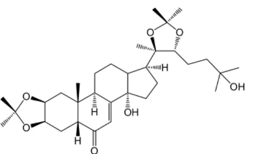
Fig. 1. The chemical structure of 20-hydroxyecdysone 2,3;20,22-diacetonide
O
O
H O
OH O
H OH H
O
20DA (Fig. 1) was prepared as published by Balázs et al. [13]. 20E was isolated previously from the plant Serratula wolffii Andrae [14].
Animals and treatment
Male Wistar rats weighing 200 ± 1.6 g were purchased from Toxicoop (Budapest, Hungary). The experiments conformed to 86/509/EEC regulation on the well-being of experimental animals. The experimental protocol has the permission number: 1810/003/2004 ANTSZ, Budapest.
Animals' treatment
Rats (three groups, n = 2, in each group) were (1) controls, treated with water, sacrificed after 15 min; (2) treated with 20E, sacrificed after 15 min;
and (3) treated with 20DA, sacrificed after 15 min.
Treatment was done by an i.p. injection of 0.2 mL of 250 mg mL−1 of either 20DA or 20E, freshly dissolved in double-distilled water. This way, the resulting dose was 50 mg 20E or 20DA per kilogram of the animal body weight. Following the 15 min of treatment, diethylether-anesthetized rats were exsanguinated through the canthus and cerebrospinal fluid (CSF) was withdrawn by cisternal punction. Brains and liver were dissected and placed on a 0 °C aluminum surface. The tissue samples were stored at
−80 °C until analysis.
Preparation of biological samples for analysis
Dissected brain and liver tissues from both untreated or treated animals were homogenized in a 4× w/v amount of acetonitrile using an Ultra Turrax T25 (Janke & Kunkel homogenizer IKA Labortechnik, Staufen, Germany) at 20,000 rpm for 30 s at room temperature. The homogenates were centri- fuged in an Eppendorf centrifuge (A. Hettich, Tuttlingen, Germany) with 14,000 rpm for 20 min at 4 °C. The supernatants were collected for analysis.
Serum and CSF samples (200 μL) were mixed with 800 μL methanol and vortexed for 1 min, followed by centrifugation at 13,500 rpm for 5 min.
The 100-μL aliquots of supernatant were diluted with 100 μL water and submitted for quantitative analysis as such (20DA treatment) or evaporated under N2 stream and redissolved in 20% aqueous acetonitrile prior to testing (20E treatment).
Quantitative determination by high-performance liquid chromatograph connected to diode-array detector (HPLC–DAD)
A Jasco HPLC–DAD gradient system (Jasco Co., Tokyo, Japan, purchased from ABL&E Jasco, Budapest, Hungary) was utilized with a Kinetex XB-C18 (5 μm, 4.6 × 250 mm; Phenomenex, Torrance, CA, USA) analytical column and isocratic solvent systems of 20% (20E) or 80% aqueous acetonitrile (20DA) at a flow rate of 1 mL min−1. 20E and 20DA were dissolved in 20 and 80% aqueous acetonitrile, respectively, at concentrations of 1.00 mg mL−1.
Fig. 2. An eight-point calibration curve for the determination of 20-hydroxyecdysone 2,3;20,22-diacetonide
Fig. 3. An eight-point calibration curve for the determination of 20-hydroxyecdysone
A 100-fold dilution of each (10 μg mL−1) with a respective solvent was utilized as stock solution, which served as a basis for further dilutions. An eight-point calibration was performed for 20DA from standard solutions of 0.5, 0.67, 1.0, 1.43, 2.0, 5.0, 10.0 and 20.0 μg mL−1 (Fig. 2). For 20E, an eight- point calibration was performed from standard solutions representing 0.5, 0.67, 0.83, 1.0, 1.43, 2.0, 5.0, and 10.0 μg mL−1 concentrations (Fig. 3). In each case, 10-μL volumes were injected in triplicates, and manual integration was applied at λ = 254 nm.
Results and Discussion
The results of the HPLC analysis of 20DA and 20E are shown in Figs. 4–6.
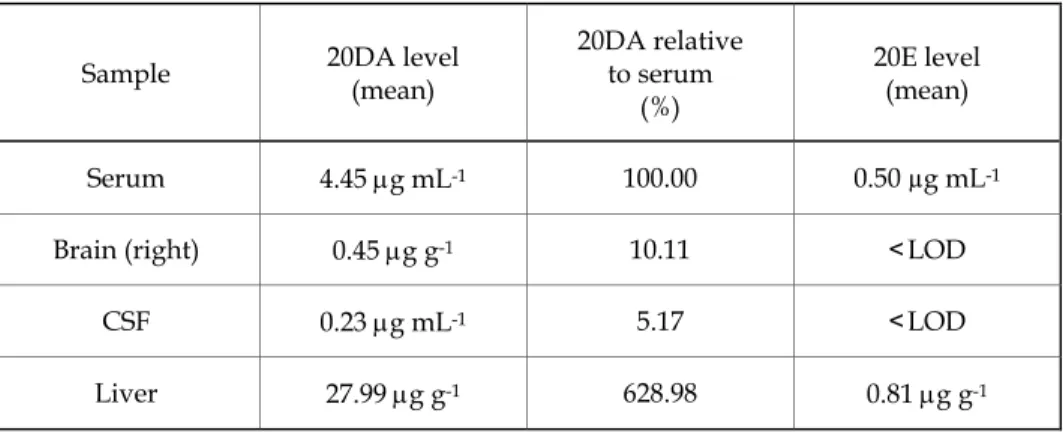
The HPLC analysis of 20DA shows an almost 5 μg mL−1 level of this compound in the serum of rats 15 min after the i.p. administration (Table I).
The serum level corresponds to almost 40% 20DA getting into circulation, while 10% of the circulating 20DA level penetrated into the brain.
Elimination of 20DA apparently takes place mainly through the liver, where its level is approximately seven times higher than in the serum. The HPLC analysis of 20E in the circulation revealed its levels by one magnitude order lower than those of 20DA, and the brain level of 20E was under the limit of detection (<LOD).
20DA shows much higher lipophilicity than either polypodine B or 20E (not shown here). Its consequence is a much higher absorption and distribution in rats.
Table I. 20DA and 20E levels in various organs and body fluids of rats following 15 min of i.p. injection
Sample 20DA level
(mean)
20DA relative to serum
(%)
20E level (mean)
Serum 4.45 μg mL-1 100.00 0.50 µg mL-1
Brain (right) 0.45 μg g-1 10.11 <LOD
CSF 0.23 μg mL-1 5.17 <LOD
Liver 27.99 μg g-1 628.98 0.81 μg g-1
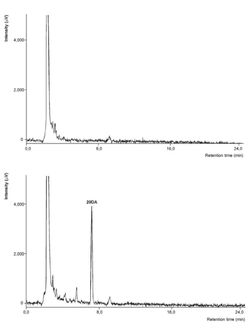
Fig. 4. HPLC chromatograms of plasma samples for the analysis of 20-hydroxyecdysone 2,3;20,22-diacetonide on a Kinetex XB-C18 (4.6 × 250 mm, 5 μm) column.
Top chromatogram plasma from untreated animals, bottom chromatogram:
plasma from animals treated with 20DA
Fig. 5. The HPLC–DAD spectrochromatograms of liver samples for the analysis of 20-hydroxyecdysone 2,3;20,22-diacetonide on a Kinetex XB-C18 (4.6 × 250 mm, 5 μm)
column. Top spectrochromatogram: liver sample from untreated animals; bottom spectrochromatogram: liver sample from animals treated with 20DA
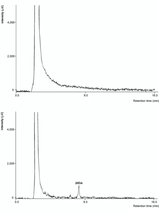
Fig. 6. HPLC chromatograms of brain samples for the analysis of 20-hydroxyecdysone 2,3;20,22-diacetonide on a Kinetex XB-C18 (4.6 × 250 mm, 5 μm) column.
Top chromatogram: brain sample from untreated animals; bottom chromatogram: brain sample from animals treated with 20DA
Acknowledgments
The Hungarian National Scientific Granting Agency (with OTKA 100155) and the Kalász Education and Research Co. (Budapest, Hungary) sponsored this project. A.H. acknowledges support from the János Bolyai Fellowships of the Hungarian Academy of Sciences. Advice and help of Dr. Mária Báthori, Mr. János Horváth, and Ms. Bogi Szalacsi as well as technical assistance from Ms. Györgyike Guth and Ms. Ibolya Hevérné-Herke are appreciated.
References
[1] A. Butenandt and P. Karlson, Z. Naturforsch., 9b, 389 (1954) [2] M. Báthori, Mini Rev. Med. Chem., 2, 285 (2002)
[3] L. Dinan and R. Lafont, J. Endocrinol., 191, 1 (2006) [4] L. Dinan, Arch. Insect Biochem. Physiol., 72, 126 (2009)
[5] R. Lafont, J.P. Giraults, and U. Kerb, Biochem. Pharmacol., 37, 1174 (1988) [6] R. Lafont and L. Dinan, J. Insect Sci., 3, 7 (2003)
[7] I.O. Ishola, C.O. Ochieng, S.O. Olayemi, M.O. Jimoh, and S.M. Lawal, Pharmacol.
Biochem. Behav., 127, 90 (2014)
[8] A. Palandri, D. L'hôte, J. Cohen-Tannoudji, H. Tricoire, and V. Monnier, Hum. Mol.
Genet., 24, 2615 (2015)
[9] H. Kalász, M. Báthori, and T. Csermely, Am. Lab., 32, 28 (2000)
[10] A. Martins, N. Tóth, A. Ványolós, Z. Béni, I. Zupkó, J. Molnár, M. Báthori, and A.
Hunyadi, J. Med. Chem., 55, 5034 (2012)
[11] A. Martins, J. Csábi, A. Balázs, D. Kitka, L. Amaral, J. Molnár, A. Simon, G. Tóth, A.
Hunyadi, Molecules, 18, 15255 (2013)
[12] A. Martins, P. Sipos, K. Dér, J. Csábi, W. Miklos, W. Berger, A. Zalatnai, L. Amaral, J. Molnár, P. Szabó-Révész, and A. Hunyadi, Biomed Res. Int., ID 895360 (2015) [13] A. Balázs, A. Hunyadi, J. Csábi, N. Jedlinszki, A. Martins, A. Simon, and G. Tóth,
Magn. Reson. Chem., 51, 830 (2013)
[14] A. Hunyadi, A. Gergely, A. Simon, G. Tóth, G. Veress, and M. Báthori, J.
Chromatogr. Sci., 45, 76 (2007)