Pázmány Péter Catholic University
Faculty of Information Technology and Bionics
Bálint Péter Kerekes
COMBINED TWO-PHOTON IMAGING,
ELECTROPHYSIOLOGICAL AND ANATOMICAL
INVESTIGATION OF THE HUMAN NEOCORTEX IN VITRO
Theses of the Ph.D Dissertation Supervisor: István Ulbert
Budapest 2015
1 Introduction
Epilepsies are one of the most common neurological disorders in humans. According to the definition of the WHO, epilepsy is a chronic brain disorder, with different etiology, characterized by spontaneous recurrent seizures emerging from the excessive and pathologically hypersynchronous firing of a large amount of neurons.
Though a large variety of antiepileptic drugs are available nowadays, a significant number ofpatients are pharmacoresistant. In those cases, where the epileptic focus can be precisely localized epilepsy surgery is a possible solution for blocking seizures.
Healthy neocortical tissue is also routinely removed due to surgical technical reasons from patients with tumor but without epilepsy, when the pathological mass is localized in the subcortical areas. Comparing the morphology and activity of epileptic and non-epileptic human brain tissue offers an excellent possibility to investigate the normal and impaired neuronal mechanisms at the network, single cell and subcellular levels.
Spontaneous synchronous population activity (SPA) can be observed in vitro during extracellular electrophysiological recording of local field potentials (LFP) in epileptic human neocortical slice preparations in physiological bathing medium. These synchronous population bursts consist of rhythmically recurring extracellular LFP deflections associated with high frequency oscillations and an increased neuronal firing. Both glutamatergic excitatory and GABAergic inhibitory signaling is involved, pyramidal cells show either depolarizing or hyperpolarizing and even mixed responses during SPA (according to our unpublished observations).
Calcium imaging of neurons is widely used to monitor cellular activity in animal slice preparations however, we have only limited knowledge about Ca2+ concentration changes in human neurons. Calcium imaging of human neurons was investigated in cells differentiated from induced pluripotent stem cell lines, and in cultured neurons of the enteric nervous system. Furthermore, a recent study shows spontaneous Ca2+ elevations in human neocortical and hippocampal astrocytes, but nothing is known about the intracellular Ca2+ properties of neurons derived from native human tissue of the central nervous system.
While two-photon Ca2+ imaging technique has high spatial resolution (<1 µm), it can cover only a relatively small area of interest (<1x1 mm). On the other hand, multiple channel extracellular electrophysiology can cover large cortical areas (3-4 mm) at the expense of its low spatial resolution (100 µm). The activity of neurons restricted to one or two cortical layers (<1 mm) can be monitored with two-photon imaging, whereas multiple channel extracellular electrophysiology is needed to record the activity of neurons in the entire depth (3-4 mm) of the human neocortex. The temporal resolution of the two techniques is also different: electrophysiological changes reflecting neuronal activity are considerably faster (<1 ms) than changes in intracellular Ca2+ (usually more than 100 ms). Combining
these two methods has several advantages. First, it helps us to gain more information on the role of different neurons in the emergence of population activity. Recording with the aid of the linear multielectrode gives information about the fast electrophysiological properties of SPA, detected in all neocortical layers, whereas Ca2+ imaging reveals the activity of a relatively large group of neighboring neurons (tens of bulk loaded cells), and their contribution to the generation of SPA. In addition, two- photon microscopy can detect inactive neurons, which are unnoticed in extracellular electrophysiological recordings. Second, the simultaneous use of Ca2+ imaging and whole cell patch clamp recording helps us to correlate electrophysiological activity and Ca2+ signals in human neurons.
One can simultaneously observe and manipulate the membrane potential fluctuations of neurons with intracellular patch clamp recordings and relate to changes in their Ca2+ concentrations. Completing these measurements with the detection of extracellular activity we can relate electrophysiological and Ca2+
signals of neurons active during SPA. In addition to Ca2+ imaging, two-photon uncaging can be used to investigate neuronal input-output functions and postsynaptic signal integration. Cell filling and anatomical reconstruction at the light and electron microscopic level may add important morphological information about the subcellular, cellular and network properties of human neocortical neurons.
2 SPA and interictal activity
For patients with pharmacoresistant focal epilepsy, resective surgery provides a good treatment alternative. The possibility of examining the removed epileptogenic zone revolutionized epilepsy research, as it raised the opportunity of measuring the activity of single neurons in a physiological or quasi-physiological state. In the experiment described in the manuscript, this is important, because our aim was not just to record and analyze LFP changes caused by the cells, we also wanted to know the underlying mechanisms involved in SPA generation. For this purpose, it was examined how all the separate cells are responding to the activity that is recorded using a laminar microelectrode.
Distinct from these pathologic interictal events, spontaneously occurring synchronous population activity could be detected in vitro in brain slices obtained from resected human epileptic neocortices, subiculum, and hippocampus. The emergence of synchronized events in the neocortex is probably based on the complex interactions between and within the neural network’s inhibitory and excitatory components.
The work of Köhling et al. involved the investigation of human neocortical tissues resected during epilepsy surgery. They investigated the role of glutamatergic and GABAergic synaptic transmission, as well as the role of voltage gated calcium channels in the generation of the spontaneous activity they describe. In their work, the extracellular field potential gradient was measured in the cortical Layers II and V. However, Köhling et.al. measured this activity only in tissue obtained from epilepsy patients.
While some have argued that SPA is distinct from the pathologic interictal events occurring in epilepsy patients, it is still controversial whether SPA is epileptic, or whether it can be found in physiological conditions.
Our group’s preliminary results indicate that an activity similar to interictal spikes (as in Köhling et. al. 1998) is detectable in non-epileptic tissue (derived from deep brain tumor patients, non-epileptic part not infiltrated by the tumor.
In the present study, I want to further investigate the origins of this SPA. Measurements performed with an extracellular laminar multielectrode provide the desired spatial information on how the different cortical layers respond. However, this approach does not yield extensive information on single cell activity. Thus, it is difficult to address the question of cellular mechanisms, as it is not feasible to patch each of the cells to obtain cell specific information. Question addressed in the present study are:
How are the cells involved in the generation of SPA? What proportion of cells is active during SPA?
Which types of cells are active (neurons, interneurons, glial cells) during SPA? When are they most active (before/during/after the LFP transient)?
Since SPA can occur both in epileptic and in healthy tissues, I decided to investigate the differences in how the healthy and versus the pathological tissue generate a very similar activity. To be able to answer the questions stated above, our research group used 2-photon microscopy. In addition,
histological analysis of the tissue is included (cell labeling and staining, followed by light and electron microscopy and 3D reconstruction) to address the question of morphological differences between epileptic and non-epileptic tissue.
3 New scientific results
SPA can be detected with electrophysiological methods in cortical slices of epileptic patients, maintained in physiological medium in vitro. I wanted to gain additional spatial information about the network mechanisms involved in the SPA generation, so I needed a new methodology.
The aim was to develop a method combining multiple channel extracellular electrophysiology, simultaneous intracellular recording, and two-photon Ca2+ imaging and uncaging supplemented by fine scale morphological analysis.
Thesis I.: I have developed a method for the two-photon Ca2+ imaging of human neocortical tissue.
Here I report for the first time the two-photon Ca2+ imaging of human neocortical neurons derived from epileptic and non-epileptic brain tissue.
Neocortical slices prepared from postoperative tissue of epileptic and tumor patients were maintained in a dual perfusion chamber in physiological incubation medium. SPA was recorded with a 24 channel extracellular linear microelectrode covering all neocortical Layers.
After identifying the electrophysiologically active regions of the slice, bulk loading of neuronal and glial markers was applied on the tissue.
SPA related Ca2+ transients were detected in a large population of neighboring neurons with two-photon microscopy, simultaneously with extracellular SPA and intracellular whole cell patch clamp recordings.
The intracellularly recorded cells were filled for subsequent anatomy. The cells were reconstructed in three dimensions and examined with light- and transmission electron microscopy.
This complex method -combining high spatial resolution two-photon Ca2+ imaging techniques and high temporal resolution extra- and intracellular electrophysiology with cellular anatomy- is suitable to reveal subcellular, cellular and network properties of human neocortical neurons engaged in spontaneous population activity and may permit a deeper understanding of the structural and functional properties of the human neocortex. The methodological difficulties I faced during the experiments will also be described.
Thesis II.: I have successfully combined the extracellular recording system with the two- photon microscope system. This way I could compare the epileptic and non-epileptic human neocortical neurons Calcium responses during SPA.
I recorded the spontaneous network activity in epileptic and non-epileptic human tissue by simultaneous Ca2+ imaging and field-potential measurements
The local field potential (LFP) was recorded in 69 human neocortical slices from 17 patients (32 slices from 8 tumor patients, 17 slices from 5 epileptic patients, and 20 from 4 tumor associated epileptic patients). SPA was detected in 15 slices (9 slices from 4 tumor patients, 4 slices from 3 epileptic patients, and 2 slices from 1 tumor associated epileptic patient) by using the following procedure. The multielectrode was placed on the surface of the slice, perpendicular to the pial surface, allowing electrophysiological recording from all neocortical Layers. The slices were mapped to localize the areas generating SPA by recording every 300-400 µm from one end of the slice to the other end.
After mapping the neocortical slices with the laminar multielectrode, regions where SPA could be detected with LFPg recording were chosen for further two-photon Ca2+ imaging and intracellular patch clamp recordings. Bulk loading was performed on the sites where SPA had the largest LFPg amplitude, and additional extracellular local field potential (LFP) signals were recorded with a glass patch pipette filled with ACSF at the site of the bulk loaded cells. This way I could effectively record the SPA generation associated Ca2+ signals with two-photon imaging in human neuronal populations (Figure 1.). The multielectrode array measured neuronal activity in the entire width of the examined neocortical region, near at the site of the bulk loading.
I simultaneously recorded the LFP signal of SPAs and the Ca2+ signals of the loaded neurons.
In the slices with detectable SPA a frame scan was taken after bulk loading, then cells were selected for fast measurement and were measured using the multiple line scanning method.
Figure 1. A) Average of LFP from a whole measurement of a tumor patient. B) The connected cells Ca2+ responses, from a whole measurement of a tumor patient. There are two of the cells that had good responses in the whole experiment. The LFP is filtered (Bessel 30 Hz low pass), and the Ca2+ responses are smoothed (Gaussian average). Dashed orange line is the 2SD.
Because of the averaging the second cell seems here not to pass the 2SD limit, but by the reliably responding cell definition below, the cell was in this group.
C) Average of LFP from a whole measurement of an epileptic patient.D) The connected cells Ca2+ responses, from a whole measurement of an epileptic patient. There are 3 cells that had good responses in the whole experiment. The LFP is filtered (Bessel 30 Hz low pass), and the Ca2+ responses are smoothed (Gaussian average). Dashed orange line is the 2SD. Because of the averaging the third cell seems here not to pass the 2SD limit, but by the reliably responding cell definition below, the cell was in this group.
I simultaneously recorded the LFP signal of SPAs and the somatic Ca2+ signal of 31 neurons in 2 slices from tumor patients, and 55 neurons in 4 slices from epileptic patients.
A relative increase in Ca2+ signal from the baseline larger than 2x standard deviation (SD) of the baseline was taken as significant Ca2+ response. The neurons showing at least one significant Ca2+
response were taken as responding cells. Occasionally responding cells showed increased Ca2+ signal during <20% of the SPA events, non-reliably responding cells responded to 20-40% of the SPA events, whereas reliably responding cells showed Ca2+ responses to >40% of the SPA events. With this method, 22 silent cell (68%), 4 occasionally (13%), 1 non-reliably (3%) and 5 reliably responding (16%) cells were identified in the tumor tissue. The distribution of the responding cells was considerably different in epileptic tissue: 19 silent cells (35%), 20 occasionally (36%), 11 non-reliably (20%) and 5 reliably (9%) responding cells were found (Table 1).
Table 1.
Examination of cellular activity during SPA with two-photon Ca2+ imaging in epileptic and non-epileptic tissue.
Patient/Slice Number of SPA
Number of recorded cells
Number of silent cells (0%)
Number of occasionally responding cells (<20%)
Number of non-reliably responding cells (20-40%)
Number of reliably responding cells (>40%) Pt 1 (tumor) slice
1
8 13 7 2 1 3
Pt 2 (tumor) slice 1
15 18 14 2 0 2
Pt 4 (epileptic) slice 2
79 26 4 16 3 3
Pt 4 (epileptic) slice 3
13 15 5 3 6 1
Pt 5 (epileptic) slice 1
15 4 2 0 2 0
Pt 5 (epileptic) slice 2
12 10 8 1 0 1
Pt 8 (tumor associated epileptic) slice 4
7 23 11 6 3 3
Tumor 31 21 (68%) 4 (13%) 1 (3%) 5 (16%)
Epileptic 55 19 (35%) 20 (36%) 11 (20%) 5 (9%)
T asszociated E 23 11 (48%) 6 (26%) 3 (13%) 3 (13%)
Thesis III.: I have demonstrated the functional coupling of LFP, Calcium responses and intracellular activity in human neocortical interneurons and pyramidal cells during SPA.
Based on the Ca2+ responses of the cells within region of interest, I chose non-reliably or reliably responding neurons for further intracellular recording. Whole cell (n=7 neurons) or loose patch clamp (n=2 neurons) recordings were made to reveal the electrophysiological activity of the given cell. In these cases LFP, intracellular recordings and Ca2+ signals of the patched and the neighboring cells were simultaneously detected (Figure 2, 3). Based on the morphology revealed by the fluorescent dyes, electrophysiological recording was made from 3 pyramidal cells and 6 interneurons.
Figure 2. Left) Simultaneous recording of LFP, Ca2+ imaging, and IC from the same slice and time. * marks the SPA. Right the bulk loaded slice and the patched cell.
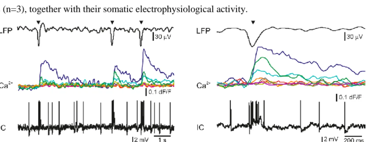
I examined the somatic and dendritic Ca2+ responses of both interneurons (n=4) and pyramidal cells (n=3), together with their somatic electrophysiological activity.
Figure 3. Left) Simultaneous LFP, Ca2+ signal (Ca2+) and loose patch clamp recording during three successive spontaneous SPA events (black triangles). Ca2+ transients show the responses of eight neurons from the eighteen recorded shown Figure 2.
Different colors of the Ca2+ signals represent different cells (middle traces). Note that three cells were responding to SPAs, but the other cells did not show increased Ca2+ levels. The intracellularly recorded cell (IC), shown in Figure 2 was burst firing during SPA, which is also reflected in a simultaneous increase in the intracellular Ca2+ level (green line). Note the trial-to-trial variability in relative Ca2+ responses between neurons.
Right) LFP signal of a spontaneous SPA event (black triangle) on an enlarged view with the corresponding Ca2+ responses recorded from the neuronal population shown in Figure 2. Bottom, simultaneously recorded loose-patch signal (IC). Note the large Ca2+ signal during the somatic AP burst associated to the SPA event (green line in the middle).
As it has been described in animal tissue positive correlation between the number of somatic action potentials and the amplitude of the dendritic Ca2+ signal was observed. Briefly, bursts of action potentials generated in pyramidal cells (n=2 cells) and multiple action potentials detected in interneurons (n=2 cells) resulted in larger dendritic Ca2+ increase than single action potentials. A detailed future study is needed to exactly correlate somatic electrophysiological recording with the somatic and dendritic Ca2+
signal of both human pyramidal cells and interneurons.
Measurement of input-output functions of cortical pyramidal cells and interneurons is important to understand dendritic integration and neuronal computation. As human neurons have more complex dendritic branching compared to animals, (see the dendritic length of the reconstructed pyramidal cell, a more complex human dendritic arithmetic were expected. Two-photon uncaging is widely used to investigate neuronal input-output functions and postsynaptic signal integration. As in animal models, I could use spatially and temporally clustered input pattern to activate short dendritic segments via glutamate uncaging and measured the postsynaptic Ca2+ response using free line scanning and somatic whole cell recording.
Thesis IV.: I successfully combined the electrophysiological and imaging measurements with anatomical reconstruction of the intracellularly loaded cells to gain more information of the morphology of the loaded cells.
Intracellularly recorded cells were filled with biocytin (n=6) and were processed for anatomy.
The successfully filled neurons showed the morphology of either pyramidal cells (n=2) or interneurons (n=2). The pyramidal cells displayed a long and thick apical dendrite and numerous thin basal dendrites (Figure 4), the interneurons appeared as small multipolar cells with shorter smooth dendrites. The whole dendritic and axonal arbor of one well filled neocortical Layer III. pyramidal cell was chosen to be reconstructed in three dimensions. Out of the 4 filled cells, this was the only neuron having an apparently complete (and well filled) dendritic arbor, as well as filled axons. The apical dendrite of the reconstructed cell was 4310 µm long, the sum of the length of its basal dendrites was 13478 µm and the length of all the axonal segments was 3875 µm long. It far exceeds the dendritic length of pyramidal cells in monkey temporal cortex, even though they were labelled in vivo. Pyramidal cells of the rodent neocortex also possess considerably shorter dendritic lengths (see www.neuromorpho.org,).
50 µm 1
2 3
4 5
6 7
50 µm 50 µm
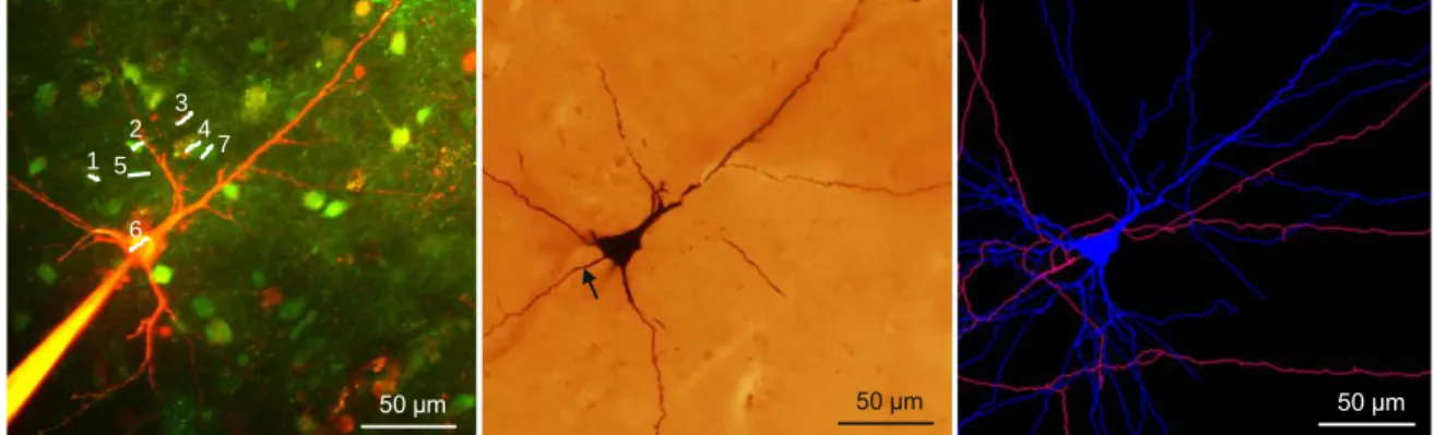
Figure 4. Left) Maximum intensity z-projection of a population of human neurons loaded with OGB-1-AM dye. The neuron corresponding to region #6 was whole-cell recorded and loaded through the recording pipette with the green Ca2+ dye OGB-1, the red Alexa594 and biocytin. Middle) Light micrograph of the cell #6 shown left, processed for anatomy. The axon initial segment is marked with arrow. Right) The dendritic (blue) and axonal (pink) arbor of the pyramidal cell #6 was reconstructed in three dimensions.
Thesis V.: I described the electron microscopic ultrastructure of the filled and reconstructed pyramidal cell at electron microscopic level.
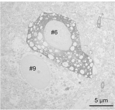
Large vacuoles were found in the cell body and the dendrites of the cell (Figure 5), while outside these areas mitochondria and other organelles such as endoplasmic reticulum seemed to be intact.
Numerous axon terminals forming either asymmetrical (presumably excitatory) or symmetrical (presumably inhibitory) synapses on the dendrites of the filled cell were found. I could not find synapses innervating the cell body of this pyramidal cell, but observed several symmetrical synapses terminating on its axon initial segment. The axon terminals of the filled cell formed asymmetrical synapses with non-stained dendrites and spines.
We hypothesized that the presence of vacuoles is the result of our methodological procedure.
1. First, applying OGB-AM and SR-101 for bulk loading may change the structure of the neurons.
2. Second, the long time (several hours) spent in the recording chamber might also affect the survival of the cells.
3. Third, patch clamp procedure (mechanical damage caused by the pipette, as well as the intracellular use of a high concentration of the fluorophores Alexa594 and OGB-AM) might also trigger changes in cellular ultrastructure.
To test these hypotheses further electron microscopic examinations were made. First, 62 non- filled cells were examined (45 neurons and 17 glial cells) in the vicinity of the biocytin-filled cell. Based on the low magnification frame scan taken during the two-photon experiment, these cells were located within the region of bulk loading. I could not see large vacuoles in any of the bulk loaded cells. Next, 61 cells were checked (43 neurons and 18 glial cells) in the same slice, in a region where bulk loading was not performed. Both blocks were re-embedded from neocortical Layer III. of Pt. 7, with a distance of ~5mm between them. None of the non-loaded cells displayed similar vacuoles in their somata. Further
experiments were made to test the hypothesis that several hours of in vitro conditions might induce the formation of somatic vacuoles. Therefore I re-embedded one block from Pt. 4, from a slice which spent 6 hours in the recording chamber and an other block from the same tissue sample (from the same part of the gyrus) which was fixed immediately after the cutting procedure. 35 neurons and 22 glial cells from the recorded tissue slice and 43 neurons and 25 glial cells from the immediately fixed tissue sample were examined. Large vacuoles were not observed in these cells.
Figure 5. The electron microscopic investigation of the same cell (Figure 18, 28 #6) showed large empty spaces (vacuoles) in the cell body. The neighboring neuron (identified on Figure 18 as #9) is a healthy pyramidal cell without large somatic vacuoles.
4 Publications
Papers
Combined two-photon imaging, electrophysiological, and anatomical investigation of the human neocortex in vitro.
Bálint Péter Kerekes, Kinga Tóth, Attila Kaszás, Balázs Chiovini, Zoltán Szadai, Gergely Szalay, Dénes Pálfi, Attila Bagó, Klaudia Spitzer, Balázs Rózsa, István Ulbert, Lucia Wittner
Neurophotonics 1:(1) pp. 111. (2014)
In vivo validation of the electronic depth control probes
Balázs Dombovári, Richárd Fiáth, Bálint Péter Kerekes, Emília Tóth, Lúcia Wittner, Domonkos Horváth, Karsten Seidl, Stanislav Herwik, Tom Torfs, Oliver Paul, Patrick Ruther, Herc Neves and István Ulbert*
Biomed Tech, 2013, DOI 10.1515/bmt-2012-0102
A novel multisite silicon probe for high quality laminar neural recordings
Grand L, Pongrácz A, Vázsonyi E, Márton G, Gubán D, Fiáth R, Kerekes B P, Karmos G, Ulbert I, Battistig G
Sensors and Actuators A: Physical 166:(1) pp. 1421. (2011)
Torfs T, Aarts A A A, Erismis M A, Aslam J, Yazicioglu R F, Seidl K, Herwik S, Ulbert I, Dombovari B, Fiáth R, Kerekes B P, Puers R, Paul O, Ruther P, Van Hoof C, Neves H P
Twodimensional multichannel neural probes with electronic depth control IEEE Transactions on Biomedical Circuits and Systems 5:(5) pp. 403412. (2011) Posters
Torfs T, Aarts A, Erismis M A, Aslam J, Yazicioglu R F, Puers R, Van Hoof C, Neves H, Ulbert I, Dombovari B, Fiath R, Kerekes B P, Seidl K, Herwik S, Ruther P, Two-dimensional multichannel neural probes with electronic depth control; 2010 IEEE Biomedical Circuits and Systems Conference, BioCAS 2010
Kerekes B P, Kaszás A, Tóth K, Chiovini B, Szalay G, Pálfi D, Spitzer K, Ulbert I, Lucia W, Rózsa B, Simultaneous Electrophysiology and Ca-imaging of human cortical synchronous population activity in vitro, IBRO Workshop 2012. 2012. Jan. 19-21, Szeged, Hungary
Bálint Péter Kerekes, Kinga Tóth, Attila Kaszás, Balázs Chiovini, Gergely Szalay, Zoltán Szadai, Dénes Pálfi, Klaudia Spitzer, Balázs Rózsa, István Ulbert, Lucia Wittner, Analysis of the human cortical spontaneous synchronous population activity in vitro based on multimodal experiments, 8th FENS Forum of European Neuroscience, 2012. Jul. 14-18., Barcelona, Spain
Kerekes B P, Kaszás A, Tóth K, Chiovini B, Szalay G, Pálfi D, Spitzer K, Ulbert I, Lucia W, Rózsa B, Multimodal analysis of the human cortical sinchronous population activity in vitro, Magyar Idegtudományi Társaság XIV. Konferenciája, 2013. Jan. 17-19., Budapest, Hungary
Bálint Péter Kerekes, Kinga Tóth, Attila Kaszás, Balázs Chiovini, Gergely Szalay, Zoltán Szadai, Dénes Pálfi, Klaudia Spitzer, Balázs Rózsa, István Ulbert, Lucia Wittner, Simultaneous Electrophysiology and Ca-imaging of human cortical population activity in vitro, Neuroscience 2013, Society for Neuroscience, 43nd Annual Meeting, 2013. Nov. 5-15., San Diego, USA
Kerekes B P, Kaszás A, Tóth K, Chiovini B, Szalay G, Pálfi D, Spitzer K, Ulbert I, Lucia W, Rózsa B, Simultaneous Electrophysiology and Ca-imaging of human cortical sinchronouspopulation activity in vitro, IBRO Workshop 2014. 2014. Jan. 16-17, Debrecen, Hungary
Kerekes B P, Kaszás A, Tóth K, Chiovini B, Szalay G, Pálfi D, Spitzer K, Ulbert I, Lucia W, Rózsa B, Simultaneous Electrophysiology and Ca-imaging of human cortical sinchronouspopulation activity in
vitro, From Medicine to Bionics 2nd European Ph.D. Conference 2014, 2014 május -10, Budapest, Hungary
Bálint Péter Kerekes, Kinga Tóth, Attila Kaszás, Balázs Chiovini, Gergely Szalay, Zoltán Szadai, Dénes Pálfi, Attila Bagó, Balázs Rózsa, István Ulbert, Lucia Wittner, Analysis of the human cortical spontaneous synchronous population activity in vitro based on multimodal experiments, 8th FENS Forum of European Neuroscience, 2014. Jul. 5-9., Milano, Italy
Bálint Péter Kerekes, Kinga Tóth, Attila Kaszás, Balázs Chiovini, Gergely Szalay, Zoltán Szadai, Dénes Pálfi, Attila Bagó, Balázs Rózsa, István Ulbert, Lucia Wittner, Spontán populációs aktivitás vizsgálata kombinált két foton és elektrofiziológiai módszerekkel, A Magyar Idegsebészeti Társaság 2014. évi Nemzeti Kongresszusa, 2014.nov.20-22 Budapest, Hungary
Kerekes B P, Kaszás A, Tóth K, Chiovini B, Szalay G, Pálfi D, Spitzer K, Ulbert I, Lucia W, Rózsa B, A method to analyze the human cortical spontaneous synchronous population activity in vitro with multimodal experiments, Magyar Idegtudományi Társaság XV. Konferenciája, 2015. Jan. 22-23., Budapest, Hungary
Bálint Péter Kerekes, Kinga Tóth, Attila Kaszás, Balázs Chiovini, Gergely Szalay, Zoltán Szadai, Dénes Pálfi, Attila Bagó, Balázs Rózsa, István Ulbert, Lucia Wittner Multimodal method and analysis of the in vitro human cortical spontaneous synchronous population activity, 31st International Epilepsy Congress, 2015 szeptember 5-9, Istanbul, Turkey
Presentations
Simultaneous Electrophysiology and Ca-imaging of human cortical synchronous population activity in vitro
Kerekes BP, Kaszás A, Tóth K, Chiovini B, Szalay G, Pálfi D, Spitzer K, Ulbert I, Lucia W, Rózsa B Neuronus 2014 IBRO & IRUN Neuroscience Forum, 2014.ápr.25-28 Krakkow, Lengyelország A method to analyze the human cortical spontaneous synchronous population activity in vitro
Kerekes BP, Kaszás A, Tóth K, Chiovini B, Szalay G, Pálfi D, Spitzer K, Ulbert I, Lucia W, Rózsa B Kálmán Erika Doktori Konferencia 2014, 2014. december 10-12. Budapest Magyarország
Else
Multimodal analysis of the human cortical spontaneous synchronous population activity in vitro Bálint Péter Kerekes
Pázmány Péter Catholic University PhD Proceedings 8: pp. 133136. (2013)
Towards combining cortical electrophysiology, fMR measurements and 2photon microscopy Bálint Péter Kerekes
Pázmány Péter Catholic University PhD Proceedings pp. 8588. (2012) Towards combining cortical electrophysiology and fMR measurements Bálint Péter Kerekes
Pázmány Péter Catholic University PhD Proceedings pp. 912. (2011)