Acta Acad. Paed. Agriensis, Sectio Biologiae XXV (2004) 9 9 - 1 2 6
Tree-dimensional Organization of Thylakoid System in Granal Chloroplasts and its
Functional Role
Mustárdy, L.
Institute of Plant Biology, Biological Research Center, Hungarian Academy of Sciences, H-6701 Szeged, Hungary
Abstract. In the chloroplasts of mesophyll cells of C3 and C4 plants the thylakoid membranes shows multiple helices of stroma lamellae around the cylindrical grana. Although this spatial relationship has been known for more than two decades, most textbooks and research papers continue to contain erroneous 3D models and simplified schemes. These reviews presents a detailed computer model, based on electron microscopic observations and explain the ontogenetic formation and functional importance of this unique structure.
Development of 3-dimenzionaI view
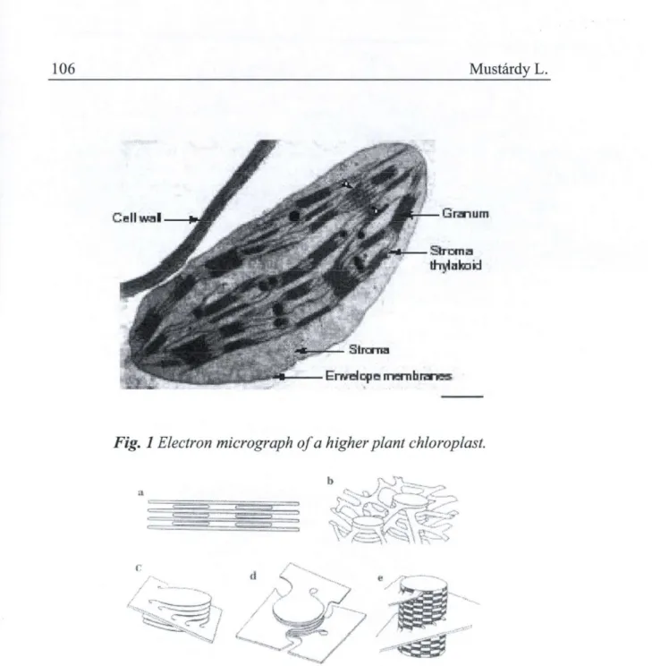
The electron microscopic investigations of thin section of chloroplast showed numerous flattened membrane sacs, called thylakoid. Thylakoid membranes are the internal membranes of chloroplasts, which accommodate all light-harvesting and energy-transducing functions. They are embedded in an aqueous matrix, called the stroma, which is surrounded by double envelope membrane (Fig 1). The thylakoids fell into two general size ranges.
The smaller sizes around 0.5 |im in diameter are appressed and form grana stacks. The larger sizes, called stroma thylakoids, are continuation of every second or third grana and interconnect the grana stacks. From this view was constructed the first simple model by Menke (1960). Weier et al., constructed a tubular model in 1961, which shows an anastomosing system of membranous channels. This concept was an artifact caused by KMn04
instead of glutaraldehyde fixation (Falk and Sitte, 1963).
The further detailed studies on the sections revealed, that in some cases the stroma thylakoid is not continued from one granum disc but interconnect
two of them. From this arrangement it was concluded, that the stroma thylakoids could intersect the granum at an angle. In the three-dimensional interpretation by Heslop-Harrison (1962) showed, that one stroma lamella interconnects all the grana compartments. The consequence of this arrangement was that all the intertylakoidal space (loculus or lumen) might be a continuum in the lamellar system. This idea was further persuade by Wehrmeyer (1964), who proposed that a single stroma lamella could exist in a spiral or helical arrangement around the granum being connected a few individual granum compartment. This general idea was elaborated by Paolillo (1970), who proposed that more than one stroma lamella could be helically attached and confirmed with the aid of a complete series of sections (Mustárdy and Brangeon 1978; Brangeon and Mustárdy 1979). The helical organization of the stroma lamellae around the grana can also be seen in other, more conventional (i.e. non-serial-section) electron micrographs. For instance, in thin sections the helices become evident when the section plane crosses the marginal region of the granum (Fig. 3a). In this relatively rare case, the thin section exposes the stroma membranes in their tilt positions and the granum stacks remain masked, or not included in the section.
The helical arrangement of the stroma thylakoids can also be seen on freeze etching replicas (Staehelin, 1986) (Fig. 3b) and it is most clear in scanning electron micrographs of isolated grana obtained by sonication and differential centrifugádon (Mustárdy and Jánossy, 1979) (Fig. 3c). This figure shows that a helical strip of the stroma membranes covers the granum 'pillar'.
Based on these electronmicroscopical observations a detailed computer model was constructed, showing the striking 3D structure with the stroma membrane helically wound around the granum (Mustárdy and Garab, 2003) (Fig. 4).
These helical granum-stroma assemblies are fused together as it can be seen on the serial section electron micrographs, i.e. the cylindrical granum of stacked membranes surrounded by multiple helices of the stroma thylakoids, are interconnected by fusion at the 'outer edges' of stroma membranes (Fig.
5). Evidently, this can be extended to the entire chloroplast, which lends a substantial stability to the thylakoid membrane system. It is shown by sequential thin sections that - as a consequence of the helical organization - the stroma membranes originating from one level of the multilamellar granum run to different levels on the 'opposite side' (i.e. on the interconnected granum) (Fig. 5a-c). The electron micrographs also show that the width of the pillars, i.e.
the relative lengths of the granum membranes with respect to the stroma thylakoids, can vary inside a single granum. This probably reflects local variations in the composition during the build-up of the granum structure.
Clearly, transient de-stacking of membranes in the marginal sections can also
Three Dimensional Organisation of Thylakoid System. 101 induce such variations, and thus the membrane system must therefore be considered to exhibit substantial structural flexibility.
Functions based on ultrastructure
More than 30 years ago Boardman and Anderson (1964) have revealed that the differentiation of thylakoids into granum and stroma membrane regions reflects a compositional and functional difference. The two photosystems are spatially separated: photosystem II (PSII) and its main chlorophyll a/b light-harvesting complex, LHCII, are found predominantly in the stacked membranes; this region is largely deficient in photosystem I (PSI) and LHCI, which are enriched in the stroma membranes (Andersson and Anderson, 1980). Separation of the two pigment systems is probably important in preventing unregulated excitation energy flow between the two photosystems (Anderson and Andersson, 1988; Trissl and Wilhelm 1993).
Without this, PSI, which is much faster than PSII, would disturb the balance of the energy distribution between the two photosystems. It is known that LHCII has large self-aggregation ability. Indeed, LHCII has been shown to stabilize the granum ultrastructure, and to participate in the cation-mediated stacking of the membranes (Arntzen, 1978; Duniec et al., 1981; Barber, 1982). The abundance of LHCII in the granum suggests that these antenna complexes also play a structural role to separate the two photosystems.
These light-harvesting complexes have also been shown to be involved, via electrostatic and osmotic forces, in the lateral organization of the membranes (Garab et al. 1991). Separation of the two photosystems (the LHCII-PSII and LHCI-PSI supercomplexes) between stacked and unstacked regions evidently requires a lateral force, such as that which governs the formation of LHCII-containing macrodomains (Garab and Mustárdy, 1999). The fact that in all LHCII-containing chloroplasts the two photosystems are segregated between the granum and stroma membranes suggests that LHCII may be involved not only in stacking but also in the lateral separation of PSII and PSI.
The helical arrangement of the stroma thylakoids around the cylindrical granum stacks has several functions. The continuity of the membrane is obviously warranted within the same granum-stroma assembly. The fusion of two (or more) stroma membranes at the edges ensures the continuity between the two types of membrane for the entire chloroplast. The continuum of the membrane system is evidently important for the diffusion of mobile components between the two regions (like plastocyanin). The 3D organization of the membrane may have special significance in the PSII repair cycle, during which the damaged particles are transferred from the granum to the stroma membranes where they are repaired and reassembled and then transferred back to the stacked region (cf. Melis 1991; Russel et al.
1995). It is tempting to speculate that the repair, which is a complex process, can be performed only at certain sites where both the key enzymes and the ribosomes are available, e.g. on the 'top' of the stroma membranes which is fully exposed to the stroma liquid. The spiral organization of these membranes and the multiple connections to the granum can facilitate the diffusion of the components from these special sites to any target. A similar mechanism may apply virtually to all components of the granum membranes, which are inserted in the stroma membranes and then 'slide' to the stacked region.
At the same time the openings on the 'sacs' ensure ionic conductance in a lateral direction between the inner aqueous phases of granum and stroma thylakoids. Further, stroma thylakoids by spiralling up and around the stacks ensure that all inner compartments of a granum are electrically connected.
Finally, fusions of adjacent stroma thylakoids wound around neighboring stacks interconnect adjacent grana (cf. Mustárdy 1996). By this means the inner aqueous phases of all thylakoids in the chloroplast become electrically connected to each other. The electrochemical gradients are rapidly 'delocalised' for the entire thylakoid system, and thus the transmembrane pH and electric potential gradients become essentially uniform (Junge 1977;
Mitchell 1977).
Ontogenetic assembly of thylakoids
The spatial relationship of grana and stroma thylakoids during chloroplast differentiation can be most successfully studied by serial section analysis at different levels of ontogenesis. This approach was taken with Lolium multiforum seedlings (Mustárdy and Brangeon, 1978; Brangeon and Mustárdy, 1979) in which four developmental stages of leaves were selected to follow the ontogenetic assembly of thylakoids by analysis and reconstitution of serial sections of developing chloroplasts.
In meristematic tissues the intrachloroplastic lamellae show a few pro- thylakoids of varying length distributed apparently at random within the proplastids (Fig. 6a). However, views of reconstituted serial sections reveal a single lamellar entity rather than separate and independent units. Thylakoids can be traced to a „parent entity" which has apparently split and given rise to several branches of different length. In some cases, the pro-thylakoid form is flattened and funlike, although apparently flexible and capable of changing orientation (Fig. 6b). In crossing this lamellar sheet, sharp cross-sectional views deteriorate into fuzzy tangentional views. Some of the pro-thylakoids show a balloon-or barrel-like form with several marginal splits in the reconstituted serial sections (Fig. 6c). In cross section, these figures appear as roughly circular thylakoids with interruptions and/or as short separate lamellae, depending upon the section plane. A few rudimentary double
Three Dimensional Organisation of Thylakoid System. 103 stacks are present at this stage. Within one proplastid, bubble-like invaginations of the inner membrane of the envelope are evident (see inset Fig. 6a) but the large, flattened lamellae are never connected to the inner envelope. Although, the possibility that these vesicles elaborate into thylakoid sheets was proposed by several authors (Hodge et. al., 1956;
Mühlethaler and Frey-Wyssling, 1959; Bradbeer et. al., 1974), it has still not been proven.
The developing chloroplasts increase in size and become flattened. In the newly formed thylakoids most of the chlorophyll is present in the reaction centers. The light-harvesting complex, which facilitates stacking of membranes (Mullet and Arntzen, 1980), becomes associated only later with the reaction centers.
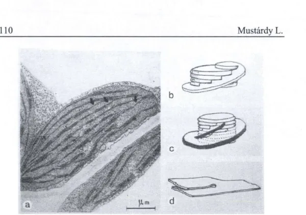
In this stage of chloroplast development the newly synthesized thylakoids are disposed parallel to one another and to the equatorial plane of chloroplasts (Fig. 7a). In the partial serial section the aligned thylakoids are revealed as individual broad sheets often traversing the width of the plastid (Fig. 7b). However, when the sections cross in a right angle, lamellar connections are encountered at the plastid ends (Fig. 7a arrow-heads).
During this differentiation step there is an occurrence of multiple perforations dispersed throughout the lamellar sheets. This phenomenon has been sited in a variety of membrane systems, particularly in a rapidly expanding membrane (Dalton and Hagueneau, 1968). The interruptions give a beaded appearance to the thylakoids in the cross-section (Fig. 7a inset) but in the reconstituted view these are revealed as slits or tears (Fig. 7c). In this developmental stage several double or triple stacks can be observed which overlap on the top of and beneath of the lamellar sheet (Fig. 7a, arrows). The three-dimensional reconstitutions show discs of varying diameter in the course of expansion and reveal an overgrowing phenomenon (simultaneous grow and apress). These reconstitutions sometimes disclose remnants of slits alongside the overlaps which suggests that the perforations may be the sites of grana initiation. It is very likely that the edges of the lamellae are the growth points, while at the periphery of the holes, the overlapping growth pattern above and below the single lamellar sheet gives rise to stacking.
Very probable, that the appearance of light-harvesting complex at the growth points associated with PSII by its high self-aggregation capability initiates the granum formation. The lateral-aggregation of PSII particles via adhesion of peripheral light-harvesting complexes can lead to the formation of nearly homogenous region which are enriched in PSII and exclude PSI to a large extent. These so-called „macrodomains" through their head-to-head aggregations ensure the granal stacking (Garab et .al., 1991; Barzda et. al.,
1994) and cause the separation of the two photosystems.
The parent lamellar sheet, interrupted in the vicinity of each nascent granum, constitutes a fret attachment at different levels to the stack. This arrangement maintains a continuity and consequently connects all grana that have been initiated on the same sheet. The „insertion" of the stacked discs into a lamellar sheet introduces a twist in the lamellae surrounding the developing granum, an orientation that would be the primary helix around the granum. Three-dimensional reconstitution of developing grana often reveal incomplete disc at the end of the stacks that have only a single fret connection. This shows that the granum can be increased by „overtopping"
of the overgrown fret (Fig. 8b).
As the number of granum compartments increases, the fretwork is built into multiple layers. There are two types of configurations contributing to the fretwork increase. Firstly, the primary helix divide and directly give rise to a secondary helix (Fig. 8c). Secondly, numerous thylakoid „splittings" can be reconstituted during this stage (Fig. 8d). After ramifications the cleaved thylakoids extend and link into the existing helical system of neighboring granum or give an additional helix to the stack. This can happen not only in the same lamellar sheet but the growing thylakoid can also extend towards the lower (or upper) plane (Fig. 8a, arrows). From this developmental stage the parallel „parent" lamellae have been replaced by a network which interconnects at different levels.
These multiple mechanisms - lamellar expansion, perforation, overgrowth; thylakoid splitting, branching, and bridging via fusion - guide the morphogenesis of the intrachloroplastic network formation and lead to the mature chloroplast.
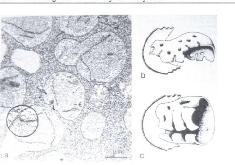
Obsolete models still in use
The first model, proposed by Wilhelm Menke still used in some books and homepages, postulates that every second granum thylakoid is continued in sheets that intersect several grana (Fig. 2a). This oversimplified scheme does not satisfy the requirement of membrane continuity, and is inconsistent with the single intrathylakoidal (lumenal) space. This model was modified by Lexa and Crang (1997) (Virtual Cell
http:/www.life.uiuc.edu/plantbio/cell/).
In a more frequently used, dangerously attractive model the grana are interconnected by tubes, which form a fret (Fig. 2b). This model is based on electron micrographs that suffer from a serious fixation artifact. Although tubular frets would in principle be consistent with most functions, they simply do not exist. Hence, this model should not be used, even for purposes of illustration, it is still found in many textbooks.
The folded membrane model (Fig. 2c) is most commonly used in research papers as a close-up view of the granum-stroma connection. This was
Three Dimensional Organisation of Thylakoid System. 105 originally proposed for the illustration of the lateral heterogeneity. In this model, the stacking is thought to originate from invaginations. According to the model, stroma thylakoids do not directly continue the granum thylakoids.
Instead, they form a 'fork': their 'upper' and 'lower' membranes are considered to originate from a layer 'above' and a layer 'below' the stacked pair of membranes. Such an arrangement, i.e. the interconnection of two granum compartments by a stroma thylakoid, can in fact, albeit rarely, be seen in electron micrographs. The reason for the appearance of 'forks' will be clear from the model based on serial sections. The main problem with this model is that, as ultrastructural studies on developing chloroplasts have revealed, the initiation of grana can be accounted for by the formation of multiple perforations, and by the overlapping of thylakoids at these slits, rather than invagination. It is still the model that is most frequently used in reviews and research papers; also available in a more elaborated form as a 3D computer model (Arvidsson and Sundby 1999).
Acknowledgement
This work was supported by research grant OTKA T042696.
Fig. 1 Electron micrograph of a higher plant chloroplast.
Fig. 2 Development of models on higher plant chloroplast lamellar arrangement. (Redrawn after different authors) (a) First model proposed by
Menke (I960) showing every second granum lamellae continues into the stroma and extends though several grana; (b) Cylindrical grana are interconnected by an anastomosing system of lamellar channels (Weier et
al., 1963); (c) Stroma lamella intersect the granum compartments at an angle connecting them together (Heslop-Harrison, 1963); (d) Single stroma
lamella interconnect the granum compartments in a helical manner (Wehrmeyer, 1964); (e) Continuous, large helices of stroma lamellae around
the granum (Paolillo, 1970).
Three Dimensional Organisation of Thylakoid System. 107
Fig. 3 Evidences of helical arrangement of stroma membranes around the granum by different electron microscopic techniques. (A) Part of a 'conventional' thin section of a chloroplast. On the right hand side, the micrograph shows a granum-stroma assembly with a near-median section, i.e. cut close to the margin; hence, virtually only the stroma thylakoids are to
be seen, tilted at an angle of approx. 20° with respect to the main axis of the granum. (B) On freeze-fractured chloroplast when the fractured plane is
parallel with the granum thylakoid (G), stroma thylakoidjunctions (arrowheads) are arranged in a circular fashion that surrounds the granum
membrane. (C) Scanning electron micrograph of an isolated granum; after sonication, the stroma thylakoids are broken down from the more rigid granum body; the narrow strips of remaining stroma lamellae exhibit a
helical arrangement, (bars 0.2 /urn.)
Fig. 4 'Computerizedform' of the 3D model constructed from electron micrographs offull serial sectioning of a granum-stroma assembly. In the model, some stroma thylakoids are removedfrom the granum stacks to show
the slits where the two types of thylakoids are connected.
Fig. 5 Thin sections from a full series (a-c), and different views of the 3D computer model of adjacent granum-stroma assemblies (d-e). It can be seen
that the stroma thylakoids 'continue' the granum lamellae. However, for their helical arrangement the stroma thylakoids in different sections do not always run to the same thylakoid of the granum (indicated by the arrows). It
is also clear (a-c) that there is a significant variation in the diameter of the stacks; this is not reflected in the schematic views in (d) and (e) which merely illustrate the fusion at the outer edges of the stroma membranes of
the two grana, i.e. of the two granum-stroma units, ( f ) The key structural element, the 'unit cell' of the mathematical model of the granum-stroma
assembly.
Three Dimensional Organisation of Thylakoid System. 109
Fig. 6 Lolium proplastids from meristematic tissue (a) and two reconstituted 3-dimensional views of lamellae (b and c). Circular inset shows invagination
of inner plastid envelope.
Fig. 7 Plastid profile from early stage of differentiation (a) showing parallel lamellar array with lamellar interruptions (inset-arrows) and juvenile grana
stacks (arrows). At the right end of the plastid lamellar connections are evident as „parent entity " (arrow-heads). In spatial view these lamellae are
independent through several sections (b) and the lamellar interruptions are revealed as slits or holes, (c) 3-dimensional reconstitution of a juvenile
granum.
Fig. 8 Plastid profde from young chloroplast (a) showing fret connections between parent lamellar sheets (arrows). Three-dimensional reconstitutions of developing granum showing new compartment formation via overtopping
(b), additional helix formation via splitting ofprimary helix (c) and thylakoid splitting on the lamellar sheet (d).
References
ANDERSON, J. M. & ANDERSSON, B. (1988): The dynamic photosynthetic membrane and regulation of solar energy conversion. Trends Biochem.Sci. 13, 351-355.
ANDERSSON, B. & ANDERSON, J. M. (1980): Lateral heterogeneity in the distribution of chlorophyll-protein complexes of the thylakoid membranes of spinach chloroplasts. Biochim.Biophys.Acta 593, 427-440.
ARNTZEN, C. J. (1978) Dynamic structural features of chloroplast lamellae. Current Topics in Bioenergetics 8, 111-160.
ARVIDSSON, P. O . & SUNDBY, C . ( 1 9 9 9 ) : A model for the topology of the chloroplast thylakoid membrane. Aust.J.Plant Physiol.2 6 , 6 8 7 - 6 9 4 .
BARBER, J. (1982): Influence of surface charges on thylakoid structure and function.
Annu.Rev.Plant Physiol. 33, 261-295.
BARZDA, V . , MUSTÁRDY, L. & GARAB. G. ( 1 9 9 4 ) : Size dependency of circular dichroism in macroaggregates of photosynthetic pigment-protein complexes.
Biochemistry3 3 , 1 0 8 3 7 - 1 0 8 4 1 .
BOARDMAN, N . K.& ANDERSON, J. M. (1964): Isolation from spinach chloroplasts of particles containing different proportions of chlorophyll a and chlorophyll
Three Dimensional Organisation of Thylakoid System. 111 b and their possible role in the light reactions of photosynthesis. Nature 206,
166-167.
BRADBEER, J. W . , IRELAND, H . M . M., SMITH, J. W., REST, J. & EDGE, H. J. W .
(1974): Plastid development in primary leaves of Phaseolus vulgaris. VII.
Development during growth in continuous darkness. New Phytol. 73, 263-270.
BRANGEON, J. & MUSTÁRDY, L. (1979): The ontogenetic assembly of intra- chloroplastic lamellae viewed in 3-dimension. Biol.Cellulaire 36, 71-80.
DALTON, A. J. & HAGUENEAU, F. (1968): In: The Memranes. Academic Press, N.Y., London.
DUNIEC, J. T. , ISRAELACHVILI, J. N . , NINHAM, B. W . , PASHLEY, R. M ., & THORNE,
S. W. (1981): An ion-exchange model for thylakoid stacking in chloroplasts.
F E B S Letters 129, 193-196.
FALK, R. H. & SLTTE, P. (1963): Zellfeinbau bei Plasmolyse. I. Der Feinbau der Elodea-Blattzellen. Protoplasma 57, 290-303.
GARAB, G. and MUSTÁRDY, L., Role of LHCII-containing macrodomains in the stucture function and dynamics of grana. Aust.J.Plant Physiol. 26, 649-658 (1999).
GARAB, G., KIELECZAWA, J., SUTHERLAND, J. C ., BUSTAMANTE, C. & HIND, G .
(1991): Organization of pigment-protein complexes into macrodomains in thylakoid membranes of wild-type and chlorophyll b-less mutant of barley as revealed by circular dichroism. Photochem Photobiol 54, 273-281.
HESLOP-HARRISON, J. (1963): Structure and morphogenesis of lamellar system in grana-containing chloroplasts. I. Membrane structure and lamellar architecture. Planta 60, 243-260.
HODGE, A. J., MCLEAN, J. D. & MERCER, F. V. ( 1956): A possible mechanism for the morphogenesis of lamellar system in plant cells. J. Biophys. Biochem. Cytol. 2,
5 9 7 - 6 0 8 .
JUNGE, W. (1977): Membrane potentials in photosynthesis. Annual Review of Plant Physiology 28, 503-536.
MELIS, A.( 1 9 9 1 ) : Dynamics of photosynthetic membrane composition and function.
Biochimica et Biophysica Acta 1058, 8 7 - 1 0 6 .
MENKE, W., Das allgemeine Bauprinzip des Lamellarsystems der Chloroplasten.
Experientia 16, 537-538 (1960).
MITCHELL, P., Vectorial chemiosmotic processes. Annual Review of Biochemistry 46, 996-1054(1977).
MULLET, J. E . & ARNTZEN, C . J., Stimulation of grana stacking in a model membrane system. Mediation by a purified light-harvesting pigment-protein complex írom chloroplasts. Biochim Biophys Acta 589, 100-117 (1980).
MUSTÁRDY, L., Development of thylakoid membrane stacking. In 'Advances in Photosynthesis, Vol. 4, Oxygenic Photosynthesis: The Light Reactions.' (Eds D. R. Ort and C.F. Yocum.) pp. 58-68. (Kluwer Academic Publisher, Dordrecht.) (1996).
MUSTÁRDY, L. & BRANGEON, J., 3-dimensional chloroplast infrastructure:
Developmental aspects. In 'Chloroplast Development'. (Ed G.
Akoyonoglou.) pp. 489-494. (Elsevier North-Holland Biomedical Press, Amsterdam.) (1978).
MUSTÁRDY, L. & GARAB, G. (2003): Granum revisited. A three-dimensional model - where things fall into place. Trends in Plant Science 8, 117- 122.
MUSTÁRDY, L. & JÁNOSSY, A . G . ( 1 9 7 9 ) : Evidence of helical thylakoid arrangement by scanning electron microscopy. Plant Sci.Lett. 16, 2 8 1 - 2 8 4 . MUHLETHALER, K. & FREY-WISSLING, A. (1959): Entwicklung und Struktur der
Proplastiden. J. Biophys. Biochem. Cytol. 6, 507-512.
PAOLILLO, D. J. (1970): The three-dimensional arrangement of intergranal lamellae in chloroplasts. Journal of Cell Science 6, 243-255.
RUSSEL, A . W . , CRITCHLEY, C., ROBINSON, S. A . , FRANKLIN, L . A . , SEATON, G . G . R., CHOW, W. S., ANDERSON, J. M., &OSMOND, C. B. (1995): Photosystem- II regulation and dynamics of the chloroplast D1 protein in Arabidopsis leaves during photosynthesis and photoinhibition. Plant Physiology 107, 943-952.
STAEHELIN, L. A. (1986): Chloroplast structure and supramolecular organization of photosynthetic membranes. In Photosynthesis III (Staehelin, L. A. and Arntzen, C.J. eds.) 1-72, Springer-Verlag .
TRISSL, H. W . & WILHELM, C . ( 1 9 9 3 ) : Why do thylakoid membranes from higher plants form grana stacks? Trends Biochem. Sci. 18, 4 1 5 - 4 1 9 .
WEHRMEYER, W . (1964): Zur Klárung structurellen Variability der Chloroplastengrana des Spinats in Profil und Aufsicht. Planta 62, 272-293.
WEIER, T. E., STOCKING, C. R., THOMSON, W. W. & D REVER, H. (1963): The grana as structural units in chloroplasts of mesophyll of Nicotiana rustica and Phaseolus vulgaris. J. Ultrastruc. Res. 8, 122-143.