doi: 10.3389/fpls.2020.00196
Edited by:
Julian Eaton-Rye, University of Otago, New Zealand Reviewed by:
Koichi Kobayashi, Osaka Prefecture University, Japan Maciej Z. Garstka, University of Warsaw, Poland
*Correspondence:
Katalin Solymosi katalin.solymosi@ttk.elte.hu;
solymosikata@hotmail.com
Specialty section:
This article was submitted to Plant Cell Biology, a section of the journal Frontiers in Plant Science
Received:24 August 2019 Accepted:11 February 2020 Published:03 March 2020
Citation:
Böszörményi A, Dobi A, Skribanek A, Pávai M and Solymosi K (2020) The Effect of Light on Plastid Differentiation, Chlorophyll Biosynthesis, and Essential Oil Composition in Rosemary (Rosmarinus officinalis) Leaves and Cotyledons.
Front. Plant Sci. 11:196.
doi: 10.3389/fpls.2020.00196
The Effect of Light on Plastid Differentiation, Chlorophyll
Biosynthesis, and Essential Oil Composition in Rosemary
(Rosmarinus officinalis) Leaves and Cotyledons
Andrea Böszörményi1, Adrienn Dobi2, Anna Skribanek3, Melinda Pávai2and Katalin Solymosi2*
1Department of Pharmacognosy, Semmelweis University, Budapest, Hungary,2Department of Plant Anatomy, ELTE Eötvös Loránd University, Budapest, Hungary,3Department of Biology, ELTE Savaria University Centre, Szombathely, Hungary
It is unclear whether light affects the structure and activity of exogenous secretory tissues like glandular hairs. Therefore, transmission electron microscopy was first used to study plastid differentiation in glandular hairs and leaves of light-grown rosemary (Rosmarinus officinalis “Arp”) plants kept for 2 weeks under ambient light conditions. During our detailed analyses, among others, we found leucoplasts with tubuloreticular membrane structures resembling prolamellar bodies in stalk cell plastids of peltate glandular hairs. To study the effect of darkness on plastid differentiation, we then dark-forced adult, light-grown rosemary plants for 2 weeks and observed occasionally the development of new shoots with elongated internodes and pale leaves on them. Absorption and fluorescence spectroscopic analyses of the chlorophyllous pigment contents, the native arrangement of the pigment-protein complexes and photosynthetic activity confirmed that the first and second pairs of leaf primordia of dark- forced shoots were partially etiolated (contained low amounts of protochlorophyll/ide and residual chlorophylls, had etio-chloroplasts with prolamellar bodies and low grana, and impaired photosynthesis). Darkness did not influence plastid structure in fifth leaves or secretory tissues (except for head cells of peltate glandular hairs in which rarely tubuloreticular membranes appeared). The mesophyll cells of cotyledons of 2- week-old dark-germinated rosemary seedlings contained etioplasts with highly regular prolamellar bodies similar to those in mesophyll etio-chloroplasts of leaves and clearly differing from tubuloreticular membranes of secretory cells. Analyses of the essential oil composition obtained after solid phase microextraction and gas chromatography-mass spectroscopy showed that in addition to light, the age of the studied organ (i.e., first leaf primordia and leaf tip vs. fifth, fully developed green leaves) and the type of the organ
(cotyledon vs. leaves) also strongly influenced the essential oil composition. Therefore, light conditions and developmental stage are both important factors to be considered in case of potential therapeutic, culinary or aromatic uses of rosemary leaves and their essential oils.
Keywords: dark-forcing, etiolation, leucoplast, peltate glandular hair, prolamellar body, rosemary, tubular complex
INTRODUCTION
Peculiar cubic phase membrane organizations, also termed tubular complexes or tubuloreticular inclusions have been observed in several animal, human or plant cells. In the former, these are thought to represent the modification of the (rough) endoplasmic reticulum, are located inside its cisternae or in the perinuclear space [e.g., Boor et al. (2018), reviewed in Luu et al. (1989), Almsherqi et al. (2006, 2009), Deng et al. (2010)] and are rich in acidic glycoproteins. In animal and human cells, the presence of tubuloreticular structures is thought to be associated with stressful or diseased conditions, however, other works suggest that they may be involved in regeneration processes of endothelial cells in wounded tissues (Eady and Odland, 1975) or are the result of altered cholesterol homeostasis induced by viral infection (Deng et al., 2010) but such structures have been also described in the aqueous lipid-protein film of lung surfactants (Larsson and Larsson, 2014). Their formation has been associated with special protein- protein interactions, overexpression of some membrane resident proteins, alterations in lipid (Almsherqi et al., 2009) or carotenoid (Park et al., 2002) composition and membrane symmetry (Larsson and Larsson, 2014), however, their exact cellular function is not known.
The formation of such cubic phase membrane structures has been also observed in semi-autonomous organelles such as mitochondria and plastids (Almsherqi et al., 2009). In the mitochondria of amoeba cells, cubic membrane formation was induced by starvation and/or autophagy processes, where they were slowing the degradation of the organelle and thus contributed to the survival of the cell upon stress conditions (Chong et al., 2017). The prolamellar bodies present in the etioplasts or etio-chloroplasts of dark-grown plants represent another well-known group of cubic phase membranes present in nature [reviewed in Solymosi and Schoefs (2010),Solymosi and Aronsson (2013), Kowalewska et al. (2019)]. Due to their very high surface-to-volume ratio, prolamellar bodies are thought to serve as a membrane reserve that can be quickly converted into a large photosynthetic membrane network upon light- dark transition of for instance seedlings germinating in the soil (Ferroni et al., 2009; Kakuszi et al., 2017) or leaf primordia of buds after bud break (Solymosi and Böddi, 2006; Solymosi et al., 2006, 2012). It has to be outlined, that prolamellar bodies represent one subtype of tubuloreticular membrane structures with highly regular spatial organization consisting of tetrahedral units of membrane tubules arranged in strictly geometric manner [reviewed in Solymosi and Schoefs (2010), Solymosi and Aronsson (2013)].
In addition to prolamellar bodies present in chlorenchyma tissues developing under light deprivation, plastids of light- grown and light-exposed tissues can also contain such peculiar membranes. Regularly arranged vesicle clusters present in chloroplasts may also form non-linear membrane organizations (Lindquist et al., 2016), and prolamellar body-like structures have been induced by UV irradiation of fruit plastids (Kovács and Keresztes, 2002), and several cells with secretory function have been reported to contain prolamellar body-like tubuloreticular membrane structures.
Such leucoplasts or secretory plastids with prolamellar body- like structures have been observed in extrafloral nectaries of Passiflora (Schnepf, 1961), and in plastids of glandular hairs of several genera belonging to various families [e.g., Cannabis– (Hammond and Mahlberg, 1978;Kim and Mahlberg, 1997; Solymosi and Köfalvi, 2017), Artemisia – (Ascensao and Pais, 1982), Chrysanthemum – (Vermeer and Peterson, 1979), Centrolobium– (Matos and Paiva, 2012), Platanthera– (Stpiczynska et al., 2005)] including Lamiaceae [e.g., Mentha piperita– (Amelunxen, 1965;Turner et al., 2000, 2012),Perilla ocymoides– (Kashina and Danilova, 1993)]. InM.piperitasuch structures were observed only in the stalk cells of the peltate glandular hairs (Amelunxen, 1965; Turner et al., 2000, 2012) and were observed as tubular-vesicular structures obtained only after osmium-tetroxide fixation (Amelunxen, 1965) or resembled rather plastoglobuli clusters both in conventionally chemically fixed and cryofixed material (Turner et al., 2000, 2012). In case of Nepeta species prolamellar body-like structures were described for the stalk cells of the peltate glandular hairs but were not shown on micrographs (Kolalite, 1998). In glandular hairs of P. ocymoides prolamellar body-like structures were reported when plants were transferred from continuous light to short-day illumination conditions, and were thus associated with processes occurring during the dark phase of growth (Kashina and Danilova, 1993). On the other hand, no cubic phase membrane structures were observed inLavandulaglandular hairs both when grown in light or in prolonged darkness (20 days) (Huang et al., 2008).
Our aim was to check whether such tubuloreticular structures were characteristic for the plastids of the peltate glandular hairs of other medicinally and economically important Lamiaceae species such as for example rosemary (Rosmarinus officinalis) (Borges et al., 2018;De Oliveira et al., 2019) and whether their presence or structure was influenced by darkness. This question is evenmore important because data about the structural and developmental plasticity of secretory plastids are rather scarce (Solymosi and Keresztes, 2012) and the role of light in the biosynthesis of the essential oil components also needs to be elucidated. This
latter may have industrial or practical relevance, because during winter in colder climates rosemary plants are often brought to warmer rooms with less light but higher temperatures, while both the leaves (Rosmarini folium) and the essential oils (Rosmarini aetheroleum) of rosemary are widely used as drugs or spices.
MATERIALS AND METHODS
Plant Material and Growth Conditions
Light-grown rosemary (R. officinalis “Arp”) plants with 20–40- cm-long shoots were purchased from the local market. Some plants were placed in complete darkness for 2 weeks at room temperature (20◦C) – a procedure to which we will refer later to as dark-forcing because it involves adult light-grown plants which were later placed into darkness for longer periods. Other plants were kept under ambient light conditions at room temperature.
Plants were watered with tap water every 4–5 days. Those being in the dark were watered using dim green safelight which was previously tested and did not induce photosynthesis or chlorophyll biosynthesis in plants. Plants purchased at the end of winter and also after the end of the long summer drought period and transferred to the dark often developed relatively large new shoots the leaves of which were used for further analyses. Rosemary has decussate leaf arrangement with a leaf pair developing at each node, but for simplicity of description later we will only refer to these pairs of leaf primordia of same developmental age and found on the same node as “first leaves”
(for the youngermost pair of leaf primordia located closest to the shoot tip, on the first node) or “fifth leaves” (in case of the leaf pair located on the fifth node downward from the shoot tip), etc. It should be noted, however, that dark-forcing of rosemary plants was in other cases often unsuccessful and did not yield new shoots.
For comparison, rosemary fruits (i.e., four-nutlets) were also purchased from commercial sources (Gála, Romero alecrim, and Kerting Réde). The fruits were germinated at room temperature on wet cotton in closed nylon bags for 2 weeks either under ambient light conditions or in complete darkness. Cotyledons of such seedlings were later used for further analyses.
Fluorescence Spectroscopy at 77 K
Youngermost and older leaves of light-grown as well as dark- forced shoots, and cotyledons of dark-grown or light-grown seedlings were separately sampled and cooled to 77 K in liquid nitrogen. To test the photoactivity of the protochlorophyll/ide pigments in the samples, the studied organs were first cooled to 77 K, measured, and then warmed up to approx. 253 K, illuminated with white light of 20µmol s−1 m−2 photon flux density and cooled to liquid nitrogen 10 s after illumination.
Low-temperature (77 K) fluorescence emission spectra were recorded using a Fluoromax-3 spectrofluorometer (Jobin Yvon- Horiba, France) with 440 nm excitation wavelength, 0.2 s integration time, 2 and 5 nm excitation and emission slits, respectively. Three spectra were recorded and automatically averaged for each sample. Emission signals were corrected for the wavelength-dependent sensitivity of the detection; when
necessary, baseline correction (to eliminate light scattering effects) and 3-point and 5-point linear smoothing were also performed using SPSERV V3.14 program (copyright: C.
Bagyinka, Institute of Biophysics, Biological Research Center of the Hungarian Academy of Science, Hungary). Samples were collected from at least 3 different plants, in different independent biological replicates, and representative spectra were chosen for presentation.
Determination of Chlorophyllous Pigment Contents
The fresh mass of selected leaves or cotyledons were measured.
Plant organs were then homogenized in mortar with pistil in 80% (v/v) acetone. Pigment contents of the acetonic extracts were measured using absorption spectroscopy or in some cases (e.g., samples with low pigment concentrations or simultaneous presence of protochlorophyll/ide and chlorophyllous pigments) with fluorescence spectroscopy using calibration curves.
Measurements of fresh mass and pigment extraction of dark- grown or dark-forced samples were carried out under dim green safelight. The absorption of acetonic pigment extracts was determined using a UV-2101 PC (Shimadzu Corp., Japan) spectrophotometer. Spectra were recorded between 400 and 800 nm, with 0.5 nm sampling interval, 1 nm optical slits and medium speed. Spectra were used for calculation of chlorophyll a and b contents using standard equations (Porra et al., 1989).
Protochlorophyll/ide quantification using calibration curve for fluorescence measurements was based on extinction coefficients as in Brouers and Michel-Wolwertz (1983), and calibration was prepared using acetonic extracts of protochlorophyll obtained from hull-less pumpkin (Cucurbita pepo subsp.pepo var.styriaca).
Measurement of Photosynthetic Activity
Photosynthetic activity of various leaves of light-grown and dark- forced shoots was studied by measuring variable chlorophyll fluorescence. Due to the size of the leaves and also in order to characterize eventual spatial heterogeneity in the parameters, fluorescence images were taken with an Imaging PAM-M series Chlorophyll Fluorometer (Heinz Walz GmbH, Germany). Dark- forced samples were positioned into the instrument and the camera focus was set under dim green safelight. Prior to the measurements light-grown samples were dark adapted for 15 min. The photosynthetic parameter Fv/Fm = (Fm-F0)/Fm (Björkman and Demmig, 1987) was calculated in at least four biological replicates as inSolymosi et al. (2007). Average values and standard deviations are provided. It is important to note that these parameters are based on optical properties of green leaves and are probably different in dark-forced tissues with lower chlorophyll content, therefore, these values should be regarded only as an estimation.
Transmission Electron Microscopy (TEM)
Leaves were cut into 1 mm × 2 mm or 1 mm × 1 mm pieces at the central leaf blade region (i.e., avoiding leaf base and leaf tip regions, but also the midrib) with sharp razor
blade in drops of the primary fixative, 2.5% glutaraldehyde in 70 mM Na2HPO4-KH2PO4buffer (pH = 7.2). Intact cotyledons were simply separated from the hypocotyl and put in the same primary fixative. After 3 h of fixation in glutaraldehyde, pieces of the plant material were rinsed three times for 15 min in the same phosphate buffer, and were then post-fixed for 1 h in 1% osmium tetroxide dissolved in the same buffer. After rinsing with the phosphate buffer for 15 min three times, samples were dehydrated in ethanol series, transferred to propylene- oxide and embedded in Durcupan ACM resin (Fluka Chemie AG, Switzerland). Ultrathin sections (70 nm) were prepared on a Reichert Jung Ultracut-E ultramicrotome (Reichert Jung AG, Austria) and were stained with uranyl acetate and Reynold’s lead citrate. Transmission electron microscopic (TEM) analyses were carried out with a Hitachi 7100 TEM (Hitachi, Japan) at 75 kV accelerating voltage and a JEOL JEM 1011 (JEOL Ltd., Japan) at 80 kV accelerating voltage. Digital images were taken using Olympus Morada CCD cameras (Olympus Optical Co.
Ltd., Japan). Fast Fourier transformation (FFT) on the selected region of interest of particular micrographs was performed using ImageJ software.
Solid Phase Microextraction (SPME) of the Essential Oil
Plant material (pooled samples of 0.5–1 g fresh mass of either several fully developed, i.e., mature leaves, or young leaves and shoot tips or cotyledons as indicated) was put into vials (20 mL headspace) sealed with a silicon/polytetrafluoroethylene septum prior to the SPME gas chromatography/mass spectrometry (SPME-GC/MS) analysis. Sample preparation using the static headspace solid phase microextraction (sHS-SPME) technique was carried out with a CTC Combi PAL (CTC Analytics AG, Switzerland) automatic multipurpose sampler using a 65 µM StableFlex polydimethyl siloxane/carboxene/divinyl benzene (CAR/PDMS/DVB) SPME fiber (Supelco, United States). After an incubation period of 5 min at 100◦C, extraction was performed by exposing the fiber to the headspace of a 20 mL vial containing the sample for 10 min at 100◦C. The fiber was then immediately transferred to the injector port of the GC/MS, and desorbed for 1 min at 250◦C. Injections were made in splitless mode. The SPME fiber was cleaned and conditioned in a Fiber Bakeout Station in pure nitrogen atmosphere at 250◦C for 15 min.
Gas Chromatography-Mass
Spectroscopy (GC/MS) Conditions
The analyses were carried out with an Agilent 6890N/5973N GC/MSD (Agilent, United States) system equipped with an Supelco (Sigma-Aldrich) SLB-5MS capillary column (30 m ×250µm×0.25µm). The GC oven temperature was programmed to increase from 60◦C (3 min isothermal) to 250◦C at 8◦C/min (1 min isothermal). High purity helium (6.0) was used as carrier gas at 1.0 mL/min (37 cm/s) in constant flow mode.
The mass selective detector (MSD) was equipped with a quadrupole mass analyzer and was operated in electron ionization mode at 70 eV in full scan mode (41–500 amu at 3.2 scan/s).
The data were evaluated using MSD ChemStation D.02.00.275 software (Agilent). The identification of the compounds was carried out by comparing retention data and recorded spectra with literature data, and the NIST 2.0 library was also consulted.
The percentage evaluation was carried out by area normalization.
Averaged data and their standard errors are from at least 3 independent biological replicates as indicated.
Statistical Analyses
The data in Tables 1, 2 represent the mean ± the standard error of the mean of the number of independent biological replicates obtained on pooled samples from different experiments as indicated. The datasets obtained in different samples and treatments were compared using the software GraphPad InStat (GraphPad Software Inc., United States) using 1-way ANOVA followed by Tukey-Kramer multiple comparisons test as posterior test where significant differences were found.
Significant differences are described in the text and designated with different letters in theTables 1,2atP<0.05.
RESULTS AND DISCUSSION
Ultrastructural Analyses of the Glandular Hairs of Light-Grown Rosemary Leaves
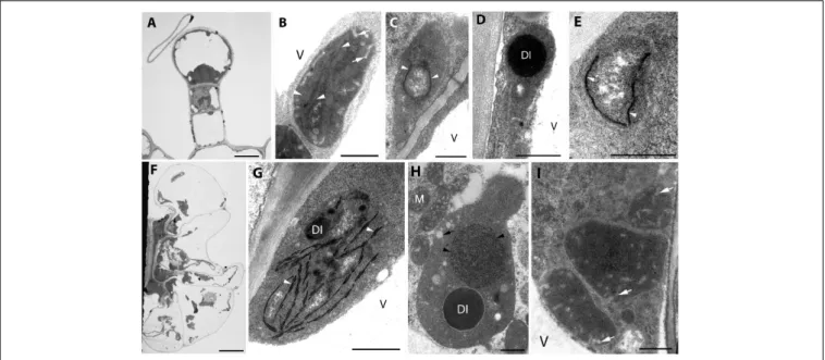
Fully differentiated capitate (Figure 1A) and peltate (Figure 1F) glandular hairs were present and studied both on mature and on young green leaves as reported earlier (Werker et al., 1985; Marin et al., 2006;Boix et al., 2011). Below we describe plastid ultrastructure in these secretory structures in rosemary.
Plastid stroma was in general very electron-dense, and the contrast between the cytoplasm and the plastid stroma was often low. Plastids of basal cells of capitate glandular hairs resembled typical epidermal chloroplasts, they contained few stacked thylakoids, i.e., grana with so-called “inverse contrast”
corresponding in fact to lightly staining membranes containing dense luminal substance (Keresztes and Sárvári, 2001), and several plastid invaginations indicating active material transport with the cytoplasm (Figure 1B). Stalk and head cell plastids were leucoplasts and had similar structure (Figures 1C–E) in capitate glandular hairs: they contained only few single thylakoid-like structures, and sometimes electron-dense dark inclusions.
The basal cells of the fully developed peltate glandular hairs had chloroplasts with electron-dense thylakoids, low grana and sometimes dark inclusions (Figure 1G). The plastids in the stalk cell contained in general one large electron-dense inclusions, and electrontransparent ovale bodies resembling plastoglobuli (Figure 1H). Often electron-dense tubuloreticular membranes in oval structures with average diameter of approx. 1.2µm were also present (Figure 1H). Plastids of the disc cells were leucoplasts with electron-dense stroma and several invaginations during the secretory phase (Figure 1I).
The observed plastid morphological features (dense inclusions, electron-dense stroma, etc., summarized in Supplementary Table S1) are consistent with literature data about the plastids of capitate trichomes [Lavandula– (Huang et al., 2008),Leonotis– (Ascensao and Pais, 1998),M.piperita–
TABLE1|Pigmentcontentsofdifferentorgansoflight-grownadultrosemary7.7pcplantsgrownfor2weeksincompletedarkness(“dark-forced”)orunderambientlightconditions(“light-grown”). Light-grown(n=3–6)Dark-forced(n=5–6) Chlorophylla(µg/g)Chlorophyllb(µg/g)Chlorophylla/bChlorophylla(µg/g)Chlorophyllb(µg/g)Chlorophylla/b Shoottipandfirstleafprimordia663.85±95.62a203.70±33.79a3.35±0.12a66.42±14.47b21.38±4.32b3.08±0.11ab Secondleafprimordia700.60±10.72a207.36±6.70a3.38±0.09a286.14±19.47c82.63±6.5c3.47±0.06a Fifthleaf728.09±88.87a243.25±29.49a2.99±0.04b623.14±46.19a211.48±13.70a2.95±0.07b Averagepigmentcontentsandtheirstandarderrorsareprovidedonafreshmassbasisforpooledsamplesandthenumberofparallelsasindicated.Datawerecalculatedfromabsorptionmeasurementsof80% acetonicextractsoftheleaves.Incaseofdark-forcedplantsthenewlydevelopedpaleshootswerestudied.Differentsuperscriptlettersindicatesignificantdifferenceinthechlorophyllaorchlorophyllborchlorophyll a/bratioofthedifferentorgans(shoottipandleafprimordia,secondleafprimordiaandfifthleaf)andtreatments(light-grownordark-forced)accordingto1-way-ANOVAfollowedbyTukey-Kramermultiplecomparison test(P<0.05).
(Amelunxen, 1965),Nepeta– (Kolalite, 1998),Stachys– (Giuliani et al., 2008)] and peltate trichomes [Lavandula – (Huang et al., 2008), Leonotis – (Ascensao et al., 1997), M. piperita – (Amelunxen, 1965; Turner et al., 2000, 2012), and Nepeta – (Kolalite, 1998)] of other light-grown Lamiaceae plants. It should be noted, however, that in case of stalk cells, the observed tubuloreticular membrane structure is different from that previously reported for Mentha (Amelunxen, 1965; Turner et al., 2000, 2012), in rosemary this structure is more clearly related to tubular complexes and bears no resemblance to clusters of plastoglobuli. The observed electron-dense inclusions present in plastids may be of any osmiophilic material (e.g., phenolic or terpenoid substances), however, the fact that their surface boundary is not completely smooth and that in our case (especially in stalk cells of peltate trichomes) they seem to be surrounded by a less electron-dense layer make it unlikely that they are “large plastoglobuli” (Turner et al., 2000) or “lipid droplets” in plastids (Huang et al., 2008).
The factors influencing the formation of these tubuloreticular membranes in stalks cells remain unknown. The basal cells of the trichomes (Figure 1G) and the epidermis cells located directly below the peltate glandular hairs (not shown) contained chloroplasts, which indicates that these cells were exposed to light and could thus produce chloroplasts. In this context, it is unlikely that only the stalk cells of the peltate glandular hairs were somewhat partially or fully shaded by the head cells and their secreted material, resulting in their etiolation and consequent formation of tubuloreticular membranes. Similarly, we had no information about the structure of “real” prolamellar bodies of rosemary, therefore, we decided to check how complete darkness influenced plastid differentiation in rosemary leaves.
The Effect of Prolonged Darkness (i.e., Dark-Forcing and Etiolation) on Plastid Differentiation in Rosemary Leaves
There are literature data describing plastid developmental differences and prolamellar body formation along decreasing light gradients being present in different cells of partly shaded leaf primordia in buds (Solymosi et al., 2012) or in sunflower cotyledons covered by the pericarp (Solymosi et al., 2007).
Therefore, it may be possible that the disc cells and their secretory products may shade the stalk cell plastids and thus influence their differentiation. Literature data about glandular hair development and plastid structure under prolonged darkness are scarce and somewhat conflicting. In case of P. ocymoides the presence of tubuloreticular inclusions was suggested to be associated with cultivation under short-day conditions and thus increased dark periods (Kashina and Danilova, 1993), while in case of Lavandula pinnata no tubuloreticular structures have been observed in axillary bud-derived explants cultivated for 20 daysin vitroon Murashige-Skoog medium both in the light (low light, i.e., 10–20 µmol photons s−1 m−2) or in complete darkness for 20 days (Huang et al., 2008) and light did not influence glandular hair development and structure (Huang et al., 2008). Therefore, the question can be raised, whether the formation of the tubuloreticular membranes in the plastids of
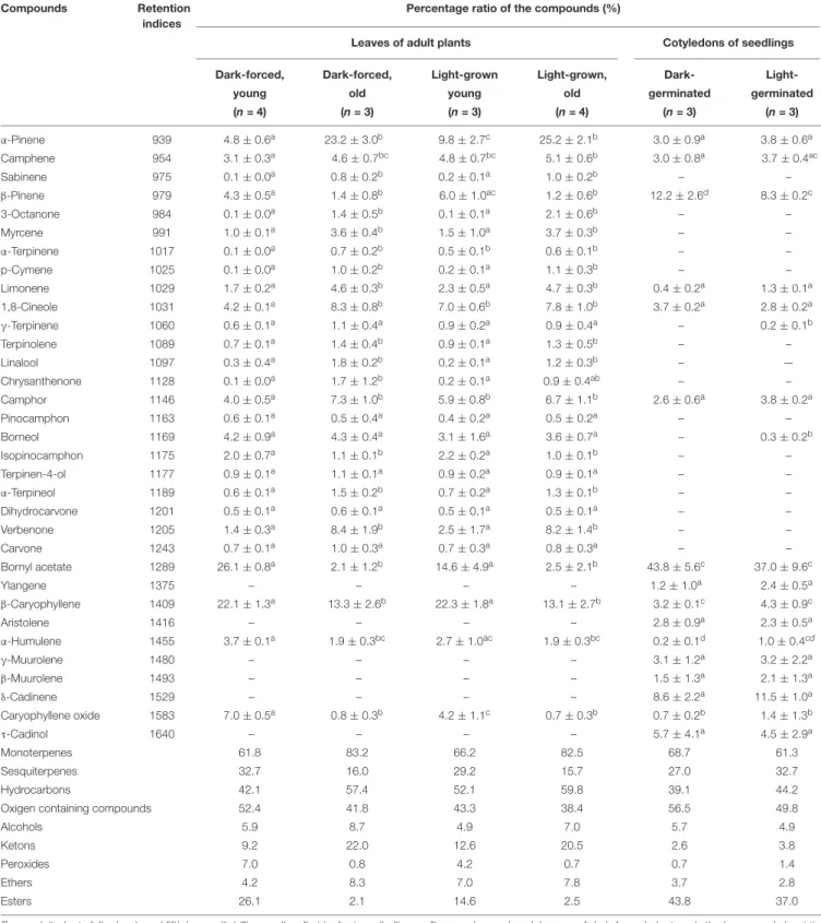
TABLE 2 |Percentage composition of the essential oils produced by the cotyledons of dark-germinated or light-germinated 2-week-old rosemary seedlings and of different leaves of adult rosemary plants kept for 2 weeks under ambient light conditions (“light-grown” plants) or in complete darkness (“dark-forced”) at room temperature.
Compounds Retention
indices
Percentage ratio of the compounds (%)
Leaves of adult plants Cotyledons of seedlings
Dark-forced, Dark-forced, Light-grown Light-grown, Dark- Light-
young old young old germinated germinated
(n= 4) (n= 3) (n= 3) (n= 4) (n= 3) (n= 3)
α-Pinene 939 4.8±0.6a 23.2±3.0b 9.8±2.7c 25.2±2.1b 3.0±0.9a 3.8±0.6a
Camphene 954 3.1±0.3a 4.6±0.7bc 4.8±0.7bc 5.1±0.6b 3.0±0.8a 3.7±0.4ac
Sabinene 975 0.1±0.0a 0.8±0.2b 0.2±0.1a 1.0±0.2b – –
β-Pinene 979 4.3±0.5a 1.4±0.8b 6.0±1.0ac 1.2±0.6b 12.2±2.6d 8.3±0.2c
3-Octanone 984 0.1±0.0a 1.4±0.5b 0.1±0.1a 2.1±0.6b – –
Myrcene 991 1.0±0.1a 3.6±0.4b 1.5±1.0a 3.7±0.3b – –
α-Terpinene 1017 0.1±0.0a 0.7±0.2b 0.5±0.1b 0.6±0.1b – –
p-Cymene 1025 0.1±0.0a 1.0±0.2b 0.2±0.1a 1.1±0.3b – –
Limonene 1029 1.7±0.2a 4.6±0.3b 2.3±0.5a 4.7±0.3b 0.4±0.2a 1.3±0.1a
1,8-Cineole 1031 4.2±0.1a 8.3±0.8b 7.0±0.6b 7.8±1.0b 3.7±0.2a 2.8±0.2a
γ-Terpinene 1060 0.6±0.1a 1.1±0.4a 0.9±0.2a 0.9±0.4a – 0.2±0.1b
Terpinolene 1089 0.7±0.1a 1.4±0.4b 0.9±0.1a 1.3±0.5b – –
Linalool 1097 0.3±0.4a 1.8±0.2b 0.2±0.1a 1.2±0.3b – —
Chrysanthenone 1128 0.1±0.0a 1.7±1.2b 0.2±0.1a 0.9±0.4ab – –
Camphor 1146 4.0±0.5a 7.3±1.0b 5.9±0.8b 6.7±1.1b 2.6±0.6a 3.8±0.2a
Pinocamphon 1163 0.6±0.1a 0.5±0.4a 0.4±0.2a 0.5±0.2a – –
Borneol 1169 4.2±0.9a 4.3±0.4a 3.1±1.6a 3.6±0.7a – 0.3±0.2b
Isopinocamphon 1175 2.0±0.7a 1.1±0.1b 2.2±0.2a 1.0±0.1b – –
Terpinen-4-ol 1177 0.9±0.1a 1.1±0.1a 0.9±0.2a 0.9±0.1a – –
α-Terpineol 1189 0.6±0.1a 1.5±0.2b 0.7±0.2a 1.3±0.1b – –
Dihydrocarvone 1201 0.5±0.1a 0.6±0.1a 0.5±0.1a 0.5±0.1a – –
Verbenone 1205 1.4±0.3a 8.4±1.9b 2.5±1.7a 8.2±1.4b – –
Carvone 1243 0.7±0.1a 1.0±0.3a 0.7±0.3a 0.8±0.3a – –
Bornyl acetate 1289 26.1±0.8a 2.1±1.2b 14.6±4.9a 2.5±2.1b 43.8±5.6c 37.0±9.6c
Ylangene 1375 – – – – 1.2±1.0a 2.4±0.5a
β-Caryophyllene 1409 22.1±1.3a 13.3±2.6b 22.3±1.8a 13.1±2.7b 3.2±0.1c 4.3±0.9c
Aristolene 1416 – – – – 2.8±0.9a 2.3±0.5a
α-Humulene 1455 3.7±0.1a 1.9±0.3bc 2.7±1.0ac 1.9±0.3bc 0.2±0.1d 1.0±0.4cd
γ-Muurolene 1480 – – – – 3.1±1.2a 3.2±2.2a
β-Muurolene 1493 – – – – 1.5±1.3a 2.1±1.3a
δ-Cadinene 1529 – – – – 8.6±2.2a 11.5±1.0a
Caryophyllene oxide 1583 7.0±0.5a 0.8±0.3b 4.2±1.1c 0.7±0.3b 0.7±0.2b 1.4±1.3b
τ-Cadinol 1640 – – – – 5.7±4.1a 4.5±2.9a
Monoterpenes 61.8 83.2 66.2 82.5 68.7 61.3
Sesquiterpenes 32.7 16.0 29.2 15.7 27.0 32.7
Hydrocarbons 42.1 57.4 52.1 59.8 39.1 44.2
Oxigen containing compounds 52.4 41.8 43.3 38.4 56.5 49.8
Alcohols 5.9 8.7 4.9 7.0 5.7 4.9
Ketons 9.2 22.0 12.6 20.5 2.6 3.8
Peroxides 7.0 0.8 4.2 0.7 0.7 1.4
Ethers 4.2 8.3 7.0 7.8 3.7 2.8
Esters 26.1 2.1 14.6 2.5 43.8 37.0
From adult plants fully developed fifth leaves (“old”) as well as first leaf primordia (“young”) were also analyzed. In case of dark-forced plants only the leaves and shoot tips of newly formed, pale shoots were studied. The experiments were carried out in three or four independent biological replicates as indicated, and for each measurement pooled samples (0.5–1 g plant material) were used. Standard errors of the mean are indicated in the Table. Different letters in each row indicate significant difference according to 1-way-ANOVA followed by Tukey-Kramer multiple comparison test (P<0.05).
FIGURE 1 |Plastid ultrastructure in capitate(A)and peltate(F)glandular hairs of light-grown rosemary (Rosmarinus officinalis). Representative plastids from the basal (epidermal) cell(B), stalk cells (lower cell:C, upper cell:D), head cell(E)of capitate glandular hairs. Plastids in the basal(G), stalk(H), and disc (i.e., head) cells (I)of peltate glandular hairs. Scale bar: 5µm(A,F), 0.5µm(B–E,G–I). DI, dense plastid inclusion; M, mitochondrium; V, vacuole; black arrow, plastoglobule-like electron-transparent inclusion; black arrowhead, tubuloreticular membrane; white arrow, invagination of the envelope; white arrowhead, thylakoid membrane.
the stalk cells of peltate glandular hairs and its absence in other secretory plastids is influenced by light or not.
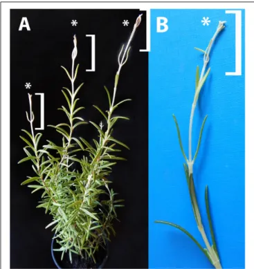
In order to check this, light-grown rosemary plants were transferred to full darkness for 2 weeks. From the shoot apical meristems or axillary buds of some plants, dark-forced shoots developed which had long internodes and smaller, pale green or whitish-yellowish leaves (Figure 2A). The original, oldest light-grown leaves had dark green color, followed by leaves with pale green color which were only partially dark-forced, and the youngest leaves close to the leaf tip were whitish-yellowish on both sides (Figure 2B).
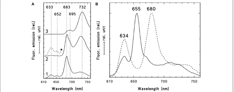
In order to check and confirm that these leaves developed in full darkness for 2 weeks, the shoot tip and different leaf primordia of dark-forced plants (Figure 2B, the leaves at the level of the asterisk or below) were collected in dim green safelight and cooled to 77 K in liquid nitrogen. The fluorescence emission spectra of the shoot tip and first leaves were recorded using 440 nm excitation and have shown major fluorescence emission bands at 633, 652 and 683 nm (Figure 3A, curve 1). When compared to the first leaves, the relative contribution of the short wavelength fluorescence bands at 633 and 652 nm decreased in the second leaf primordia, and a new band appeared at 730 nm (Figure 3A, curve 2). The third leaf primordia (Figure 3A, curve 3) only had chlorophyll fluorescence emission bands at 685, 695 and 732 nm, which are characteristic to leaves with fully developed and functional photosynthetic apparatus, and correspond to the fluorescence of the major chlorophyll-protein complexes of PSII core antenna (685 and 695 nm) and PSI light-harvesting complex (735) (Briantais et al., 1986). Similar spectra typical for green leaves were recorded in older leaves (i.e., third leaves and fully developed, fifth leaves and below)
of the dark-forced shoots as well as in the shoot tip and all different leaves of light-grown plants, and in light-germinated cotyledons (not shown).
For comparison, the fluorescence emission spectra of the cotyledons of dark-germinated seedlings were also recorded (Figure 3B, solid line). In these spectra fluorescence bands were only observed at 634 and 655 nm and no chlorophyll fluorescence emission bands were observed, indicating that these tissues developed in complete darkness and were thus fully etiolated.
Upon warming up and illumination and subsequent repeated freezing of the cotyledons, the band at 655 nm disappeared and a new band appeared at around 680 nm, indicating the transformation of photoactive protochlorophyllide pigments into chlorophyllide. Similar transformation (i.e., disappearance) of the fluorescence emission band at 652 nm was observed in the first and second leaves of dark-forced shoots upon illumination (not shown).
The fluorescence spectra of dark-forced first and second leaf primordia and the shoot tip had a chlorophyll band at 683 nm with a small shoulder at 695 nm (seen as the assymetry of the band at 683 nm), and the second leaf primordia also had a fluorescence band at around 730 nm. This indicates that these tissues were not completely devoid of chlorophylls, but probably contained remnants of chlorophylls that were present in the plastids of the light-exposed shoot apical meristem before dark-forcing and have been carried over by plastid divisions to the dark-forced tissues similarly to earlier observations in other species (Böddi et al., 1999;Solymosi et al., 2004, 2006). In addition, the dark-forced first and second leaf primordia evidently showed the presence of protochlorophyll/ide fluorescence emission at 652 and 633 nm, which originates from photoactive and non-photoactive
FIGURE 2 |Dark-forced rosemary (R. officinalis) plant(A)with new, dark-grown shoots (asterisks and square brackets) which developed during 2 weeks of growth in complete darkness. These new shoots(B)have pale, whitish-yellowish etiolated leaves and elongated stems which developed from the shoot apical meristems on the light-grown plant.
protochlorophyll/ide spectral forms, i.e., protochlorophyllide bound to NADPH:protochlorophyllide oxidoreductase (POR) or to other proteins, respectively (Böddi et al., 1989;Solymosi and Schoefs, 2010; Solymosi and Aronsson, 2013). The accumulation of these pigments, especially that of the photoactive protochlorophyllide form with emission maximum at around 652 nm that is converted to chlorophyllide uponµs time scale of illumination and thus disappears when exposed to light (Scrutton et al., 2012), proved that during dark-forcing, the previously light-grown tissues accumulated considerable amounts of a chlorophyll precursor, and had inhibited chlorophyll biosynthesis due to the lack of light (Solymosi and Schoefs, 2010;
Solymosi and Aronsson, 2013). The simultaneous presence of protochlorophyll/ide and chlorophyll pigments is typical for tissues that have been illuminated earlier during their development and were dark-forced or etiolated later only transiently (Solymosi et al., 2006, 2007, 2012; Schoefs and Franck, 2008) or completely (Böddi et al., 1999;Solymosi et al., 2004). The accumulation of photoactive protochlorophyllide confirms that these dark-forced rosemary plants developed in the absence of light during 2 weeks. The 77K fluorescence emission spectra showed clearly that dark-germinated cotyledons were fully etiolated and did not receive any light during their germination, while the leaf primordia which developed on previously illuminated (light-grown) shoot apex developed in complete darkness and got thus dark-forced for 2 weeks, but
still contained chlorophyll-protein complexes that were probably transmitted to them via plastid division.
In order to quantify the pigment contents of the various studied tissues, acetonic pigment extraction was performed and the pigment contents were determined using absorption spectroscopy (Table 1). Furthermore, fluorescence spectroscopic measurements of the acetonic extracts enabled the detection of approx. 1 µg/g protochlorophyll/ide in the leaf tip and first leaf primordia of dark-forced shoots. The protochlorophyll/ide content of the second leaf primordia was beyond the detection limit of the instrument. For comparison, the fully etiolated cotyledons of 2-week-old seedlings contained 7.87±0.78µg/g protochlorophyll/ide (n= 7), and the light-grown cotyledons of 2-week-old seedlings contained 942.57±42.69µg/g chlorophyll a, 308.22±22.23µg/g chlorophyll b, and had a chlorophyll a/b ratio around 3.11±0.12 (n= 8).
In case of light-grown shoots only a very slight (and not significant) increase in pigment contents was observed from the shoot tip and first leaf primordia toward the fully green fifth leaves, indicating that chlorophyll biosynthesis almost fully proceeded during 2 weeks in young leaf primordia. On the other hand, the pigment contents of the dark-forced shoot tip and first leaf primordia, and the second leaves were one tenth and one third of the corresponding light-grown organs, while that of the fifth leaves was only slightly, and not significantly lower in the dark than in the light. This indicates that chlorophyll biosynthesis was inhibited in the dark-forced young tissues, and the chlorophyll present in these tissues originates probably from the originally light-exposed shoot tip. In these dark-forced tissues protochlorophyll/ide accumulated to moderate levels similarly to partially or transiently etiolated or naturally dark-forced or etiolated tissues (Solymosi et al., 2004, 2006, 2007, 2012;Solymosi and Böddi, 2006; Schoefs and Franck, 2008). The relatively low chlorophyll contents of rosemary observed in this work when compared to chlorophyll contents of light-grown organs of other plants (Solymosi et al., 2004, 2006, 2007, 2012;Solymosi and Böddi, 2006; Schoefs and Franck, 2008) may be related to the presence of phenolic or terpenoid compounds which may interfere with acetonic pigment extraction (Dumbravˇa and Moldovan, 2012). Photosynthetic pigment extraction from such plant material is challenging (Dumbravˇa and Moldovan, 2012) and may thus require further methodological development and optimalization. However, using the available standard pigment extraction protocol (Porra et al., 1989), our data can be compared across treatments. The pigment content analyses confirmed that chlorophyll biosynthesis proceeded in the light-grown shoots (e.g., also in the shoot tip and first leaf primordia), while the same process was partially inhibited in the shoot tip and first and second leaf primordia of dark-forced shoots during 2 weeks of dark development.
Photosynthetic activity was also compared in the different leaves of dark-forced and light-grown shoots. The maximum quantum yield of PSII (Fv/Fm) was significantly lower in the dark-forced shoots and first leaves (0.61±0.04), while in second (0.74 ± 0.03) and fifth leaves (0.76 ± 0.01) of dark-forced shoots it was similar to values found in first (0.79 ± 0.01) and fifth leaves (0.77±0.01) of light-grown shoots, indicating
FIGURE 3 |77 K fluorescence emission spectra of the shoot tip and first leaf primordia (1), second leaf primordia (2), and third leaves (3) of 2-week-old dark-forced shoots of rosemary plants (as shown close to the asterisk inFigure 2B)(A)and 2-week-old dark-germinated cotyledons(B). After measurement at 77K, the dark-germinated cotyledon (B, solid line) was warmed to 253 K in dim green safelight, illuminated for 10 s with white light of 20µmol s−1m−2photon flux density, cooled to 77 K after 10 s in the dark and measured again (B, broken line). The spectra were normalized at their maxima and in case of the panel(A)they were shifted along theyaxis for better presentation. In case of spectrum 2(A)the spectrum region between 610 and 670 nm was multiplied by 10 and is also shown (*, broken line). Excitation wavelength: 440 nm.
impaired photosynthetic activity in the young dark-forced leaves when compared to light-grown leaves. These data show that the significantly lower chlorophyll content (Table 1) and different native organization of the photosynthetic pigments (Figure 3A) was also accompanied by significantly lower quantum efficiency of the photosynthetic apparatus only in the youngermost leaves and the shoot tip of the dark-forced shoots. However, although the more developed photosynthetic apparatus remained stable during 2 weeks of dark-forcing in older leaves, due to the absence of light photosynthesis was inhibited and thus the plants had to live heterotrophically using their food reserves. The question could be raised how this influenced plastid structure in these tissues.
Analyses of plastid ultrastructure in the first leaf primordia of these dark-forced shoots have shown that leaf mesophyll cells contained plastids with dark, electron-dense stroma, not highly regular or clearly distinguishable prolamellar bodies (average diameter: 0.7 µm), few peripheral vesicles or invaginations and thylakoids with “inverse contrast” that were sometimes stacked (mostly as bithylakoids or low grana consisting of three thylakoid layers) (Figure 4A, for summary seeSupplementary Table S1). It is noteworthy to mention that plastid sections with extremely dense stroma were sometimes observed within the same cell along with plastid sections of moderate electron density.
Plastids of leaf epidermis cells always had extremely electron- dense stroma with dark inclusions, clusters of plastoglobule- like ovale structures and peripheral vesicles or invaginations, and hardly distinguishable inner membranes (Figure 4B) which were sometimes stacked (to bithylakoids or rarely to low grana) (Supplementary Figure S1A). Plastids of the second leaf primordia of dark-forced shoots were etio-chloroplasts
with electron-dense stroma, which contained grana that were occasionally interconnected with small, hardly visible prolamellar bodies (average diameter: 0.4µm) (Supplementary Figure S1B).
The fifth leaves of dark-forced shoots contained fully developed chloroplasts and no prolamellar body was observed in them (Supplementary Figure S1C). The presence of peripheral vesicles or invaginations was characteristic for dark-forced mesophyll plastids in all leaf layers (see Supplementary Figures S1A–C) but not for light-grown leaves of the same age (not shown).
Phytoferritin inclusions were also present occasionally in the plastids of all studied leaf layers (Supplementary Figure S1D).
Capitate and peltate glandular hairs were observed already on primary leaves of the dark-forced shoots. Capitate glandular hairs had basically similar plastid structure to light-grown leaves (not shown), while some differences were observed in the plastids of fully differentiated peltate glandular hairs at the secretory stage as follows. The basal cells of the peltate glandular hairs of dark-forced leaves contained plastids with large electron-dense inclusions, and clusters of plastoglobule-like ovale structures (Figure 4C) and thus resembled plastids of basal cells in light-grown leaves (Figure 1G) and in dark-grown epidermis cells (Figure 4B). Plastid sections of the stalk cells resembled those present in light-grown plants (Figure 1H) but had in general very electron-densely stained stroma. The observed plastid sections sometimes contained only dark inclusions, but tubuloreticular membranes (diameter of approx. 0.9 µm) and clusters of plastoglobule-like ovale inclusions were also often present, as well as invaginations of the plastid envelope membrane (Figures 4D,E). Most plastid sections of the head cells of the peltate glandular hairs were similar to those observed in light-grown plants (Figure 1I) and had most typically extensive
FIGURE 4 |Plastid ultrastructure in first leaf primordia of dark-forced rosemary (R. officinalis) shoots, i.e., newly developed shoots formed on adult plants during 2 weeks of growth in complete darkness. Plastids are from leaf mesophyll cell(A), epidermis cell(B), and peltate glandular hairs(C–F).
Plastid(s) from basal cell(C), stalk cells(D,E), head cells(F,G). Scale bar:
0.5µm. DI, dense plastid inclusion; Cw, cell wall; N, nucleus; V, vacuole;
asterisk, electron-dense membrane bound inclusion; black arrow, plastoglobule-like electron-transparent inclusion; black arrowhead, tubuloreticular prolamellar body-like membrane; white arrow, peripheral vesicle or invagination of the envelope; white arrowhead, thylakoid membrane.
peripheral reticulum like structure, i.e., invaginations and vesicles at the plastid envelope (not shown). However, rarely prolamellar body-like dense membrane structures were also observed at the secretory phase (Figure 4F). Although we do not intend to provide an analysis of plastid development during glandular hair development, in this context we think it is important to mention that sometimes clusters of irregularly shaped, electron- dense vesicle like structures (average diameter: approx. 50 nm) were observed in peculiarly shaped, electron-dense plastids of the head cells at the late secretory phase (Figure 4G). This may be interesting as the enzymes involved in the biosynthesis of monoterpenes were immunolocalized to the cytoplasm of head cells and were absent from the stalk cells (Turner et al., 2012), however, the isoprenoid precursors needed for terpenoid biosynthesis may be produced by the plastids of head cells.
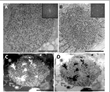
For further comparison of the tubuloreticular structures in rosemary, we provide high magnification images of the prolamellar body-like membranes of the stalk cells of peltate glandular hairs of light-grown leaves (Figure 5A) and of dark-forced leaves (Figures 5C,D) as well as of “real” and highly regular prolamellar bodies (average diameter: 1.0 µm) of the etioplasts of fully etiolated cotyledons of 2-week-old
FIGURE 5 |High magnification images of the tubuloreticular membrane structures in rosemary plastids.(A)Tubuloreticular inclusion in the stalk cell plastid of a peltate glandular hair of a light-grown leaf.(B)Prolamellar body from a mesophyll plastid of the cotyledon of 2-week-old rosemary seedling.
(C,D)Tubuloreticular membrane structure in the stalk cell plastid of a peltate glandular hair of a dark-forced first leaf.(D)Represents the inverted (i.e.,
“negative”) image of(C). Insets in(A,B)correspond to fast Fourier transformation (FFT) images of selected large central portions of the tubuloreticular membranes on pictures(A,B), respectively. Scale bar: 0.5µm.
seedlings (Figure 5B). The membranes seem to show inverse contrast [i.e., they have lightly staining membranes and electron- dense lumen – (Keresztes and Sárvári, 2001)], especially in dark-forced glandular hairs, therefore, in the latter case the
“negative” image is also shown (compare Figures 5C,D).
Unfortunately, the prolamellar bodies of mesophyll plastids of dark-forced primordia had low contrast when compared with the plastid stroma (Figure 4A), but in all cases the tubuloreticular arrangement of anastomosing membranes was evident. In order to characterize the regularity and periodicity of these membranes FFT of the image regions containing tubuloreticular structures was performed in ImageJ (see insets inFigures 5A,B). Periodic prolamellar body patterns appeared only in the FFT of prolamellar bodies of etio-chloroplasts of leaf mesophyll cells of dark-forced shoots (not shown) and in etioplasts of cotyledons of dark-germinated seedlings (Figure 5B, inset), no periodicity and regularity was observed by FFT in any tubuloreticular structure present in secretory tissues (e.g., Figure 5A, inset). The observed FFT image of the prolamellar bodies of cotyledons and etio-chloroplasts of dark-forced leaves resembled the structure observed in maize prolamellar bodies which clearly showed hexagonal symmetry (Schoefs et al., 2000).
Altogether, the tubuloreticular membranes of the etio- chloroplasts of dark-forced rosemary shoots had low contrast when compared to prolamellar bodies of etioplasts of several well characterized dark-grown model plants especially cereals (Gunning, 2001; Solymosi and Schoefs, 2008), while those in fully etiolated rosemary cotyledons clearly showed regular