Article
The Rockefeller University Press $30.00
Src family kinases are best known for their role in malignant transformation and tumor progression, as well as signaling through cell surface integrins (Parsons and Parsons, 2004; Playford and Schaller, 2004). Due to their role in cancer development and progression, Src family kinases have become major targets of cancer therapy (Kim et al., 2009;
Zhang and Yu, 2012). Src family kinases are also present in immune cells with dominant expres- sion of Lck and Fyn in T cells and NK cells; Lyn, Fyn, and Blk in B cells and mast cells; and Hck, Fgr, and Lyn in myeloid cells such as neutrophils and macrophages (Lowell, 2004).
The best known function of Src family kinases in the immune system is their role in integrin
signal transduction. Indeed, Hck, Fgr, and Lyn mediate outside-in signaling by
1and
2inte- grins in neutrophils and macrophages (Lowell et al., 1996; Meng and Lowell, 1998; Mócsai et al., 1999; Suen et al., 1999; Pereira et al., 2001;
Giagulli et al., 2006; Hirahashi et al., 2006), Lck participates in LFA-1–mediated T cell responses (Morgan et al., 2001; Fagerholm et al., 2002;
Feigelson et al., 2001; Suzuki et al., 2007), and Src family kinases are required for LFA-1–mediated
CORRESPONDENCE Attila Mócsai:
mocsai.attila@
med.semmelweis-univ.hu Abbreviations used: FcR, Fc receptor -chain; GST, gluta- thione S-transferase; ITAM, immunoreceptor tyrosine-based activation motif; KC, keratino- cyte chemoattractant; LTB4, leukotriene B4; MCP-1, mono- cyte chemoattractant protein 1;
MIP-2, macrophage inflamma- tory protein 2; MPO, myelo- peroxidase; ROS, reactive oxygen species.
The Src family kinases Hck, Fgr, and Lyn are critical for the generation of the in vivo inflammatory environment without a direct role in leukocyte recruitment
Miklós Kovács,
1,2Tamás Németh,
1,2Zoltán Jakus,
1,3Cassian Sitaru,
4Edina Simon,
1,2Krisztina Futosi,
1Bálint Botz,
5,6Zsuzsanna Helyes,
5,6Clifford A. Lowell,
7and Attila Mócsai
1,21Department of Physiology, Semmelweis University School of Medicine, 1094 Budapest, Hungary
2MTA-SE “Lendület” Inflammation Physiology Research Group of the Hungarian Academy of Sciences and the Semmelweis University, and 3MTA-SE “Lendület” Lymphatic Physiology Research Group of the Hungarian Academy of Sciences and the Semmelweis University, 1094 Budapest, Hungary
4Department of Dermatology, University Hospital Freiburg and BIOSS Centre for Biological Signalling Studies, 79104 Freiburg, Germany
5Department of Pharmacology and Pharmacotherapy, Faculty of Medicine, and 6János Szentágothai Research Centre, University of Pécs, 7624 Pécs, Hungary
7Department of Laboratory Medicine, University of California, San Francisco, San Francisco, CA 94143
Although Src family kinases participate in leukocyte function in vitro, such as integrin signal transduction, their role in inflammation in vivo is poorly understood. We show that Src family kinases play a critical role in myeloid cell–mediated in vivo inflammatory reactions. Mice lacking the Src family kinases Hck, Fgr, and Lyn in the hematopoietic compartment were completely protected from autoantibody-induced arthritis and skin blistering disease, as well as from the reverse passive Arthus reaction, with functional overlap between the three kinases. Though the overall phenotype resembled the leukocyte recruitment defect observed in
2integrin–deficient (CD18
/) mice, Hck
/Fgr
/Lyn
/neutrophils and monocytes/macrophages had no cell- autonomous in vivo or in vitro migration defect. Instead, Src family kinases were required for the generation of the inflammatory environment in vivo and for the release of proinflammatory mediators from neutrophils and macrophages in vitro, likely due to their role in Fc receptor signal transduction. Our results suggest that infiltrating myeloid cells release proinflammatory chemokine, cytokine, and lipid mediators that attract further neutrophils and monocytes from the circulation in a CD18-dependent manner. Src family kinases are required for the generation of the inflammatory environment but not for the intrinsic migratory ability of myeloid cells.
© 2014 Kovács et al. This article is distributed under the terms of an Attribution–
Noncommercial–Share Alike–No Mirror Sites license for the first six months after the publication date (see http://www.rupress.org/terms). After six months it is available under a Creative Commons License (Attribution–Noncommercial–
Share Alike 3.0 Unported license, as described at http://creativecommons.org/
licenses/by-nc-sa/3.0/).
The Journal of Experimental Medicine on September 26, 2014 jem.rupress.org Downloaded from
Published September 15, 2014
Supplemental Material can be found at:
phagocytosis of IgG-coated red blood cells is delayed but not blocked in Hck
/Fgr
/Lyn
/macrophages (Fitzer-Attas et al., 2000; Lowell, 2004). The differential requirement for Src family kinases in TCR, BCR, and Fc receptor signaling is thought to derive from the fact that Syk, but not ZAP-70, is itself able to phosphorylate ITAM tyrosines (Rolli et al., 2002), making Src family kinases indispensable for signaling by the ZAP-70–coupled TCR but not by the Syk-coupled BCR and Fc receptors.
Autoantibody production and immune complex for- mation is one of the major mechanisms of autoimmunity- induced tissue damage. In vivo models of those processes include the K/B×N serum transfer arthritis (Korganow et al., 1999) and autoantibody-induced blistering skin diseases (Liu et al., 1993; Sitaru et al., 2002, 2005), which mimic important aspects of human rheumatoid arthritis, bullous pemphigoid, and epidermolysis bullosa acquisita. Activation of neutrophils or macrophages (Liu et al., 2000; Wipke and Allen, 2001;
Sitaru et al., 2002, 2005; Solomon et al., 2005), recognition of immune complexes by Fc receptors (Ji et al., 2002; Sitaru et al., 2002, 2005), and
2integrin–mediated leukocyte recruit- ment (Watts et al., 2005; Liu et al., 2006; Chiriac et al., 2007;
Monach et al., 2010; Németh et al., 2010) are indispensable for the development of those in vivo animal models.
signal transduction and target cell killing by NK cells (Riteau et al., 2003; Perez et al., 2004).
Src family kinases also mediate TCR signal transduction by phosphorylating the TCR-associated immunoreceptor tyrosine-based activation motifs (ITAMs), leading to recruitment and activation of ZAP-70 (van Oers et al., 1996; Zamoyska et al., 2003; Palacios and Weiss, 2004). However, their role in receptor-proximal signaling by the BCR and Fc receptors is rather controversial. Although the combined deficiency of Lyn, Fyn, and Blk results in defective BCR-induced NF-B activation, receptor-proximal BCR signaling (ITAM phos- phorylation) is not affected (Saijo et al., 2003). Genetic defi- ciency of Lyn, the predominant Src family kinase in B cells, even leads to enhanced BCR signaling and B cell–mediated autoimmunity (Hibbs et al., 1995; Nishizumi et al., 1995;
Chan et al., 1997). Similarly, both positive (Hibbs et al., 1995;
Nishizumi and Yamamoto, 1997; Parravicini et al., 2002;
Gomez et al., 2005; Falanga et al., 2012) and negative (Kawakami et al., 2000; Hernandez-Hansen et al., 2004;
Odom et al., 2004; Gomez et al., 2005; Falanga et al., 2012) functions for Fyn and Lyn during Fc receptor signaling in mast cells have been reported. In addition, Hck
/Fgr
/neutrophils respond normally to IgG immune complex–induced activation (Lowell et al., 1996) and Fc receptor–mediated
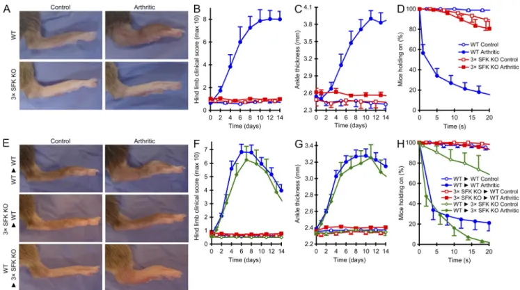
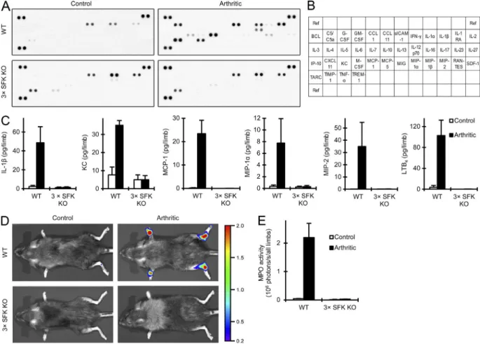
Figure 1. Myeloid Src family kinases are indispensable for autoantibody-induced arthritis. Intact (A–D) WT or Hck/Fgr/Lyn/ (3× SFK KO) mice or bone marrow chimeras (E–H) generated by transplanting WT or Hck/Fgr/Lyn/ bone marrow to WT (WT►WT and 3× SFK KO►WT, respec- tively) or Hck/Fgr/Lyn/ recipients (WT►3× SFK KO) were injected with B×N (control) or K/B×N (arthritic) serum i.p. on day 0. Arthritis development was followed by photographing on day 8 (A and E), clinical scoring of the hind limbs (B and F), ankle thickness measurement (C and G) and an articular function test (hanging on a wire grid; D and H). Images are representative of, and quantitative data show mean and SEM from, 4 control and 6 arthritic serum-treated individual mice per group from 2 independent experiments (A–D) or 4–12 control and 7–26 arthritic serum-treated mice per group from 2–8 independent experiments (E–H). D and H show results from functional test performed 6–21 times on each mouse between days 6–12.
on September 26, 2014 jem.rupress.org Downloaded from
Ar ticle
RESULTS
Hck, Fgr, and/or Lyn are required for autoantibody-induced arthritis
To determine the role of Src family kinases in autoantibody- induced arthritis, we first tested the development of K/B×N serum transfer arthritis in Hck
/Fgr
/Lyn
/triple knock- out mice. As shown in Fig. 1 A, administration of arthritogenic (K/B×N) serum triggered robust arthritis in WT but not Hck
/Fgr
/Lyn
/animals. The Hck
/Fgr
/Lyn
/mutation completely protected mice from disease develop- ment both in terms of visible clinical signs (Fig. 1 B; P = 4.2 × 10
5; n = 6) and ankle thickening (Fig. 1 C; P = 4.5 × 10
4; The role of Src family kinases in
2integrin signaling and
the requirement for
2integrins during autoantibody- induced in vivo inflammation prompted us to test the role of Src family kinases in autoantibody-induced inflammatory disease models. We found that Hck
/Fgr
/Lyn
/mice were completely protected from autoantibody-induced ar- thritis and inflammatory blistering skin disease. Surprisingly, this was not due to a cell-autonomous defect in
2integrin–
mediated leukocyte migration but to defective generation of an inflammatory microenvironment, likely due to the role of Src family kinases in immune complex–induced neutrophil and macrophage activation.
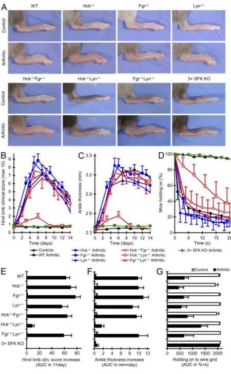
Figure 2. Overlapping role of Hck, Fgr, and Lyn during autoantibody-induced arthritis. (A) Arthritis was induced and assessed in WT, Hck/, Fgr/, and Lyn/ single, Hck/Fgr/, Hck/Lyn/, and Fgr/
Lyn/ double, and Hck/Fgr/Lyn/ (3× SFK KO) triple knockout bone marrow chimeric mice as de- scribed in the Fig. 1 legend. Controls for all genotypes were combined in B–D. E–G show cumulative data obtained from the experiments shown in B–D. Images are representative of, and quantitative data show mean and SEM from 1–12 control and 7–26 arthritic serum- treated mice per genotype from 10 independent ex- periments. The joint functional test (D and G) were performed 3–24 times on each mouse between days 6 and 12. The WT and Hck/Fgr/Lyn/ chimeric data include results presented in Fig. 1 (E–H). AUC, area under the curve.
on September 26, 2014 jem.rupress.org Downloaded from
Published September 15, 2014
both control and arthritic serum-treated mice in Fig. 2 (E–G).
Hck, Fgr, or Lyn single deficiency did not affect arthritis development in our model (p-values for clinical score, ankle thickness, and joint functional test were between 0.34 and 0.98; n = 10–11). Hck
/Fgr
/and Fgr
/Lyn
/double knockout chimeras showed no protection either (0.086 ≤ P ≤ 0.99; n = 4–10). The Hck
/Lyn
/double mutation caused substantial but incomplete reduction of arthritis development (P = 1.2 × 10
10, 1.4 × 10
9, and 0.031 for clinical score, ankle thickness, and joint functional test, respectively; n = 5–7);
however, the poor overall health status of these chimeras (Xiao et al., 2008) may have affected their response in our model.
Importantly, complete protection from arthritis development was only seen in the absence of all three kinases (P = 2.0 × 10
12, 3.5 × 10
11, and 1.2 × 10
7, respectively; n = 14–21), indicating significant functional overlap between Hck, Fgr, and Lyn during autoantibody-induced inflammation.
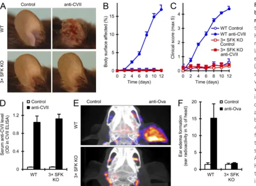
Other immune complex–induced inflammation models We also tested the effect of the Hck
/Fgr
/Lyn
/mutation on other autoantibody-induced inflammatory processes. Sys- temic administration of collagen VII–specific antibodies (a mouse model of the human blistering skin disease epidermolysis bullosa acquisita; Sitaru et al., 2005, 2007) triggered skin inflammation in WT but not Hck
/Fgr
/Lyn
/mutant animals (Fig. 3 A).
Hck
/Fgr
/Lyn
/mutants were completely protected both in terms of the body surface area affected (Fig. 3 B; P = 1.5 × 10
10; n = 11) and the overall disease severity (Fig. 3 C; P = 1.1 × 10
10; n = 13). However, circulating collagen VII–specific antibody titers were not affected by the Hck
/Fgr
/Lyn
/n = 6). We also tested the ability of the mice to hold onto the lower side of a wire grid as a measure of joint function ( Jakus et al., 2009). Although the majority of arthritic serum- treated WT mice fell off the grid within a few seconds, most of the Hck
/Fgr
/Lyn
/were able to hold onto the grid throughout the entire 20-second assay period (Fig. 1 D;
P = 9.5 × 10
6; n = 6).
We also performed arthritis experiments in bone marrow chimeras generated by transplanting Hck
/Fgr
/Lyn
/bone marrow cells to WT recipients or vice versa. As shown in Fig. 1 (E–H), bone marrow chimeras carrying Hck
/Fgr
/Lyn
/bone marrow cells in a WT recipient were completely pro- tected from all signs of K/B×N serum transfer arthritis (P = 2.0 × 10
12, 3.5 × 10
11, and 1.2 × 10
7for clinical score, ankle thickness, and joint functional assay, respectively; n = 14–21), whereas Hck
/Fgr
/Lyn
/mice transplanted with WT bone marrow cells showed a normal disease course and an even somewhat more severe functional defect (P = 0.36, 0.59, and 0.028, respectively; n = 7). The above results indicate that expression of Hck, Fgr, and/or Lyn in the hematopoietic com- partment is indispensable for the development of autoantibody- induced arthritis in experimental mice.
Overlapping role of Hck, Fgr, and Lyn during autoantibody-induced arthritis
We next tested K/B×N serum transfer arthritis in bone marrow chimeras with single or double deficiency of Hck, Fgr, or Lyn in the hematopoietic compartment. Representative hind limb images are shown in Fig. 2 A, quantitative analyses of arthritic serum-treated mice in Fig. 2 (B–D), and cumulated data for
Figure 3. Src family kinases in autoanti- body-induced skin blistering disease and the reverse passive Arthus reaction. (A–D) Blis- tering skin disease was triggered in WT or Hck/Fgr/Lyn/ (3× SFK KO) mice or bone marrow chimeras by systemic injection of col- lagen VII–specific (-CVII) antibodies. Skin dis- ease was followed by photographing on day 14 (A) and clinical assessment of the total body surface affected (B) and the overall disease se- verity (C). The serum titer of -CVII antibodies was tested on day 6 by ELISA (D). Representative images (A) or mean and SEM (B–D) from 5–7 control (1 intact and 4–6 bone marrow chimeric) and 13–14 -CVII–treated (4 intact and 9–10 bone marrow chimeric) mice per genotype from 4 independent experiments are shown. No dif- ference between intact and chimeric mice of the same hematopoietic genotype was ob- served (not depicted). (E and F) Reverse passive Arthus reaction was triggered in intact WT or Hck/Fgr/Lyn/ (3× SFK KO) mice by sys- temic administration of ovalbumin, followed by intradermal injection of normal (control) or anti- ovalbumin (anti-Ova) rabbit serum into the left and right ears, respectively. Edema formation was assessed by determining the accumulation of radioactively labeled albumin from the circulation by NanoSPECT with a reference CT scan. Representative images (E) and mean and SEM (F) from 4–8 mice per genotype from 3 independent experiments are shown.
on September 26, 2014 jem.rupress.org Downloaded from
Ar ticle
animals (P = 0.013; n = 8). Collectively, Hck, Fgr, and Lyn are required for antibody-induced tissue damage in various in vivo inflammation models.
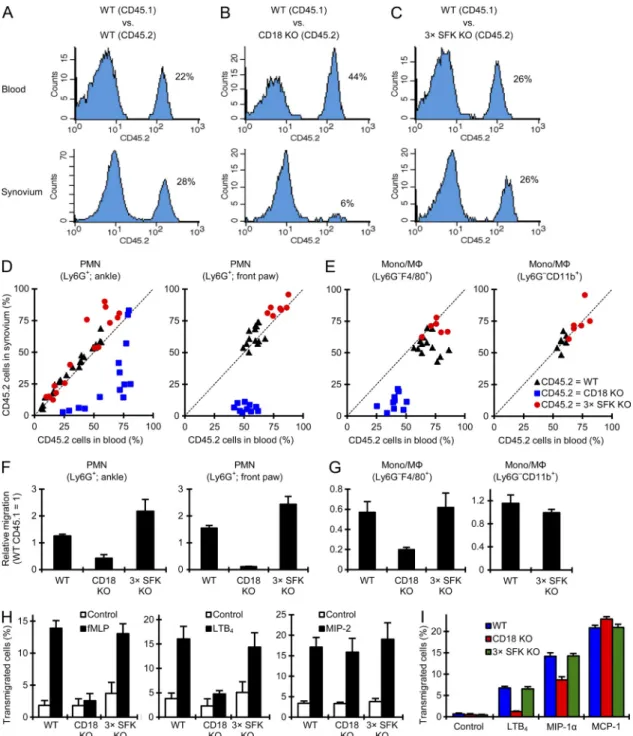
Myeloid cells fail to accumulate at the site of inflammation We and others reported complete protection of CD18
/mice from K/B×N serum transfer arthritis, likely due to a cell- autonomous role for LFA-1 in migration of myeloid cells to the site of inflammation (Watts et al., 2005; Németh et al., 2010;
Monach et al., 2010). Given the role for Src family kinases in integrin signaling in myeloid cells and in autoantibody-induced mutation (Fig. 3 D; P = 0.34; n = 2). Importantly, intact
Hck
/Fgr
/Lyn
/mice and Hck
/Fgr
/Lyn
/bone marrow chimeras were both completely protected from skin disease in our model (unpublished data).
We also tested the reverse passive Arthus reaction triggered by systemic administration of ovalbumin followed by intra- dermal injection of anti-ovalbumin into the ear. Edema forma- tion was followed by imaging the accumulation of radioactively labeled albumin from the circulation. As shown in Fig. 3 (E and F), anti-ovalbumin injection caused robust increase of vascular permeability in WT but not Hck
/Fgr
/Lyn
/Figure 4. Myeloid cells fail to accumulate at the site of inflammation. Intact WT, CD18/ (CD18 KO), or Hck/Fgr/Lyn/ (3× SFK KO) mice were subjected to K/B×N serum transfer arthritis (A, B, D, and E) or autoantibody-induced skin blistering disease (C) as described in the legends to Figs. 1 and 3. (A) Hematoxylin-eosin–stained sections of ankle joints 7 d after the serum transfer. Images are representative of 4 control and 6 arthritic serum- treated mice per genotype from 2 independent experiments. Lower images were magnified from the upper sections. Bars, 200 µm. (B–D) The ankle area or the front paw was flushed (B, D, and E) or the nasal skin was digested (C), and the number of neutrophils (PMN; B and C) or monocytes/macrophages (mono/M; D and E) was determined by flow cytometry. Graphs represent mean and SEM of data obtained from 3–7 control and 5–13 arthritic serum- treated mice per genotype from 2–5 independent experiments (B and D), 3–4 control and 6 arthritic serum-treated mice per genotype from 2 indepen- dent experiments (E), or 2 control and 4 anti-CVII–treated mice per genotype from 2 independent experiments (C). a., ankle; f., front paw.
on September 26, 2014 jem.rupress.org Downloaded from
Published September 15, 2014
animal (WT:WT, WT:CD18
/, and WT:Hck
/Fgr
/Lyn
/chimeras, respectively). We then compared the percentage of CD45.2-positive cells in the blood and synovial tissue within the same individual mouse during K/B×N serum trans- fer arthritis.
In chimeras having CD45.2-expressing WT and CD45.1- expressing WT hematopoietic tissues (WT:WT chimeras), the percentage of CD45.2-positive neutrophils was similar in the blood and synovial infiltrate in all chimeras tested (Fig. 5 A). In contrast, in WT:CD18
/chimeras (Fig. 5 B), the percentage of CD45.2-expressing (i.e., CD18
/) neutrophils in the in- flamed synovium was much lower than that in the circulation, indicating a cell-autonomous recruitment defect of CD18
/neutrophils. Surprisingly, in WT:Hck
/Fgr
/Lyn
/chi- meras (Fig. 5 C), no difference between the percentage of CD45.2-positive (i.e., Hck
/Fgr
/Lyn
/) neutrophils in the blood and the synovial infiltrate could be observed, indi- cating that Hck
/Fgr
/Lyn
/neutrophils can accumulate normally at the site of inflammation when WT cells are also present in the same animal. The results of such experiments performed on a large cohort of mice are summarized in Fig. 5 D, where each dot represents an individual mouse. Although dots of WT:WT and WT:Hck
/Fgr
/Lyn
/chimeras line up along the dotted line representing equal ratios in the blood and the synovium, dots of WT:CD18
/chimeras are shifted to the lower right, indicating reduced ability of CD18
/cells to enter the synovium from the circulation. The same data have also been used to calculate the relative migratory capacity of neutrophils of the different genotypes (Fig. 5 F). Those calcu- lations confirmed a strong cell-autonomous recruitment defect of CD18
/neutrophils (P = 2.0 × 10
12and 1.0 × 10
9for ankle and front paw, respectively; n = 10–14), whereas the accumulation of Hck
/Fgr
/Lyn
/neutrophils was even slightly higher (P = 0.021 and 0.11, respectively; n = 7–15) than that of WT cells within the same animal.
We have also tested the accumulation of monocytes/macro- phages in a similar manner. As shown in Fig. 5 (E and G), al- though CD18 deficiency caused a significant cell-autonomous reduction of the percentage of Ly6G
F4/80
+macrophages in the synovial tissue (relative to the percentage of CD18
/cells among circulating Ly6G
F4/80
+monocytes; P = 8.0 × 10
6; n = 7), the Hck
/Fgr
/Lyn
/mutation did not af- fect the accumulation of F4/80-positive cells in the inflamed synovium (P = 0.83; n = 5). The Hck
/Fgr
/Lyn
/muta- tion had no effect on macrophage accumulation when the cells were identified as Ly6G
CD11b
+cells either (Fig. 5, E and G;
P = 0.37; n = 6). Collectively, when WT cells are present in mixed bone marrow chimeras, Hck
/Fgr
/Lyn
/neutro- phils and monocytes/macrophages accumulate normally at the site of inflammation, arguing against a cell-autonomous migration defect of Hck
/Fgr
/Lyn
/myeloid cells.
Normal in vitro migration of Hck
/Fgr
/Lyn
/neutrophils and monocytes
We next tested the migration of neutrophils and mono- cytes in an in vitro Transwell assay. As shown in Fig. 5 H, in vivo inflammation (Figs. 1–3), we next tested their role in
leukocyte accumulation at the inflammatory site. As shown in Fig. 4 A, K/B×N serum transfer arthritis triggered robust leukocytic infiltration of the synovial area of WT but not Hck
/Fgr
/Lyn
/mice. Flow cytometric analysis revealed a dramatic increase of neutrophil numbers in the synovial tissue of arthritic serum-treated WT mice that was entirely depen- dent on CD18 (Fig. 4 B; P = 6.8 × 10
5and 1.6 × 10
4for ankle and front limb, respectively; n = 5). Importantly, the Hck
/Fgr
/Lyn
/mutation also abrogated neutrophil in- filtration at the synovial area (Fig. 4 B; P = 7.2 × 10
5and 1.7 × 10
4, respectively; n = 13). The defective neutrophil infiltration was not due to reduced circulating numbers of neutrophils because blood neutrophil counts were even higher in both CD18
/(10.7 ± 2.1 and 16.9 ± 5.7 thousand cells/µl before and on day 4 after K/B×N serum transfer, respectively; n = 4) and Hck
/Fgr
/Lyn
/(2.5 ± 0.6 and 4.6 ± 1.6 thousand cells/µl, respectively; n = 4) than in WT animals (0.6 ± 0.1 and 1.4 ± 0.3 thousand cells/µl, respectively; n = 4).
We also tested neutrophil infiltration in the nasal skin dur- ing autoantibody-induced skin blistering disease. As shown in Fig. 4 C, neutrophils infiltrated the skin of WT but not Hck
/Fgr
/Lyn
/animals (P = 0.0088; n = 4).
We next tested the accumulation of monocytes/macro- phages. Ly6G
F4/80
+cells infiltrated the synovial area of arthritic serum-treated WT mice, and this was again entirely dependent on the presence of CD18 (Fig. 4 D; P = 1.1 × 10
4and 1.6 × 10
4for ankle and front paw, respectively; n = 5).
The Hck
/Fgr
/Lyn
/mutation also completely abro- gated the accumulation of Ly6G
F4/80
+cells (Fig. 4 D; P = 1.1
× 10
4and 1.7 × 10
4, respectively; n = 13). Again, the defec- tive infiltration was not due to reduced number of circulating monocytes because Ly6G
F4/80
+blood monocyte counts in CD18
/(710 ± 144 and 1,591 ± 351 cells/µl on days 0 and 4, respectively; n = 4) and Hck
/Fgr
/Lyn
/mice (553 ± 154 and 244 ± 59 cells/µl, respectively; n = 4) was similar to or higher than those in WT animals (433 ± 90 and 258 ± 40 cells/µl, respectively; n = 4). No infiltration of monocytes/
macrophages identified as Ly6G
CD11b
+cells could be ob- served in the synovial area of Hck
/Fgr
/Lyn
/mice ei- ther (Fig. 4 E; P = 0.015 and 0.031 for ankle and front paw, respectively; n = 6; this could not be tested in CD18
/mice because their leukocytes do not express CD11b). Collectively, similar to the CD18 deficiency, the Hck
/Fgr
/Lyn
/mutation also abrogates the accumulation of myeloid cells at the site of inflammation.
No cell-autonomous migration defect in Hck
/Fgr
/Lyn
/myeloid cells
To reveal whether the defective leukocyte recruitment in Hck
/Fgr
/Lyn
/mice is due to a cell-autonomous migra- tion defect, we directly compared the accumulation of WT and mutant cells within the same animal. To this end, we generated mixed bone marrow chimeras carrying varying ratios of CD45.1- expressing WT, along with CD45.2-positive WT, CD18
/, or Hck
/Fgr
/Lyn
/hematopoietic cells within the same
on September 26, 2014 jem.rupress.org Downloaded from
Ar ticle
Figure 5. Normal in vitro and in vivo migration of Hck/Fgr/Lyn/ neutrophils. (A–G) Mixed bone marrow chimeras with CD45.1-expressing WT and CD45.2-expressing WT, CD18/ (CD18 KO), or Hck/Fgr/Lyn/ (3× SFK KO) hematopoietic cells were subjected to K/B×N serum transfer arthritis as described in the Fig. 1 legend. In vivo accumulation of neutrophils (PMN; A–D and F) and monocytes/macrophages (Mono/M; E and G) was determined by flushing the synovial area of arthritic serum-treated mixed bone marrow chimeras on day 4, followed by flow cytometric analysis of the ratio of CD45.1- and CD45.2-expressing cells in peripheral blood and the synovial infiltrate. A–C shows representative histograms of CD45.2 expression in blood or synovial neu- trophils. In D and E, each dot represents an individual mouse. Bar graphs in F and G show mean and SEM of relative migration. Data are representative of (A–C) or summarize (D and F) data obtained from 7–26 mice per group from 3–9 independent experiments or 7–14 mice per group from 3–5 independent experiments (E and G). (H and I) In vitro migration of CD18/ and Hck/Fgr/Lyn/ bone marrow neutrophils (H) and monocytes (I) toward the indicated chemoattractants in a fibrinogen-coated Transwell system. Data represent mean and SEM of 3–13 (H) or 3–5 (I) independent experiments.
neutrophil migration toward fMLP (which mimics bacterial/
mitochondrial-derived signals) was strongly reduced in CD18
/(P = 4.1 × 10
4; n = 4) but not in Hck
/Fgr
/Lyn
/cells
(P = 0.17; n = 4). Similarly, neutrophil migration toward the lipid chemoattractant LTB
4was dependent on CD18 (P = 1.7 × 10
6; n = 3) but was not affected by the Hck
/Fgr
/Lyn
/on September 26, 2014 jem.rupress.org Downloaded from
Published September 15, 2014
in vitro migration of neutrophils and monocytes toward major proinflammatory chemoattractants does not depend on Hck, Fgr, or Lyn irrespective of the requirement for
2integrins in the given assay.
Defective generation of the inflammatory environment The above observations could be explained by a role for my- eloid Src family kinases in the generation of the inflammatory environment. As shown in the cytokine arrays in Fig. 6 A (see array map in Fig. 6 B and quantification in Table S1), K/B×N serum transfer arthritis triggered robust accumulation of vari- ous inflammatory mediators in the synovial tissue of WT but not Hck
/Fgr
/Lyn
/animals. Prior studies suggested important roles for IL-1, keratinocyte chemoattractant (KC;
CXCL1), MIP-1, MIP-2, and LTB
4in myeloid cell infil- tration during autoantibody-induced inflammation (Kim et al., 2006; Chou et al., 2010; Jacobs et al., 2010), whereas MCP-1 is considered a major monocyte chemoattractant (Deshmane et al., 2009). ELISA assays (Fig. 6 C) confirmed mutation (P = 0.41; n = 4). Interestingly, migration of neutrophils
toward macrophage inflammatory protein 2 (MIP-2; CXCL2), a distant homologue of human IL-8, was not affected by either the CD18
/(P = 0.58; n = 9) or the Hck
/Fgr
/Lyn
/(P = 0.39; n = 7) mutation.
We have also tested the in vitro migration of freshly iso- lated bone marrow monocytes (Fig. 5 I). Monocyte migration toward leukotriene B
4(LTB
4) was abrogated by CD18 defi- ciency (P = 0.0038; n = 3) but not by the Hck
/Fgr
/Lyn
/mutation (P = 0.93; n = 5). Migration of monocytes toward MIP-1 (CCL3), one of the major monocyte chemoattrac- tants, was also moderately reduced by the CD18
/mutation with a borderline statistical significance (P = 0.057; n = 3), but it was normal in Hck
/Fgr
/Lyn
/cells (P = 0.63;
n = 5). The migration of the cells toward monocyte chemoat- tractant protein 1 (MCP-1; CCL2), another major monocyte chemoattractant, was slightly even increased by CD18 defi- ciency (P = 0.0039; n = 3), but it was not affected by the Hck
/Fgr
/Lyn
/mutation (P = 0.74; n = 5). Collectively,
Figure 6. Myeloid Src family kinases are required for the generation of an inflammatory environment in vivo. Intact WT or Hck/Fgr/Lyn/
(3× SFK KO) mice were subjected to K/B×N serum transfer arthritis as described in the legend to Fig. 1. (A and C) The synovial area was flushed on Day 4. The cell-free supernatants of the synovial infiltrates were probed using a commercial cytokine array (A) or by ELISA assays for the indicated pro-inflammatory mediators (C).
(B) Map of the position of the different analytes on the cytokine array. A shows representative images from 2 independent experiments, whereas C shows mean and SEM of 4–6 mice per group from 3–5 independent experiments. (D-E) MPO activity was determined in vivo by chemiluminescence imaging after i.p. injection of luminol. Color-coded photon flux intensity is superposed on the grayscale photo of the mouse (D) and quantitated in defined regions of interest (E). Represen- tative images (D) and mean and SEM (E) from 4 control and 6 arthritic serum-treated mice per genotype from 2 independent experiments are shown.
on September 26, 2014 jem.rupress.org Downloaded from
Ar ticle
Hck
/Fgr
/Lyn
/myeloid cells fail to respond to immune complex stimulation
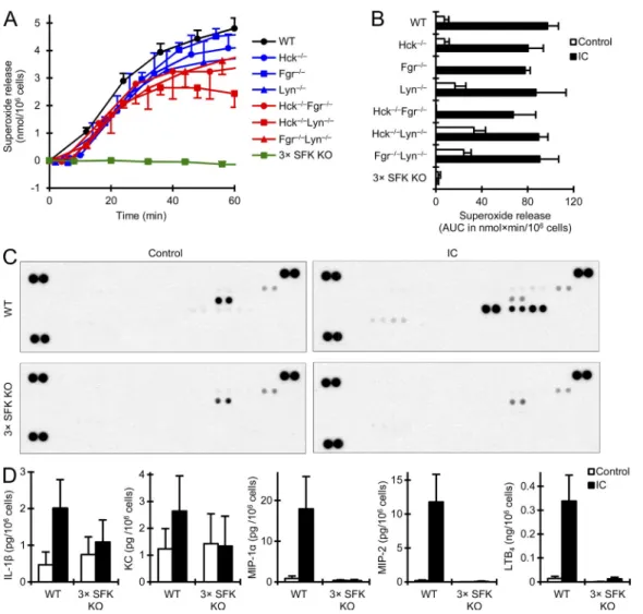
To test whether Src family kinases are directly involved in the gen- eration of an inflammatory environment, we investigated immune complex–induced in vitro responses of neutrophils and macro- phages. As shown in Fig. 7 (A and B), the Hck
/Fgr
/Lyn
/mutation completely blocked immune complex–induced su- peroxide release from neutrophils (P = 2.4 × 10
21; n = 15).
Similar to the in vivo findings with K/B×N serum transfer arthritis (Fig. 2), the Hck
/Lyn
/mutation caused a partial but statistically significant reduction (P = 5.8 × 10
4; n = 4;
this may in part be due to increased basal levels in those sam- ples), whereas the other single or double knockouts had no significant defect (Fig. 7, A and B; 0.10 < P < 0.71; n = 3).
As shown in the cytokine arrays in Fig. 7 C (see array map in Fig. 6 B and quantification in Table S1), immune complex stim- ulation also triggered the release of several cytokines and che- mokines from WT but not Hck
/Fgr
/Lyn
/neutrophils.
that the Hck
/Fgr
/Lyn
/mutation abrogated the up- regulation of IL-1 (P = 0.0026; n = 5), KC (P = 9.0 × 10
7; n = 5), MCP-1 (P = 0.0014; n = 5), MIP-2 (P = 0.023; n = 6), and LTB
4(P = 7.4 × 10
15; n = 3) in the synovial tissue and caused a substantial (though not statistically significant) reduc- tion of MIP-1 levels (P = 0.095; n = 4).
We have also tested the generation of reactive oxygen spe- cies (ROS) and the release of granule enzymes into the extra- cellular space at the inflammatory site by in vivo imaging of the activity of myeloperoxidase (MPO), a neutrophil granule- derived ROS-producing enzyme (Gross et al., 2009; Nussbaum et al., 2013). As shown in Fig. 6 (D and E), K/B×N serum transfer arthritis triggered a robust ROS bioluminescence signal localized to the ankles and front paws in WT but not Hck
/Fgr
/Lyn
/mutant animals (P = 0.010; n = 6). Col- lectively, myeloid Src family kinases are required for various aspects of the generation of a proinflammatory environment during autoantibody-induced arthritis.
Figure 7. Hck/Fgr/Lyn/ neutrophils fail to respond to immune complex stimulation. WT, Hck/Fgr/Lyn/ (3× SFK KO), and the various sin- gle and double knockout neutrophils were placed on immobilized immune complex (IC) surfaces. Superoxide release (A and B) was followed by a spectrophoto- metric assay. Cytokine, chemokine, and lipid mediator levels in cell-free supernatants were determined after an incubation for 1 h (LTB4) or 6 h (all other readouts) using a commercial cytokine array (C) or ELISA assays (D). Graphs in A and B show mean and SEM of 3–22 independent experiments per genotype.
Values at the zero time point were subtracted. C is representative of 2 independent experiments. D shows mean and SEM from 4–11 independent experiments.
on September 26, 2014 jem.rupress.org Downloaded from
Published September 15, 2014
Lowell, 1998; Mócsai et al., 1999; Suen et al., 1999; Fitzer- Attas et al., 2000; Pereira et al., 2001; Lowell, 2004). Our above observations led us to reevaluate this issue.
ELISA assays revealed that the Hck
/Fgr
/Lyn
/mutation dramatically reduced or even completely abrogated the im- mune complex–induced release of IL-1 (P = 5.1 × 10
5; n = 6), KC (P = 3.6 × 10
5; n = 6), MIP-1 (P = 5.0 × 10
6; n = 6), MIP-2 (P = 4.8 × 10
15; n = 11), and LTB
4(P = 8.8 × 10
5; n = 4) from neutrophils (Fig. 7 D), whereas no release of MCP-1 could be observed even from WT cells (not depicted).
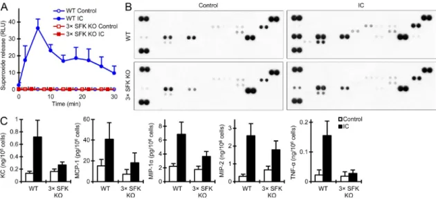
We also tested the immune complex–induced responses of bone marrow–derived macrophages. As shown in Fig. 8 A, the Hck
/Fgr
/Lyn
/mutation completely blocked im- mune complex–induced ROS production by macrophages (P = 2.8 × 10
8; n = 4). The cytokine arrays shown in Fig. 8 B (see array map in Fig. 6 B and quantification in Table S1) in- dicated immune complex–induced release of various pro- inflammatory cytokines from WT macrophages which was strongly reduced in Hck
/Fgr
/Lyn
/cultures. ELISA assays (Fig. 8 C) confirmed that the Hck
/Fgr
/Lyn
/mutation strongly inhibited the immune complex–induced re- lease of KC (P = 0.0013; n = 5), MCP-1 (P = 0.010; n = 4), and TNF (P = 4.0 × 10
4; n = 4) from macrophages, caused a partial but statistically significant inhibition of MIP-2 release (P = 0.027; n = 4), and led to an apparent reduction of MIP- 1 release which, however, did not reach statistical significance (P = 0.29; n = 4). In contrast, no release of IL-1 or LTB
4could be observed even in WT cultures (unpublished data). Collectively, the genetic deficiency of Hck, Fgr, and Lyn blocks the immune complex–induced release of various components of the inflam- matory environment from both neutrophils and macrophages.
Src family kinases are required for initial Fc receptor signaling in neutrophils
Prior studies indicated that Src family kinases are critically in- volved in integrin- but not Fc receptor–mediated functional responses of myeloid cells (Lowell et al., 1996; Meng and
Figure 8. Defective responses of Hck/Fgr/Lyn/ macrophages to immune complex stimulation. Bone marrow–derived WT or Hck/Fgr/Lyn/
(3× SFK KO) macrophages were placed on immobilized immune complex (IC) surfaces. Their superoxide release (A) was followed by a luminometric assay. Cytokine and chemokine levels from the cell-free supernatants taken after 24 h were tested by a commercial cytokine array (B) or by ELISA assays (C). A shows mean and SEM from 4 independent experiments. B is representative of 2 experiments. C shows mean and SEM of data from 4–5 independent experiments.
Figure 9. Src family kinases are required for immune complex–induced phosphorylation of FcR and Syk. (A–C) WT or Hck/Fgr/Lyn/
(3× SFK KO) neutrophils were plated on immobilized IgG immune complexes (IC). Cell lysates were subjected to a GST pulldown assay (A and B) using either WT or R41A/R194A double mutant (SH2-Dead) GST- Syk(SH2)2 fusion protein and probed for the presence of FcR by Western blotting (WB), or processed for Syk immunoprecipitation (IP), followed by immunoblotting for phosphotyrosine (PY) or Syk as a loading control (C).
Whole cell lysates (WCL) served as loading control in A and B. Blots in A–C are representative of 3 independent experiments. Note substantial mobil- ity shift caused by the tyrosine phosphorylation of FcR homodimers under the used nonreducing (NR) conditions in A and B. WB, Western blot.
on September 26, 2014 jem.rupress.org Downloaded from
Ar ticle
no such signal could be observed upon functional disruption of both SH2 domains (Mócsai et al., 2006), indicating that this assay specifically detects phosphorylation of the FcR
ITAM tyrosines. Importantly, no FcR phosphorylation could be observed in lysates of immune complex–stimulated Hck
/Fgr
/Lyn
/neutrophils (Fig. 9 B).
We first tested the phosphorylation of the Fc receptor
-chain (FcR), the ITAM-containing signaling chain of activating Fc receptors. As shown in Fig. 9 A, a glutathione S-transferase (GST) fusion protein of the tandem SH2 domains of Syk (GST-Syk(SH2)
2) was able to pull down FcR from lysates of immune complex–activated WT neutrophils, whereas
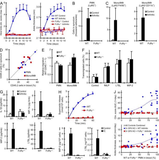
Figure 10. FcR deficiency phenocopies the deficiency of Src family kinases. (A–C) K/B×N serum transfer arthritis was induced in intact WT and FcR/ mice as described in the Fig. 1 legend, followed by analysis of the clinical disease course as described in Fig. 1 A (3 control and 5–7 arthritic mice per genotype from 3 independent experiments) or the quantification of leukocyte infiltration as described in Fig. 4 (B and C; 2–3 control and 6 arthritic mice per genotype from 3 independent experiments). (D and E) In vivo migration of neutrophils (PMN) and monocytes/macrophages (Mono/M) determined using mixed bone marrow chimeras containing CD45.1-expressing WT and CD45.2-expressing FcR/ cells as described in Fig. 5. Each dot in D represents an indi- vidual mouse. E shows mean and SEM of relative migration. (D and E) Data were obtained from 6–8 mice per group from 3 independent experiments.
(F) In vitro migration of WT and FcR/ neutrophils tested as described in Fig. 5. Data were obtained from 3 independent experiments. (G) Inflammatory mediator release from WT and FcR/ mice determined as described in Fig. 6. Data were obtained from 3–5 mice per group from 3 independent experiments.
(H and I) Superoxide (H) or cytokine/chemokine/lipid mediator (I) release from immune complex-activated WT and FcR/ neutrophils from intact mice was determined as described in Fig. 7. Data were obtained from 4 (H) or 3 (I) independent experiments. (J) Bone marrow chimeras generated by transplanting FcR/ or CD45.1-expressing WT bone marrow cells mixed with Hck/Fgr/Lyn/ cells at varying ratios were subjected to K/B×N serum transfer arthritis as described in Fig. 1. Area under the curve (AUC) values for daily clinical scoring and ankle thickness measurements are shown in relation to the percentage of WT or FcR/ (i.e., non-Hck/Fgr/Lyn/) cells in the peripheral blood. Data represent results from 8–13 control and 28–32 arthritic serum-treated mice per group from 3 independent experiments. All error bars show SEM from the indicated number of mice or experiments.
on September 26, 2014 jem.rupress.org Downloaded from
Published September 15, 2014
restored arthritis development. Obvious arthritis was clearly seen at 50% WT neutrophils in the circulation and the severity of arthritis further increased with increasing percentage of WT cells. In contrast, mixing Hck
/Fgr
/Lyn
/and FcR
/hematopoietic tissues did not result in any signs of arthritis de- velopment in the resulting chimeras at any ratio of the two geno- types in the circulating neutrophil pool. Collectively, the in vivo and in vitro phenotypes of Hck
/Fgr
/Lyn
/and FcR
/mutants are strikingly similar and FcR
/cells cannot restore arthritis development on the Hck
/Fgr
/Lyn
/back- ground, suggesting a functional overlap between Src family kinases and Fc receptors.
DISCUSSION
Our experiments have revealed complete protection of Hck
/Fgr
/Lyn
/mice from K/B×N serum transfer arthri- tis, autoantibody-induced skin blistering, and the reverse passive Arthus reaction (Figs. 1 and 3). Hck, Fgr, and Lyn were required in the hematopoietic compartment (Fig. 1, E–H) and showed significant functional overlap with each other (Fig. 2).
The overall phenotype of Hck
/Fgr
/Lyn
/mutants was very similar to that of
2integrin–deficient (CD18
/) mice (Watts et al., 2005; Liu et al., 2006; Chiriac et al., 2007;
Németh et al., 2010).
2integrins have long been known to me- diate leukocyte migration to the site of inflammation (Mizgerd et al., 1997; Scharffetter-Kochanek et al., 1998; Walzog et al., 1999; Schymeinsky et al., 2007). Indeed, we observed a complete defect of leukocyte infiltration into the synovial tissue of CD18
/animals (Fig. 4), and our mixed bone marrow chime- ric experiments (Fig. 5, A–G) indicated, in agreement with a prior report (Monach et al., 2010), that CD18
/myeloid cells had a cell-autonomous leukocyte migration defect. The simi- larities of the CD18
/and Hck
/Fgr
/Lyn
/phenotypes and the defective leukocyte infiltration in Hck
/Fgr
/Lyn
/mice (Fig. 4) suggested that Hck
/Fgr
/Lyn
/leukocytes have an intrinsic migration defect. However, Hck
/Fgr
/Lyn
/myeloid cells accumulated normally in the inflamed synovium in mixed bone marrow chimeras (Fig. 5, A–G) and showed no in vitro migration defects either (Fig. 5, H and I). In con- trast, they failed to release inflammatory mediators when ac- tivated by immobilized immune complexes in vitro (Fig. 7 and 8). Therefore, the arthritis defect in Hck
/Fgr
/Lyn
/mice is likely due to the defective generation of an inflam- matory environment rather than, as initially expected, to a cell-autonomous defect of
2integrin–mediated leuko- cyte migration.
Our results, in agreement with prior studies (Kim et al., 2006; Chen et al., 2006; Chou et al., 2010), indicate the exis- tence of positive-feedback loops acting through the release of proinflammatory mediators (including IL-1, neutrophil and monocyte/macrophage chemokines, and LTB
4) from myeloid cells that attract further myeloid cells to the site of inflammation.
2integrins and Src family kinases apparently function at dif- ferent points of the feedback loop; whereas the former are re- quired for the intrinsic migratory ability of leukocytes, the latter are involved in the release of proinflammatory mediators.
We next tested the activation of the Syk tyrosine kinase.
As shown in Fig. 9 C, immune complex–induced neutrophil activation led to tyrosine phosphorylation of Syk in WT but not Hck
/Fgr
/Lyn
/neutrophils. The above results in- dicate an indispensable role for Src family kinases in receptor- proximal Fc receptor signaling in neutrophils.
FcR deficiency recapitulates the phenotypes of Hck
/Fgr
/Lyn
/mutants
The apparent role of Src family kinases in Fc receptor signaling prompted us to test the deficiency of FcR
/mice (which lack all activating Fc receptors; Takai et al., 1994; Bruhns, 2012) in more detail. In agreement with previous studies ( Ji et al., 2002; Corr and Crain, 2002), FcR
/mice were completely protected from clinical signs of arthritis development in the K/B×N serum transfer model (Fig. 10 A; P = 0.0033 and 0.0039 for clinical score and ankle thickness, respectively; n = 5).
As shown in Fig. 10 (B and C), no accumulation of neu- trophils (P = 0.028; n = 6) or of monocytes/macrophages de- fined either as Ly6G
F4/80
+(P = 0.040; n = 6) or Ly6G
CD11b
+(P = 0.042; n = 6) cells could be observed in the ankle synovium in FcR
/mice. In contrast, in mixed bone mar- row chimeras where WT hematopoietic cells were also pres- ent (Fig. 10, D and E), the FcR
/mutation did not affect the accumulation of neutrophils (P = 0.27; n = 8) or macro- phages (P = 0.13; n = 8) in the synovial tissue (normalized to the percentage of circulating FcR
/neutrophils and mono- cytes, respectively). FcR
/neutrophils also migrated nor- mally toward fMLP, LTB
4, and MIP-2 in vitro (Fig. 10 F; 0.42 <
P < 0.46; n = 3).
We also tested typical components of the inflammatory environment. FcR deficiency prevented the increase of IL- 1 (P = 0.017; n = 5), KC (P = 0.026; n = 5), MCP-1 (P = 1.5 × 10
5; n = 4), MIP-1 (P = 0.023; n = 3), MIP-2 (P = 0.025;
n = 4), and LTB
4(P = 0.016; n = 5) in the synovial tissue of K/B×N serum-treated mice (Fig. 10 G and not depicted). As reported before (Jakus et al., 2008), FcR
/neutrophils failed to release superoxide when plated on an immobilized IgG immune complex surface in vitro (Fig. 10 H; P = 7.4 × 10
5; n = 4). FcR deficiency also completely blocked the im- mune complex–induced release of IL-1 (P = 0.0020; n = 3), KC (P = 0.038; n = 3), MIP-1 (P = 0.031; n = 3), MIP-2 (P = 5.2 × 10
4; n = 3), and LTB
4(P = 0.013; n = 3) from neutrophils (Fig. 10 I and not depicted).
All the above experiments indicated that the Hck
/Fgr
/Lyn
/and the FcR
/mutations cause similar phenotypes in our in vitro and in vivo models of autoantibody-induced inflammatory reactions. To provide further evidence for the functional overlap between Src family kinases and FcR, we tested whether FcR
/hematopoietic cells can rescue ar- thritis development in Hck
/Fgr
/Lyn
/mutants. To this end, we generated mixed bone marrow chimeras carrying Hck
/Fgr
/Lyn
/hematopoietic system mixed with WT or FcR
/hematopoietic cells, and we tested arthritis develop- ment in those chimeras. As shown in Fig. 10 J, the presence of WT hematopoietic tissues in addition to Hck
/Fgr
/Lyn
/on September 26, 2014 jem.rupress.org Downloaded from
Ar ticle
Nevertheless, due to the nature of autoamplification loops, the CD18
/and the Hck
/Fgr
/Lyn
/mutations both abrogate the entire inflammation process.
Although several prior reports proposed important roles for Src family kinases in in vitro (Fumagalli et al., 2007; Sarantos et al., 2008) and in vivo (Lowell and Berton, 1998; Meng and Lowell, 1998; Vicentini et al., 2002) migration of myeloid cells, several other studies came to the opposite conclusion (Mócsai et al., 2002; Zhang et al., 2005; Hirahashi et al., 2006; Yoo et al., 2011). The interpretation of those studies is hindered by their limited scope (Lowell and Berton, 1998; Sarantos et al., 2008), apparently contradicting results (Meng and Lowell, 1998; Mócsai et al., 2002; Zhang et al., 2005), indications of indirect effects on leukocyte migration (Vicentini et al., 2002), or defects seen only in unique in vitro assay systems (Fumagalli et al., 2007; Sarantos et al., 2008). Though our studies confirm the overall requirement for Src family kinases in leukocyte migration to the site of inflammation (Fig. 4), we conclude that this is not due to an intrinsic migration defect but to de- fective generation of the inflammatory environment. We never- theless believe that certain specific in vitro conditions (such as very narrow Transwell pore sizes, transmigration through tightly sealed endothelial monolayers, or truly two-dimensional assays) may favor leukocyte migration in a Src family–dependent manner. In this context, it is interesting to note that dasatinib, an inhibitor of Src family kinases, blocked human neutrophil migration in a two-dimensional Zigmond chamber assay but not in various three-dimensional assays (Futosi et al., 2012).
Similarly, the Hck
/Fgr
/Lyn
/mutation partially reduced (but did not abrogate) neutrophil migration toward fMLP in a two-dimensional Zigmond chamber assay but not within a three-dimensional collagen gel (Barbara Walzog, personal com- munication). Further internally controlled in vivo experiments and detailed imaging studies, as well as analysis of other (e. g.
microbial) chemotactic agents, will be needed to reveal the full spectrum of the contribution (or the lack of contribution) of Src family kinases to in vitro and in vivo migration of myeloid cells.
Several studies indicated a critical role for Src family ki- nases in
2integrin–mediated neutrophil and macrophage activation (Lowell et al., 1996; Mócsai et al., 1999; Suen et al., 1999; Giagulli et al., 2006). In agreement with those studies, both CD18
/and Hck
/Fgr
/Lyn
/neutrophils failed to adhere to an ICAM1-coated surface (unpublished data).
This is in sharp contrast to the normal
2integrin–mediated migration of Hck
/Fgr
/Lyn
/neutrophils and macro- phages (Fig. 5). Though those results suggest that Src family–
mediated outside-in signaling of
2integrins and the resulting cellular responses (e.g., cell spreading) are not required for
2integrin–mediated leukocyte migration, additional studies will be needed to fully clarify that issue.
It should also be mentioned that a prior study revealed integrin-independent leukocyte migration in three-dimensional environments (Lämmermann et al., 2008). Though it would be tempting to speculate that the normal in vivo recruitment of Hck
/Fgr
/Lyn
/myeloid cells in mixed bone marrow chimeras is due to such integrin-independent mechanisms,
the clear cell-autonomous requirement for CD18 (Fig. 5, A–G) indicates that this is not the case. Eosinophils comprised 19 ± 4% of circulating granulocytes but only 1.1 ± 0.7% of granulocytes in the synovial infiltrate (n = 5 mice per group), and eosinophil-deficient mice were even hyperresponsive in K/B×N serum transfer arthritis (Chen et al., 2014). Despite some con- troversy (Lee et al., 2002; Zhou et al., 2007; Feyerabend et al., 2011), our current understanding is that mast cells and basophils do not make a substantial contribution to autoantibody-induced arthritis either. Those points argue against a major contribution of eosinophils and mast cells/basophils to autoantibody-induced inflammation and the Hck
/Fgr
/Lyn
/phenotype.
Concerning how Src family kinases participate in the gener- ation of the inflammatory environment, our biochemical stud- ies (Fig. 9, A–C) suggest that the Hck
/Fgr
/Lyn
/mutation abrogates proximal signal transduction by activating Fc re- ceptors. This is further supported by the similarities between the Hck
/Fgr
/Lyn
/and FcR
/mutants (Fig. 10, A–I) and the lack of rescue of the Hck
/Fgr
/Lyn
/arthritis phenotype by FcR
/hematopoietic cells (Fig. 10 J). As Fc receptors are also required for autoantibody-induced skin blis- tering disease (Sitaru et al., 2002, 2005), this may also explain the protection of Hck
/Fgr
/Lyn
/mice in that model (Fig. 3, A–C).
Our results (Figs. 7–10) argue against prior assumptions that Src family kinases do not play a major role in Fc receptor signaling in myeloid cells (Lowell et al., 1996; Fitzer-Attas et al., 2000; Lowell, 2004). In case of neutrophils, Lyn ap- pears to compensate for the lack of the other two kinases in Hck
/Fgr
/cells; therefore, a dramatic effect is only observed in Hck
/Fgr
/Lyn
/triple mutants (Fig. 7, A and B). In case of macrophages, the apparent contradiction may stem from a similar compensation by another Src family kinase (e. g. Src or Yes), or by different signals required for Fc receptor–mediated phagocytosis and responses to immobilized immune complexes.
An additional level of complexity may derive from a po- tential role for
2integrins in immune complex–induced ac- tivation of myeloid cells. In agreement with a prior study (Chen et al., 2003), we observed partial reduction of immune complex–induced functional responses of CD18
/neutro- phils which, however, was not nearly as pronounced as the complete defect seen in Hck
/Fgr
/Lyn
/and FcR
/cells (unpublished data). Analysis of CD11a
/and CD11b
/mice revealed that the partial reduction of immune complex–
induced neutrophil responses was mediated by Mac-1 (CD11b/
CD18) rather than LFA-1 (CD11a/CD18), whereas, in agree- ment with prior studies (Watts et al., 2005; Monach et al., 2010), in vivo arthritis was primarily mediated by LFA-1 but not Mac-1 (unpublished data). In agreement with prior reports suggesting a negative role for Mac-1 in various neutrophil- and autoantibody-mediated inflammatory diseases (Watts et al., 2005; Rosetti et al., 2012), Mac-1–deficient (CD11b
/) mice were even hyperresponsive in the K/B×N serum trans- fer model (unpublished data). Furthermore, although
2integrin–mediated responses are primarily mediated by Hck and Fgr (Lowell et al., 1996; Mócsai et al., 1999; Suen et al.,
on September 26, 2014 jem.rupress.org Downloaded from
Published September 15, 2014