Brief scientific papers UDK 582.671-119:546.59 doi: 10.7251/COMEN1801070D
PREPARATION AND CHARACTERIZATION OF MAGNETIC IRON OXIDE NANOPARTICLES
Dijana Đeorđić1*, Marija Perović2, Marko Bošković2, Erzsébet Illés2
1 University of Belgrade, Faculty of Physics, Studentski trg 12, Belgrade, Serbia
2 Institute for Nuclear Sciences Vinča, University of Belgrade, Mike Petrovića Alasa 12−14, Belgrade, Serbia
Abstract: The presented experimental research focuses on obtaining iron oxide nanoparticles with high efficiency for magnetic hyperthermia treatments. They were prepa- red by two different synthesis methods. The first sample was prepared in the modified pro- cess of thermal decomposition of Fe precursor in a polyol solution. The second sample was prepared by coprecipitation based on mixing iron salts solution with NaOH solution. Sam- ples were characterized by X-ray powder diffraction, SQUID measurements, transmission electron microscopy and Zetasizer Nano series. The results derived from two systems obta- ined by different synthesis methods represent valuable knowledge in the fundamental rese- arch on magnetic behavior of nanoparticle systems and also a significant contribution in the developing field of application of magnetic hyperthermia.
Keywords: magnetic nanoparticles, iron oxide, hyperthermia, cancer treatment.
1. INTRODUCTION
There is a wide field of applications of iron oxide magnetic nanoparticles with sizes of a few nm in biomedical diagnosis, therapy and imaging [1].
The biocompatible ferrofluids based on iron oxide nanoparticles are of great interest in clinical applica- tion such as hyperthermia [2, 3]. Magnetic hyperthermia can be described in a following man- ner: Firstly, magnetic nanoparticles have to be injec- ted into the human body. Once these particles are situated and later disseminated in a tumor area, ex- ternal AC magnetic field is used. Nanoparticles are used to convert heat from the electromagnetic radia- tion. The produced heat causes enhancement of the temperature in the tumor area, which ruins or completely devastates cancerous cells. As opposed to cells affected by a tumor, healthy cells are more resistant to high temperatures [4]. Cancerous cells can be destructed without huge influence on a healthy tissue by increasing a local temperature of the tumor region (to about 42 - 46°C) [5]. This ena- bles very effective and localized cancer treatment avoiding harm which is caused by another medical therapies like chemotherapy or radiotherapy [6]. The fact that iron oxide particles manifest minimal toxicity is the reason why they are extremely good candidate for biomedical applications. In this rese-
arch, they were prepared by two different synthesis methods: thermal decomposition and coprecipitation method. In order to provide feasibility of the synthesis of the magnetic particles, the process has to be appropriately optimized. Crucial aspect con- cerning this optimization process is a regulation of the particle size [7, 8]. There is a variety of analytical methods which can be applied in the esti- mation of size and hydrodynamic diameter of the particle. Among them are transmission electron microscopy (TEM), dynamic light scattering (DLS), X-ray techniques as well as magnetic techniques [9].
2. SYNTHESIS OF IRON OXIDE NANOPARTICLES
Iron oxide nanoparticles were prepared by two different synthesis methods. The first sample
„Flower60” was prepared in the modified process of thermal decomposition of Fe precursor in a polyol solution. As a starting point, 1.412 g of Fe (acac)3 was dispersed in 120 ml of triethylene glycol (TEG) by ultrasonic bath. Dispersion was heated in reflux under nitrogen up to 280 °C, with a rate of 2 °C/min. After reaching TEG boiling tempe- rature, the solution was kept in reflux for the next 60 min, when the solution was poured into the glass
* Corresponding author: dijana.djeordjic@mf.unibl.org
with ethyl acetate. The synthesis product was washed five times with ethyl acetate and ethanol and separated with magnet. During the investigation, sample was kept in ethyl acetate as stable ferrofluid and only small amounts were dried to powder at 60 °C, prior to measurements [10].
The second sample „MNPD” was synthesized from iron salts and NaOH solution. Dense solutions from crystalline FeCl3 and FeCl2 were prepared and mixed in one beaker. During intensive non-magnetic stirring, iron salt solution (1-2 drops) was added to the water, and then precipitated with some drops of NaOH. The whole amount of iron salts was added to the pale yellow mixture and during intensive magne- tic stirring, half of the NaOH solution was dropped into the suspension which formed red-brown preci- pitate. The other part of the NaOH solution was added to the suspension which became dark brown and then the precipitate was poured into water. The suspension was washed by decantation assisted by magnetic separation with distilled water to reach neutral pH. The iron oxide suspension was dialysed until the conductivity of the inner and the outer field became the same. The outer solution was replaced every day. The dialysis takes at least a few days depending on the salt content of the synthesis mixture.
3. EXPERIMENTAL RESULTS
Structure and morphology of the iron oxide samples were further investigated by selected area electron diffraction (SAED) and transmission elec- tron microscopy (TEM) which shows that nanopar- ticles are spherical in shape (Figure 1, 2). TEM per- formed on a Philips microscope CM12 (W cathode, 120 kV electron beam). The size of the nanoparticles is determined by analyzing the recorded TEM mic- rographs. The TEM images of „Flower60” iron oxide nanoparticles, are shown in Figure 2.
Figure 1. SAED electron diffraction of „Flower60”
iron oxide nanoparticles
Figure 2. TEM micrographs of “Flower60” iron oxide nanoparticles
TEM micrographs of iron oxide nanoparticles are processed in ImageJ [11] program in order to analyse the morphology and determine the particle size distribution. We have derived the log
Normal distribution of particle size with the mean particle diameter estimated to be (11 ± 2) nm which is shown in Figure 3.
Figure 3. Size distribution (TEM, ImageJ) The size of Fe2O3 particles in suspension was measured by Malvern Zetasizer Nano Series by DLS principle (Figure 4, 5) [12]. The hydrodynamic diameter obtained by Zetasizer technique is 11.4 - 40.4 nm for sample obtained by thermal decomposition. Nanoparticles obtained by coprecipitation have hydrodynamic parameters in the range 15.8 - 40.3 nm.
Figure 4. Size distribution by intensity of sample obtained by coprecipitation
Figure 5. Size distribution by intensity of sample obtai- ned by thermal decomposition
The crystal structure of the nanoparticle sample was characterized by a Rigaku X-ray difractometer with CuK
1 radiation. The approximate crystallite size is evaluated using the following Scherrer’s equation
cos
d k (1)
where k is the shape factor, λ is the wavelength (λ=1.54178 Å), β is the full width at half maximum (FWHM) and θ is the diffraction angle [13].
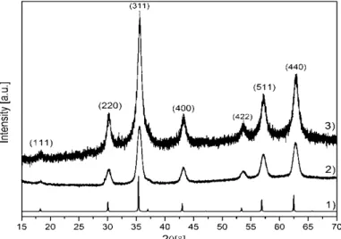
The intensity XRD data for both investi- gated samples was collected over the range 15˚- 70˚ with a step size of 0.01 and an exposure time of 16 s/step. Both patterns exhibit the characteri- stic XRD spectra of JCPDS card No. 85-1436.
The peaks appear at 2θ angle of 18.27˚, 30.05˚, 35.39˚, 43.01˚, 53.38˚, 56.88˚ and 62.46˚. Accor- ding to Scherrer formula, the mean size of
“Flower60” iron oxide nanoparticles is 11.3 nm.
Nanoparticles obtained by coprecipitation method have the size of 10.9 nm. The XRD images of samples are shown in Figure 6.
Figure 6. X-ray diffraction spectra of 1) FindIt, 2) Thermal decomposition method, 3) Precipitation method
The value of mean particles diameter obtained by Zetasizer is higher than the diameter estimated from TEM and XRD because the hydrodynamic diameter includes the diameter of the nanoparticle or groups from ferrofluid carrier attached or absorbed on surface [14].
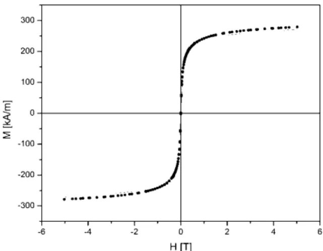
Magnetic nanoparticles were also characteri- zed by SQUID. Figure 7, 8 depict the M-H curve measured at 300 K on iron oxide nanoparticles. The measured M-H curve is fitted by Langevin function [15]. The median diameter of particles, obtained by thermal decomposition method, 10.2 nm was deter- mined from fitting parameters. Nanoparticles obtai- ned by coprecipitation have diameter 9.8 nm. The approximate diameter is evaluated using the following equation
6
3 s p
M m
d (2) where mp is magnetic moment and Ms is satura- tion magnetization [16].
Figure 7. Magnetization histeresis loop of „Flower60”
nanoparticles with fitted Langevin function
Figure 8. Magnetization histeresis loop of „MNPD”
nanoparticle with fitted Langevin function
4. CONCLUSION
In the presented experimental research, we have obtained and characterized magnetic nanopartic- les which can be used as efficient agent in magnetic hyperthermia. We have synthesized iron oxide nano- particles by two different chemical methods, deter- mined their size distributions as well as hydrodyna- mic diameters of single-core iron oxide nanoparticle samples applying different analysis techniques. The properties of the iron oxide nanoparticles and nano- particle-based ferrofluids were studied by X-ray dif- fraction, transmission electron microscopy, SQUID measurments and DLS technique. According to Scherrer formula, the mean XRD size of the iron oxide nanoparticles is about 11 nm. The microscope studies indicated the formation of nanosized partic- les nearly spherical in shape, whereas the average particle size for both samples is found to be about 11 nm. Zeta Sizer is used to determine hydrodynamic diameter which is in range between 15.8 – 40.3 nm for sample synthesized by coprecipitation and between 11.4 − 40.4 nm for the sample obtained by thermal decomposition. The median diameter of nanoparticles, determined from Langevin fitting parameters, is about 10 nm. The results have proven that synthesized nanoparticles have very promising properties for application in magnetic hyperthermia.
5. REFERENCES
[1] B. Issa, I. M. Obaidat, B. A. Albiss, Y.
Haik, Magnetic Nanoparticles: Surface Effects and Properties Related to Biomedicine Applications, Int J Mol Sci., Vol. 14−11 (2013) 21266–21305.
[2] R. Hiergeist, W. Andria, N. Buske, R.
Hergt, I. Hilger, U. Richter, W. Keiser, Application of magnetite ferrofluids for hyperthermia, Journal of magnetism and magnetic materials, Vol. 201 (1999) 420−422.
[3] N. Iacob, G. Schinteie, P. Paladem C. M.
Ticos, V. Kuncser, Stepped heating procedure for experimental SAR evaluation of ferrofluids, The European Physical Journal E, Vol. 38 (2015 ) 57.
[4] M. Banobre-López, A. Teijeiro, J. Rivas, Magnetic nanoparticle-based hyperthermia for can- cer treatment, Reports of practical oncology and radiotherapy, Vol. 18 ( 2013 ) 397–400.
[5] E. A. Perigo, G. Hemery, O.Sandre, D.
Ortega, E. Garaio, F. Plazaola, F. J. Teran, Funda- mental and advances in magnetic hyperthermia, Applied Physics Reviews, Vol. 2 (2015) 041302.
[6] Emil Pollert and Karel Zaveta, Nanocrystaline oxides in magnetic fluid hyperthermia, Praha, 2012, 3−11.
[7] Z. Nemati, S. M. Salili, J. Alonso, A. Ata- ie, R. Das, M. H. Phan, H. Srikant, Superparamag- netic iron oxide nanoparticle for hyperthermia therapy: Does size matter?, Journal of Alloys and Compounds, Vol. 714 (2017) 709−714.
[8] V. Patstula, M. Moskvin, S. Dutz, D.
Horak, Size-dependent magnetic properties of iron oxide nanoparticles, Journal of Physics and Chemistry of Solids, Vol. 88 (2016) 24−30.
[9] F. Ludwig, C. Balceris, T.Viereck, O.
Posth, U. Steinhoff, H. Gavilan, R. Costo, L. Zeng, E. Olsson, C. Jonasson, C. Johansson, Size analysis of single-core magnetic nanoparticles, Journal of magnetism and Magnetic Materials, Vol. 427 (2017) 19−24.
[10] M. R. Gao, S. R. Zhang, J. Jiang, Y. R.
Zheng, D. Q. Tao, S. H. Yu, One-pot synthesis of hierarchical magnetite nanochain assemblies with complex building units and their application for
water treatment, Journals of Materials Chemistry, Vol. 21 (2011) 16888-16892
[11] https://imagej.nih.gov/ij/
[12]http://www.-
biophysics.bioc.cam.ac.uk/files/
Zetasizer_Nano_user_manual_Man0317- 1.1.pdf
[13] S. Junaid S. Qazi, Adrian R. Rennie, Jeremy K. Cockcroft, Martin Vickers, Use of wide- angle X-ray diffraction to measure shape and size of dispersed colloidal particles, Journal of Colloid and Interface Science 338 (2009) 105–110.
[14] J. Stetefeld, S. A. McKenna, T. R. Patel, Dynamic light scattering: a practical guide and applications in biomedical sciences, Biophys Rev., Vol. 8−4 (2016) 409–427.
[15] S. Blundell, Magnetism in Condensed Matter, Oxford University Press, London, 2001, 24–
25.
[16]http://www.df.uns.ac.rs/files/200/marin_ta di___-_doktorska_disertacija_pdf_(f1-25).pdf.
ДОБИЈАЊЕ И КАРАКТЕРИЗАЦИЈА МАГНЕТНИХ НАНОЧЕСТИЦА ГВОЖЂЕ-ОКСИДА Сажетак: Магнетна хипертермија је нова метода лијечења канцера којa би упо- требом магнетних наночестица могла да унаприједи постојеће терапијске методе.
Хипертермијски третман подразумијева загријавање ткива помоћу магнетних наноче- стица што омогућава уништавање ћелија канцера, уз минимално оштећење здравог ткива. Загријавање се остварује примјеном магнетног поља наизмјеничне струје. Циљ истраживања је синтеза наночестица оксида гвожђа погодних за примјену у хипер- термији. Наночестице су добијене примјеном двије различите методе. Први узорак је добијен у процесу термалне декомпозиције у раствору полиола, гдје је као прекурсор коришћена со гвожђа. Други узорак добијен је примјеном методе преципитације, која се базира на мијешању раствора гвожђе-оксида са раствором NaOH. Припремљени узорци испитивани су дифракцијом рендгенских зрака, SQUID магнетометром, тран- симисионим електронским микроскопом и Zeta Sizer уређајем за карактеризацију нано- честица. Добијени резултати су важни за проучавање фундаменталног понашања маг- нетних наночестичних система, али такође дају значајан допринос у развоју методе магнетне хипертермије.
Kључне ријечи: магнетне наночестице, гвожђе-оксид, хипертермија, третман карцинома.