M E T H O D O L O G Y A R T I C L E Open Access
A novel family of expression vectors with multiple affinity tags for wheat germ cell- free protein expression
Szilvia Krisztina Nagy†, Brigitta Margit Kállai†, Judit András and Tamás Mészáros*
Abstract
Background:Cell-free protein expression has become a widely used alternative of in vivo, cell-based systems in functional and structural studies of proteins. The wheat germ-based method outstands from the commercially available eukaryotic in vitro translation systems by its flexibility, high translation efficiency and success rate of properly folded eukaryotic protein synthesis. The original T7 promoter containing pEU3-NII vector was improved previously by addition of a ligation-independent cloning site, His6- and GST-tags, and a TEV protease cleavage site to facilitate the creation of recombinant plasmids, permit affinity purification, and enable production of purified, tag-free target proteins, respectively.
Results:Here, we describe a further development of pEU3-NII vector by inserting the rare-cutting, NotI restriction enzyme cleavage site to simplify vector linearization step prior to in vitro transcription. Additionally, His12, FLAG, and Halo affinity tag coding vectors have been created to increase detection sensitivity, specificity of interaction studies, and provide covalently linkable ligands for pull-down assays, respectively. Finally, the presented GST-His6, and GST- biotin double-tagging vectors could broaden the range of possibilities of protein-protein interaction studies.
Conclusions:The new generation of pEU3-NII vector family allows a more rapid production of translationally active mRNA and wheat germ cell-free expression of target proteins with a wide variety of affinity tags thus enables designing flexible and diverse experimental arrangement for in vitro studies of proteins.
Keywords:Cell-free, Protein expression, Wheat germ, TEV, Affinity tag, FLAG, Halo
Background
Although straightforward and cost-effective production of properly folded proteins is a general starting point of protein studies, the accomplishment of this step could be a challenging task, especially in the case of eukaryotic proteins. Various cell-free in vitro translation systems have been developed to subdue this obstacle and amongst them, the wheat germ-based approach systems
seem to be the choice of method for high-throughput eukaryotic protein production [1,2].
The eukaryotic translation machinery requires an exten- sively modified mRNA, namely 5′methylguanosine cap- ping and 3′ poly(A) tailing. In order to evade these complex posttranscriptional modifications, a wheat germ in vitro translation compatible vector has been con- structed with the cap replacing, translational enhancerΩ sequence from the tobacco mosaic virus and an additional GAA triplet at the 5′-end (GAAΩ) [3]. The same group also demonstrated that translation did not strictly depend on the presence of poly(A) tail, it could be replaced by a 1626 nucleotide long sequence, thus any 3′-UTR se- quence of minimum length could substitute the poly(A)
© The Author(s). 2020Open AccessThis article is licensed under a Creative Commons Attribution 4.0 International License, which permits use, sharing, adaptation, distribution and reproduction in any medium or format, as long as you give appropriate credit to the original author(s) and the source, provide a link to the Creative Commons licence, and indicate if changes were made. The images or other third party material in this article are included in the article's Creative Commons licence, unless indicated otherwise in a credit line to the material. If material is not included in the article's Creative Commons licence and your intended use is not permitted by statutory regulation or exceeds the permitted use, you will need to obtain permission directly from the copyright holder. To view a copy of this licence, visithttp://creativecommons.org/licenses/by/4.0/.
The Creative Commons Public Domain Dedication waiver (http://creativecommons.org/publicdomain/zero/1.0/) applies to the data made available in this article, unless otherwise stated in a credit line to the data.
* Correspondence:tamas.meszaros.su@gmail.com
†Szilvia Krisztina Nagy and Brigitta Margit Kállai contributed equally to this work.
Department of Medical Chemistry, Molecular Biology and Pathobiochemistry, Semmelweis University, 37-47 Tűzoltó Street, Budapest H-1094, Hungary
tail. To produce a linear DNA template for T7 RNA poly- merase [4], the vector construct is cut by a restriction endonuclease at a specific site, forming a 3′-UTR of the appropriate length. The above-mentioned observations re- sulted in the creation of pEU3-NII and pEU-E01 vectors harbouring T7 and SP6 promoters, respectively, that could be efficiently applied for in vitro production of translation- ally active mRNAs.
However, their application was hindered by restriction endonuclease-based insertion of the coding sequence of interest and lack of protein purification aiding affinity tags.
To increase their practicality, we have redesigned the ori- ginal vectors and replaced the multicloning site by a ligation-independent cloning (LIC) compatible sequence and inserted tobacco etch virus (TEV) protease cleavable hexahistidine (His6), and glutathione S-transferase (GST) coding sequences to aid the construction of recombinant plasmids and the purification of in vitro translated pro- teins [5]. The improved vectors (pEU3-NII-HLIC and pEU3-NII-GLIC) could shorten the timeline of ‘from DNA to purified functional protein’to a week and we suc- cessfully applied them for synthesizing dozens of eukaryotic proteins of various organisms [6–12].
The emergence of recombinant affinity tags was a sig- nificant improvement of the heterologous protein produ- cing systems since they streamlined purification of the protein of interest and augmented the solubility of expressed proteins in many cases. Since the first applica- tion of affinity tags several protein labelling techniques have been invented mainly to meet the demands of downstream applications of synthesized proteins [13].
In order to extend the versatility of wheat germ in vitro translation systems, we set out to expand the pEU3-NII vector family by providing various protein affinity tags, which could aid pull-down assays, interaction analysis, and enzymatic functional studies of synthesized target proteins. Firstly, a rare-cutting restriction endonuclease (NotI) cleavage site [14] was inserted into the pEU3-NII vectors to allow the straightforward plasmid linearization prior to in vitro mRNA transcription. We demonstrate that protein detection sensitivity is elevated by using our double-His6label coding vector [15]; while the FLAG-tag offering plasmid ensures a highly selective detection of la- belled protein [16, 17]. Additionally, we show that our HaloTag [18] encoding vector is optimal for producing proteins that could be covalently linked to affinity beads of pull-down assays. Finally, we provide the vectors with GST-His6and GST-biotin affinity tags with TEV cleavable GST to ease the purification of labelled recombinant proteins.
Results and discussion
The pEU3-NII-HLIC and pEU3-NII-GLIC vectors were extensively used for synthesizing a great variety of
recombinant proteins by the wheat germ in vitro transla- tion, therefore a next improvement was implemented to make the procedure even more straightforward and adaptable.
The vector constructs are linearized by restriction endonuclease in advance of in vitro mRNA synthesis by T7 RNA polymerase. Cleavage sites present in the ORF of the genes of interest are not suitable, thus a generally usable, rare-cutting restriction endonuclease cleavage site, NotI (GCGGCCGC) was inserted by in vitro muta- genesis in both vectors 2424 bp downstream from the LIC site (pEU3-NII-HLICNot and pEU3-NII-GLICNot).
That resulted in a more straightforward sample handling in case of multiple plasmid constructs and mRNAs with identical, appropriate size 3′-UTR were produced.
Probably, the most popular label of recombinant pro- teins is the amino acid stretch of six histidines (His6);
however, its usability is hindered by the relatively low binding affinity of coordinating histidine residues to metal ions and promiscuity of His6selective antibodies.
To mitigate these drawbacks, a double-His6 holding E.
coli expression vector was constructed by Khan et al.
[15]. The authors demonstrated that application of double-His6improved the binding affinity of proteins to nickel-nitrilotriacetic acid (Ni-NTA) modified surfaces and increased the detectability of overexpressed proteins.
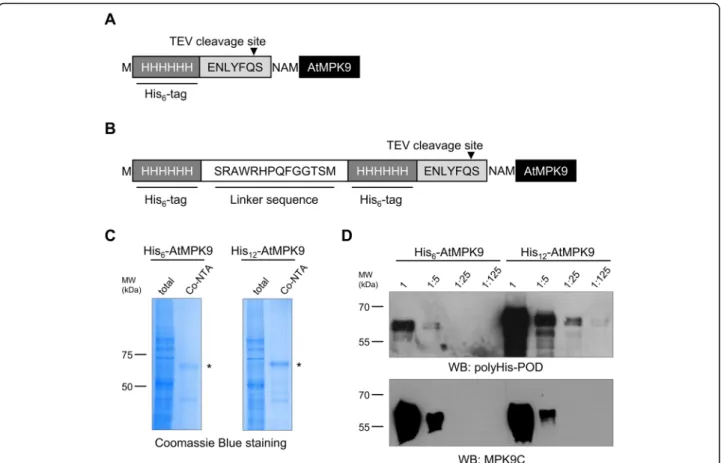
Considering that characterization of weak protein- protein interactions requires sensitively detectable pro- teins, we constructed a vector for in vitro translation of N-terminally double-His6 (His12) labelled recombinant proteins. In order to assess the advantage of the novel plasmid construct, the coding sequence of anArabidop- sis thaliana mitogen-activated protein kinase (AtMPK9) was inserted into the created pEU3-NII-HxHLICNot vector by LIC method. His6-AtMPK9 (Fig. 1a) and His12-AtMPK9 (Fig. 1b) were produced by wheat germ in vitro translation, purified by immobilized metal affin- ity chromatography (IMAC) with Co-NTA beads and analysed by Coomassie Blue staining. The results of af- finity purification demonstrated that both recombinant proteins were effectively synthesized and purified by Co- NTA beads (Fig. 1c). To test the sensitivity of protein detection, we prepared five-fold dilutions of total translation mixtures containing His6-AtMPK9 and His12-AtMPK9 proteins and analysed them by Western blot. The results showed that doubling the number of histidine residues significantly elevated the sensitivity of Western blot analysis; at least one order of magnitude smaller amount of His12-AtMPK9 was sufficient for the detection with anti-polyHistidine antibody in compari- son to the traditionally His6-tagged AtMPK9 (Fig. 1d).
To rule out unequal loading of the differently tagged AtMPK9 proteins, immunodetection with anti-MPK9C antibody was also performed. (Fig.1d).
Due to its small size, hydrophilic nature, and tyrosine content, the FLAG epitope tag commonly resides on the surface of the fusion protein and provides an excellent antibody binding site [17]. These properties of FLAG-tag resulted in the generation of greatly discriminating com- mercial antibodies and wide-spread application of this labelling system. FLAG-tagging is a rational choice of protein labelling when highly selective detection rather than purification of the synthesized protein is the prior- ity. To assess the performance of the FLAG system in comparison to other antibodies in the wheat germ pro- tein extract, total translation mixtures were separated by SDS-PAGE, blotted and analysed by using anti- polyHistidine, anti-GST, and anti-FLAG antibodies.
Detection of protein-bound antibodies by enhanced chemiluminescence (ECL) demonstrated that anti- polyHistidine and anti-GST IgG could bind to several endogenous wheat germ proteins, while anti-FLAG anti- body remarkably did not produce any signal (Fig. 2a).
The undetectable background signal of FLAG antibodies with wheat germ protein extract encouraged us to de- velop a pEU3-NII vector with N-terminal FLAG-tag. In
order to examine the superiority of the novel vector, AtMPK9 was synthesized with His6- and FLAG-tag (Fig.
2b) and the total translation mixtures were studied by Western blot analysis. In accordance with the previous results, the FLAG selective antibody decorated a single band of FLAG-AtMPK9 at the expected molecular weight (Fig.2c). Although the polyHistidine antibody ef- fectively detected His6-AtMPK9, an additional, endogen- ous protein of wheat germ extract was also clearly recognized by this antibody (Fig.2d).
Elucidating protein function involves the understand- ing of dynamic cellular protein networks and the pull- down technique is a valuable tool for revealing the underlying protein-protein interactions. It is equally suit- able for identification of novel interacting partners and confirmation of putative interactions. The validity of pull-down assay provided data is greatly determined by the appropriately chosen binding and washing condi- tions; thus, the strong ligand-bead interaction is an inev- itable component of successful assays. To address the above issue, HaloTag, a 297 amino acid tag derived from a bacterial haloalkane dehalogenase was designed that
Fig. 1IMAC purification and Western blot analysis of His6-AtMPK9 and His12-AtMPK9 proteins.a,bAmino acid sequence of the tagging regions of His6- and His12-labeled AtMPK9 vector constructs.cThe in vitro translated His6-AtMPK9 and His12-AtMPK9 were purified with Co-NTA affinity beads, and the total translation mixtures and purified fractions were separated with SDS-PAGE and visualized by Coomassie Blue staining. The purified AtMPK9 proteins are indicated by asterisks.dSerial five-fold dilutions of 1μl of total translation mixtures were studied by Western blotting using anti-polyHis-POD and anti-MPK9C. The molecular weight markers are shown in kDa
offers an important advantage by formation of a highly specific covalent bond with its synthetic ligands [18].
This property allows immobilization and manipulation of bait proteins even under stringent buffer conditions.
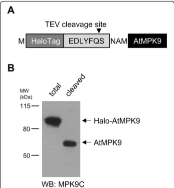
In order to offer a HaloTag bait protein-coding vector, the Halo protein coding sequence was inserted upstream of the TEV protease cleavage recognition motif and the LIC site (Fig. 3a). To check the functionality of the cre- ated vector, Halo-AtMPK9 protein was translated and purified by HaloTag affinity beads. Since the strong co- valent bond formed by the dehalogenase enzyme was not cleavable even by boiling the sample in SDS loading buffer (data not shown), AtMPK9 was released from the beads by TEV protease treatment. Western blot analysis of total translation mixture and supernatant of TEV treated beads by AtMPK9 selective antibody demon- strated the success of the vector construction: a single protein of total translation mixture was decorated at the expected molecular weight, and according to the West- ern blot results, the covalently bead-bound Halo- AtMPK9 was recovered from the beads by TEV protease cleavage since a single protein with the molecular weight of AtMPK9 was detected in the supernatant by anti- MPK9C antibody (Fig.3b).
The most generally used method to produce purified and tagged recombinant proteins is the elution of
proteins by solutions of ligand binding site competing compounds. However, the presence of added com- pounds in the eluate could often hinder the downstream processes; hence, they have to be removed by dialysis or desalting. The above-mentioned steps necessarily result in some loss of protein of interest making purification of protein at low concentration extremely challenging.
Double-labelled recombinant protein coding vectors could be valuable tools in the above scenarios assuming that one of the tags can be cleaved by protease digestion.
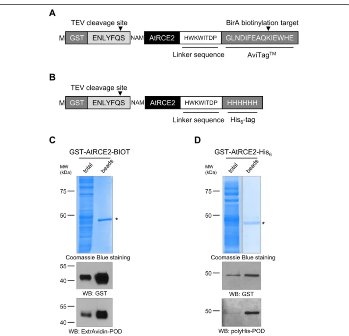
In consideration of this advantage of double-labelled proteins, we set out to further modify our GST-tag con- taining vector by the insertion of His6-tag or AviTag bio- tin labelling sequence [19] downstream of the LIC site (Fig. 4a and b). The created novel vectors were assessed by applying them to produce AtRCE2, an Arabidopsis thaliana ubiquitin-conjugating enzyme. The proteins were in vitro translated as described above and the Avi- Tag biotinylation was accomplished co-translationally.
To this end, the translation mixture was supplemented with biotin and BirA biotin ligase enzyme. Following 20 h of translation, the proteins were purified by using glutathione affinity beads. PAGE separation and Coo- massie Blue staining of bead-bound fraction showed a single protein of estimated size implying successful GST-tagging and effective purification of AtRCE2 (Fig.
Fig. 2Western blot analysis of crude wheat germ protein extract and FLAG-tagged AtMPK9.aTotal translation mixture of wheat germ extract (W7240) was separated by TGX Stain-Free gel and imaged by UV light exposure (bottom panel). Following the transfer of proteins to PVDF membrane samples were analysed by Western blotting using anti-polyHis-POD (lane H), anti-GST (lane G), and anti-FLAG (lane F) antibodies.b Amino acid sequence of the tagging region of FLAG-AtMPK9.cTotal translation mixture containing FLAG-AtMPK9 was separated by SDS-PAGE and analysed by Western blotting using anti-FLAG antibody.dTotal translation mixture containing His6-AtMPK9 was separated by SDS-PAGE and analysed by Western blotting using polyHis-POD antibody. The most abundant non-specific protein signal detected with anti-polyHis-POD is marked by an asterisk. The molecular weight markers are shown in kDa
4c and d). To testify the presence of C-terminal His6
and biotin label extensions of synthesized AtRCE2, both total translation mixtures and glutathione bead-bound fractions were analysed by using peroxidase conjugated anti-polyHistidine antibody and ExtrAvidin. The perox- idase generated chemiluminescence signal provided a single band at the expected molecular weight with both tags indicating successful creation of double-tagging vec- tors (Fig.4c and d).
Conclusions
Here, we presented the improvement of the previously constructed pEU3-NII ligation-independent cloning plas- mids and the creation of a wheat germ cell-free translation compatible vector family with a variety of affinity tags. In- sertion of NotI - a rare-cutting restriction enzyme - recog- nition site to the 3′-UTR region evades extended restriction endonuclease site analysis of vector constructs thus allows general handling of multiple constructs simul- taneously. The application of developed vector family pro- vides various labelling opportunities for the protein of interest. The N-terminal His6- or FLAG-tag coding vec- tors could be respectively applied in experimental arrange- ments when sensitivity or selectivity has the highest priority. The HaloTag possessing vector could be a
rational choice for synthesizing proteins for pull-down as- says since the Halo provided covalent linkage of bait pro- tein permits application of stringent washing conditions.
The double-labelled GST-His6 and GST-biotin holding vectors have two advantageous features. First, the N- terminally localized GST- tag helps folding of the protein of interest; thus, it could help the production of challen- ging proteins. Second, due to TEV cleavability of GST, the affinity-tagged proteins could be purified without using buffers that could hinder the downstream application of the protein of interest. Summarily, the described vector family could present a useful toolkit for in vitro protein studies by providing a straightforward approach for pro- ducing proteins of various labels.
Methods Cloning
The pEU3-NII vector backbone was previously modified by Bardóczy et al. [5] to encode for His6-tag (pEU3-NII- HLIC) and GST-tag (pEU3-NII-GLIC). The NotI restric- tion endonuclease was introduced into both vectors using QuikChange Site-Directed Mutagenesis Kit (Agi- lent) according to the provided manual with the 5′- ATTCTGAGAATAGTGTATGCGGCcgCCGAGTT GCTCTTGCCCGG-3′ and 5′-CCGGGCAAGAGCAA CTCGGcgGCCGCATACACTATTCTCAGAAT-3′
primers (NotI recognition sequence underlined, mutated bases lowercase). The resulting pEU3-NII-HLICNot and pEU3-NII-GLICNot vectors were further manipulated to produce novel modified vectors suitable for wheat germ cell-free protein expression.
In general, the previously constructed pEU3-NII- HLICNot and pEU3-NII-GLICNot vectors were cut with the appropriate FastDigest restriction endonucleases (Thermo Scientific), treated with FastAP Thermosensi- tive Alkaline Phosphatase (Thermo Scientific) and used in ligation reactions. The colonies containing the new fragments were selected by colony PCR with pEU3-NII sequencing primers pEU3-NII-forward (5′-CACTAT AGGGTACACGGAATTCGC-3′) and pEU-rev (5′- TATAGGAAGGCCGGATAAGACG-3′). Colony PCR positive clones were propagated, and the plasmid DNAs were isolated with Zippy Plasmid Miniprep kit (Zymo Research) for sequencing. See Supplementary Figure 1, Additional file 1 for the schematic representations of vector constructs.
pEU3-NII-HxHLICNot
The SpeI-linker-hexaHistidine-SpeI encoding fragment was generated by annealing oligonucleotides: 5′- CGGACTAGTATGCATCATCATCATCATCATTCTC GTGCTTGGCGTCACCCGCAGTTCGGTGGTACT AGTCCG-3′and 5′-CGGACTAGTACCACCGAACTGC GGGTGACGCCAAGCACGAGAATGATGATGATG
Fig. 3Western blot analysis of translated Halo-AtMPK9 protein.a Amino acid sequence of tagging region of HaloTag-labelled AtMPK9 vector construct.bHalo-AtMPK9 was purified from total translation mixture with HaloTag affinity beads and AtMPK9 was released from the beads by TEV protease cleavage. The samples were separated by SDS-PAGE and AtMPK9 was detected by immunoblotting with anti- MPK9C antibody. The molecular weight markers are shown in kDa
ATGATGCATACTAGTCCG-3′. The created fragment was digested with SpeI and inserted into SpeI-digested pEU3-NII-HLICNot.
pEU3-NII-FLAGLICNot and pEU3-NII-HaloLICNot
The FLAGTEVLIC encoding cassette harbouring SpeI and SspI recognition sites was generated by annealing oligonucleotides 5′-GGACTAGTATGGATTATAAGG ACGACGACGACAAAGAGAACCTGTACTTCCAATC CAATATTGG-3′ and 5′-CCAATATTGGATTGGAAG
TACAGGTTCTCTTTGTCGTCGTCGTCCTTATAAT CCATACTAGTCC-3′. The HaloTag sequence including the TEV protease recognition site was amplified with forward primer 5′-TAACTAGTATGGCAGAAATCGG TACTG-3′ and reverse primer 5′-ATTGGATTGG AAGTACAGATCCTCAGTGG-3′ from the pHTN HaloTag® CMV-neo Vector (Promega) so that SpeI re- striction endonuclease recognition sequence and SspI blunt end sequence were appended to both ends of the PCR product. Both FLAG and Halo coding DNA
Fig. 4Western blot analysis of purified GST-AtRCE2-BIOT and GST-AtRCE2-His6double-tagged proteins.aThe amino acid sequence of the tagging region of GST-AtRCE2-BIOT.bThe amino acid sequence of the tagging region of GST-AtRCE2-His6.c,dThe double-tagged proteins (GST- AtRCE2-BIOT and GST-AtRCE2-His6) were purified by glutathione affinity beads. The samples were separated by SDS-PAGE, visualized by
Coomassie Blue staining and Western blotting with anti-GST, and polyHis-POD antibodies or ExtrAvidin-POD. Affinity purified proteins are marked by asterisks. The molecular weight markers are shown in kDa
sequences were digested with the appropriate enzymes and inserted into SpeI and SspI digested pEU3-NII- GLICNot vector.
pEU3-NII-GLICNot-C-BIOT and pEU3-NII-GLICNot-C-his The C-terminal AviTag and His6-tag encoding cassettes with BamHI and SmaI overhangs were generated by an- nealing the following oligonucleotides:
AviTag
5′-AAGGATCCCGGCCTCAACGACATCTTCGAG GCCCAGAAGATCGAGTGGCACGAGTGACCCGG GT-3′and 5′-ACCCGGGTCACTCGTGCCACTC GATCTTCTGGGCCTCGAAGATGTCGTTGAGGC CGGGATCCTT-3′;
His6-tag
5′-AAGGATCCCCATCATCATCATCATCATTGA CCCGGGT-3′and 5′-ACCCGGGTCAATGATGAT GATGATGATGGGGATCCTT-3′.
The obtained dsDNA fragments were treated with the appropriate enzymes and inserted into BamHI and SmaI digested pEU3-NII-GLICNot vector.
Ligation-independent cloning
Protein coding sequences were inserted into the appropri- ate vectors by ligation-independent cloning (LIC) accord- ing to the previously described method [20]. AtMPK9 (AT3G18040) was inserted into pEU3-NII-HLICNot, pEU3-NII-HxHLICNot, pEU3-NII-FLAGLICNot, and pEU3-NII-HALOLICNot. AtRCE2 (AT2G18600) was inserted into pEU3-NII-GLICNot-C-BIOT and pEU3- NII-GLICNot-C-His.
Briefly, the inserts were amplified by PCR with their specific forward and reverse primers (See Supplementary Table 1, Additional file 2) and purified by polyethylene glycol and ethanol precipitation. The vectors were line- arized with SspI and purified from agarose gel following electrophoresis. The SspI cut vectors were incubated with dGTP (Bioline) and the inserts were incubated with dCTP (Bioline) in the presence of T4 DNA polymerase (Roche). The T4 treated vectors and inserts were mixed at a 1:3 molar ratio in the presence of EDTA and the resulting ligation mixtures were transformed into XL10- Gold competent cells. Colony PCR positive clones were propagated, and the plasmid DNAs were isolated with Zippy Plasmid Miniprep kit (Zymo Research).
In vitro transcription and translation
The vector constructs were used for in vitro transcription and translation as described previously [20]. Briefly, the vectors were linearized by NotI restriction endonuclease enzyme, purified by polyethylene glycol and ethanol pre- cipitation; 1μg of linearized plasmid was used as a
template of in vitro transcription reaction performed by TranscriptAid T7 High Yield Transcription Kit (Thermo Scientific) according to the manufacturer’s protocol. The mRNA was purified by ammonium acetate and ethanol precipitation and resuspended in 20μl of 1X SUB-AMIX solution (CellFree Sciences). 5μl WEPRO wheat germ extract (CellFree Sciences, W7240G except for the His- tagged proteins that were translated with W7240H or W7240 extract, as indicated), 2.5μl SUB-AMIX, 2.5μl mRNA, 0.4μl 1 mg/ml creatine kinase (Roche) were gen- tly mixed and carefully underlaid to 103μl SUB-AMIX solution in a 96-well plate. AviTag containing proteins were biotinylated during the translation reactions by add- ing 25 ng Trx-BirA biotin ligase enzyme [21] to the bot- tom layer, and 0.5μM (final concentration) of D-biotin (Supelco) to both layers [22]. The translation reactions were carried out for 20 h at 20 °C, that yielded approxi- mately 5–10μg protein of interest in 113.4μl total reac- tion mixture. Expected molecular weights of expressed proteins: His6-AtMPK9 60.4 kDa, His12-AtMPK9 62.8 kDa, FLAG-AtMPK9 60.6 kDa, Halo-AtMPK9 93.5 kDa, GST-AtRCE2-BIOT 50.8 kDa, GST-AtRCE2-His6 49.8 kDa.
Purification of expressed proteins
Purification of His6-AtMPK9 and His12-AtMPK9
One hundred microliter translation mixture was diluted two times with 2X Lysis Buffer (100 mM NaH2PO4, 600 mM NaCl, 20 mM imidazole, pH 8), mixed with 13μl equilibrated PureCube Co-NTA Agarose (Cube Biotech) and incubated on a rotator for 1 h at room temperature.
After washing the beads three times with Wash Buffer (50 mM NaH2PO4, 300 mM NaCl, 20 mM imidazole, pH 8), the beads were resuspended in 50μl PBS.
Purification of HALO-AtMPK9
Twenty microliter translation mixture was diluted two times with HALO Equilibration buffer (PBS with 0.005%
NP-40), mixed with 0.5μl equilibrated Magne HaloTag Beads (Promega) and incubated on a rotator for 1 h at room temperature. After washing the beads three times with HALO Equilibration buffer, the beads were resus- pended in 10μl 1X TEV buffer (Sigma) containing 0.1μl TEV protease (Sigma, T4455) and 1 mM DTT, and the cleavage reaction was conducted for 1 h at 30 °C. The supernatant containing the untagged AtMPK9 with two extra amino acids at its N-terminal (expected molecular weight 58.7 kDa) was mixed with SDS-DTT sample buf- fer and boiled for 5 min at 95 °C.
Purification of GST-AtRCE2-BIOT and GST-AtRCE2-His6
One hundred microliter translation mixture was diluted six times with Binding/Wash Buffer (125 mM Tris-HCl pH 8, 150 mM NaCl, 0.5% Triton-X), mixed with 12.5μl
equilibrated Pierce Glutathione Magnetic Beads (Thermo Scientific) and incubated on a rotator for 1 h at room temperature. The beads were washed three times with Binding/Wash buffer and resuspended in 50μl PBS.
SDS-PAGE and Western blot
The samples were mixed with SDS-DTT sample buffer and boiled for 5 min at 95 °C. If not indicated otherwise, 5μl of total translation mixture and 10μl of bead-bound proteins were prepared for Coomassie Blue staining, and 1–3μl of total translation mixture and 2–5μl of bead- bound proteins were prepared for immunoblotting. The protein samples were separated on 8%/10%/15% SDS- PAGE with Laemmli buffer or 4–12% Bis-Tris gradient gels (Expedeon) with MES buffer (Expedeon) and the proteins were either stained directly in the gel by Coo- massie Blue or transferred to PVDF membrane by semi- dry blotting. The protein samples, which were separated on 10% TGX Stain-Free Precast gels with Laemmli buf- fer, were activated and imaged with Gel Doc XR+ Gel Documentation System (Bio-Rad) and subsequently transferred to PVDF membrane by semi-dry blotting.
The membranes were blocked with 5% non-fat milk powder in PBS/TBS containing 0.05% Tween-20. The proteins were detected either directly by anti- polyHistidine-POD (Sigma) diluted 1:2000 or by rabbit anti-GST (Upstate) diluted 1:2000, rabbit anti-MPK9C (raised in-house) diluted 1:2000, mouse anti-FLAG® M2 (Sigma) diluted 1:2000 and secondary antibodies goat anti-rabbit-HRP (Cell Signaling) and horse anti-mouse- HRP diluted 1:5000. To detect biotinylated proteins, the membranes were blocked with 5% BSA in PBS contain- ing 0.05% Tween-20 and challenged with 1:5000 diluted ExtrAvidin-POD (Sigma). The peroxidase activity was detected by ECL (Thermo Scientific) in all cases.
Accession numbers
The created vectors are available via Addgene by using the following ID numbers:
pEU3-NII-HLICNot (ID 140181), pEU3-NII-GLICNot (ID 140182), pEU3-NII-HxHLICNot (ID 140183), pEU3- NII-FLAGLICNot (ID 140184), pEU3-NII-HaloLICNot (140185), pEU3-NII-GLICNot-C-BIOT (ID 140186), pEU3-NII-GLICNot-C-His (140187).
Supplementary information
Supplementary informationaccompanies this paper athttps://doi.org/10.
1186/s12896-020-00610-5.
Additional file 1 : Supplementary Figure 1. Schematic illustrations of modified pEU3-NII plasmids. Utilized restriction endonucleases (the unique cutters are shown in bold), plasmid names and sizes are indicated on the maps. Ampicillin resistance gene (AmpR), origin of replication (ori), T7 promoter, glutathione-S-transferase (GST) gene, tobacco etch virus protease recognition site (TEV site with amino acid sequence E(N/
D)LYFQS), LIC (ligation-independent cloning) site. Plasmid maps produced by using SnapGene software (from GSL Biotech; available atsnapgene.
com).
Additional file 2 Supplementary Table 1.Sequences of primers used for ligation-independent cloning. Underlined letters represent the start and stop codons, bold letters the gene-specific sequences.
Abbreviations
AtMPK9:Arabidopsis thalianamitogen-activated protein kinase 9;
AtRCE2:Arabidopsis thalianarelated to ubiquitin-conjugating enzyme 2;
GST: Glutathione S-transferase; His12: Double-His6; His6: Hexahistidine;
IMAC: Immobilized metal affinity chromatography; LIC: Ligation-independent cloning; Ni-NTA: Nickel-nitrilotriacetic acid; PVDF: Polyvinylidene fluoride;
TEV: Tobacco etch virus
Acknowledgments
We are grateful for the contribution of András Merényi, Anna Gyurkovics, and Anna Petschner to the experimental work.
Authors’contributions
BKM, SKN, JA and TM designed the experiments. BKM, SKN and JA carried out the practical work. BKM, SKN and TM drafted and finalized the manuscript. All authors read and approved the final manuscript and are personally accountable for the author’s own contributions.
Funding
This study was supported by TM’s 111085 and SKN’s 129083 grant of the National Research, Development and Innovation Office, Hungary and by BMK’s EFOP-3.6.3-VEKOP-16-2017-00009 grant. The funders had no role in study design, data collection and analysis, decision to publish, or preparation of the manuscript.
Availability of data and materials
All data generated or analysed during this study are included in this published article and Additional files1and2.
Ethics approval and consent to participate Not applicable.
Consent for publication Not applicable.
Competing interests
The authors declare that they have no competing interests.
Received: 10 January 2020 Accepted: 2 March 2020
References
1. Goshima N, Kawamura Y, Fukumoto A, Miura A, Honma R, Satoh R, et al.
Human protein factory for converting the transcriptome into anin vitro- expressed proteome. Nat Methods. 2008;5:1011–7.https://doi.org/10.1038/
nmeth.1273.
2. Harbers M. Wheat germ systems for cell-free protein expression. FEBS Lett.
2014;588:2762–73.https://doi.org/10.1016/j.febslet.2014.05.061.
3. Sawasaki T, Ogasawara T, Morishita R, Endo Y. A cell-free protein synthesis system for high-throughput proteomics. Proc Natl Acad Sci U S A. 2002;99:
14652–7.https://doi.org/10.1073/pnas.232580399.
4. Milligan JF, Groebe DR, Witherell GW, Uhlenbeck OC. Oligoribonucleotide synthesis using T7 RNA polymerase and synthetic DNA templates. Nucleic Acids Res. 1987;15:8783–98.https://doi.org/10.1093/nar/15.21.8783.
5. Bardóczy V, Géczi V, Sawasaki T, Endo Y, Mészáros T. A set of ligation- independentin vitrotranslation vectors for eukaryotic protein production.
BMC Biotechnol. 2008;8:1–7.https://doi.org/10.1186/1472-6750-8-32.
6. Kálmán FS, Lizák B, Nagy SK, Mészáros T, Zámbó V, Mandl J, et al. Natural mutations lead to enhanced proteasomal degradation of human Ncb5or, a novel flavoheme reductase. Biochimie. 2013;95:1403–10.https://doi.org/10.
1016/j.biochi.2013.03.004.
7. Nagy SK, Darula Z, Kállai BM, Bögre L, Bánhegyi G, Medzihradszky KF, et al.
Activation of AtMPK9 through autophosphorylation that makes it
independent of the canonical MAPK cascades. Biochem J. 2015;467:167–75.
https://doi.org/10.1042/BJ20141176.
8. Kohoutová L, Kourová H, Nagy SK, Volc J, Halada P, Mészáros T, et al. The Arabidopsismitogen-activated protein kinase 6 is associated withγ-tubulin on microtubules, phosphorylates EB1c and maintains spindle orientation under nitrosative stress. New Phytol. 2015;207:1061–74.https://doi.org/10.
1111/nph.13501.
9. Németh CE, Marcolongo P, Gamberucci A, Fulceri R, Benedetti A, Zoppi N, et al. Glucose transporter type 10—lacking in arterial tortuosity syndrome—facilitates dehydroascorbic acid transport. FEBS Lett. 2016;590:
1630–40.https://doi.org/10.1002/1873-3468.12204.
10. Horvath BM, Kourova H, Nagy S, Nemeth E, Magyar Z, Papdi C, et al.
ArabidopsisRETINOBLASTOMA RELATED directly regulates DNA damage responses through functions beyond cell cycle control. EMBO J. 2017;36:
1261–78.https://doi.org/10.15252/embj.201694561.
11. Dory M, Hatzimasoura E, Kállai BM, Nagy SK, Jäger K, Darula Z, et al.
Coevolving MAPK and PID phosphosites indicate an ancient environmental control of PIN auxin transporters in land plants. FEBS Lett. 2017;592:89–102.
https://doi.org/10.1002/1873-3468.12929.
12. Kállai BM, Kourová H, Chumová J, Papdi C, Trögelová L, Kofroňová O, et al.
γ-Tubulin interacts with E2F transcription factors to regulate proliferation and endocycling inArabidopsis. J Exp Bot. 2019;71:1265–77.https://doi.org/
10.1093/jxb/erz498.
13. Kimple ME, Brill AL, Pasker RL. Overview of affinity tags for protein purification. Curr Protoc Protein Sci. 2013;73:9.9.1–9.9.23.https://doi.org/10.
1002/0471140864.ps0909s73.
14. Lambert AR, Sussman D, Shen B, Maunus R, Nix J, Samuelson J, et al.
Structures of the rare-cutting restriction endonuclease NotI reveal a novel metal binding fold involved in DNA recognition. Structure. 2008;16(4):558– 69.https://doi.org/10.1016/j.str.2008.01.017.
15. Khan F, He M, Taussig MJ, Khan F, He M, Taussig MJ. Double-hexahistidine tag with high-affinity binding for protein immobilization, purification, and detection on Ni-nitrilotriacetic acid surfaces. Anal Chem. 2006;78:3072–9.
https://doi.org/10.1021/ac060184l.
16. Knappik A, Pluckthun A. An improved affinity tag based on the FLAG peptide for the detection and purification of recombinant antibody fragments. Biotechniques. 1994;17:754–61.
17. Einhauer A, Jungbauer A. The FLAG™peptide, a versatile fusion tag for the purification of recombinant proteins. J Biochem Biophys Methods. 2001;49:
455–65.https://doi.org/10.1016/s0165-022x(01)00213-5.
18. Los GV, Encell LP, McDougall MG, Hartzell DD, Karassina N, Zimprich C, et al.
HaloTag: a novel protein labeling technology for cell imaging and protein analysis. ACS Chem Biol. 2008;3:373–82.https://doi.org/10.1021/cb800025k.
19. Cull MG, Schatz PJ. Biotinylation of proteinsin vivoandin vitrousing small peptide tags. Methods Enzymol. 2000;326:430–40.https://doi.org/10.1016/
s0076-6879(00)26068-0.
20. Nagy SK, Mészáros T. Cell-free protein synthesis, vol. 1118; 2014. p. 231–43.
https://doi.org/10.1007/978-1-62703-782-2.
21. Li Y, Sousa R. Expression and purification ofE.coliBirA biotin ligase for in vitrobiotinylation. Protein Expr Purif. 2012;82:162–7.https://doi.org/10.
1016/j.pep.2011.12.008.
22. Ramadan A, Nemoto K, Seki M, Shinozaki K, Takeda H, Takahashi H, et al.
Wheat germ-based protein libraries for the functional characterisation of theArabidopsisE2 ubiquitin conjugating enzymes and the RING-type E3 ubiquitin ligase enzymes. BMC Plant Biol. 2015;15:275.https://doi.org/10.
1186/s12870-015-0660-9.
Publisher’s Note
Springer Nature remains neutral with regard to jurisdictional claims in published maps and institutional affiliations.