VIL 1. THE ROLE OF THIOL GROUPS IN THE STRUCTURE AND FUNCTION OF THE MITOTIC APPARATUS
Daniel Mazia*
Department of Zoology, University of California, and the Adolph and Mary Sprague Miller Institute for Basic Research in Science, University of California, Berkeley, California
I. Introduction 367 II. The Isolation of the Mitotic Apparatus 368
1. Information Obtained from Earlier Methods: The Problem of Stability . 368 2. The Direct Isolation of the Mitotic Apparatus from Living Cells . 371
III. The Fate of the Glutathione Cycle 375 IV. —SH and the Functioning of the Mitotic Apparatus : Blockage Experiments 377
V. Protein-S in Intercellular Adhesion: Experiments of Twinning . . . . 382 I. Introduction
Not many scientific ideas possess such charm as to convince us that they ought to be true, and to influence the direction of our research ac- cordingly. One of these has been the notion that the thiol groups play a unique and intimate role in the mechanism and control of cell division, an idea that has been with us for over a quarter of a century. Of course there has been evidence to support it, some of which were reviewed (1) at the time of the 1954 Glutathione Symposium, but its viability could not, until recently, have been accounted for by the evidence alone. More important has been the need for conceptual models of rapid and sensitive changes in the cell that could be valid for the energetics of division, the formation of specialized fibrous structures for carrying out the mitotic process, and the partitioning of the cell body. The cell biologist could hardly have over- looked the fact-of-life that is responsible for this conference: that when we consider proteins in action, we are likely to find clues to their action in the behavior of the thiol groups.
It must be admitted that preconceptions concerning the role of pro- tein-S in mitosis have played a large part in the studies of the mitotic
* These investigations were supported by grants from the American Cancer So- ciety and the National Science Foundation, and by a contract with the Office of Naval Research [Nonr-22(24)L
367
368 DANIEL MAZIA
apparatus which shall be reported here. The mitotic apparatus ( M A ) is the rather well-designed machine which most cells construct at the time of division to carry out the orderly and equitable distribution of the heredi- tary material. This apparatus is not a permanent organ of the cell, but has an embarrassingly teleological history. It is made when the cell is ready to divide and breaks down when its job is done. The performance of its job depends on its essential geometry. It polarizes the cell with respect to the coming division and governs the orientation of the chromosomes with respect to the poles toward which they separate. The visible expres- sion of the polarization is a system of fibers whose disposition appears to be governed by two classes of particles: the mitotic centers, which deter- mine the poles, and the kinetochores, which are specialized regions of the chromosomes which make connections to the poles. These still rather mysterious particles govern the formation of the fibrous connections ac- cording to certain simple rules. The centers behave like bodies that divide and move as far apart from each other as possible. As they do so, fibers are laid down, connecting them. Other fibers are formed connecting the kinetochore regions of the chromosomes to the poles according to the rule that sister kinetochores cannot be connected to the same pole. Once we have established such connections of centers-to-centers and kinetochores- to-centers, the chromosomes are separated by movements that may be described as an elongation of continuous fibers between the centers and a contraction of the fibers connecting each chromosome to one center.
These few descriptive facts may seem oversimplified to the cytologist and overcomplicated to the chemist, but they are introduced only to em- phasize the fact that we shall be dealing with a biochemical system whose correct functioning is accounted for by a visible ordering of its parts, whose ordering is accounted for by the behavior of certain controlling particles, and whose assembly and operation cannot really be distin- guished. B y way of analogy, you may try to imagine the sarcomeres of striated muscle as structures that are formed each time the muscle cell undertakes to contract, and that disintegrate upon relaxation.
II. Isolation of the Mitotic Apparatus
1. INFORMATION OBTAINED WITH OLDER METHODS:
T H E PROBLEM OF STABILITY
If the mitotic apparatus, which occupies a large volume in the dividing cell, is a discrete and coherent "organ," it should be possible to extricate it from the rest of the cell for direct chemical study. This was accom- plished by Dr. Katsuma D a n and myself in 1 9 5 2 , and since that time a number of improved methods have been described {2-4). For starting ma-
terial, we require large numbers of cells that are going through mitosis in synchrony, and this need has been satisfied by the use of the eggs of sea urchins or other marine invertebrates. The general principle involved is to break open the dividing cells under conditions which will disperse every- thing but the mitotic apparatus, which can then be concentrated by differ- ential centrifugation.
Ideally, one would like to isolate the mitotic apparatus directly from cells that are alive at the instant of isolation, and, of course, under condi- tions least likely to denature or alter them. Until recently, this aim had been thwarted by the instability of the native mitotic apparatus. If it was kept under microscopic observation while attempts were made to disrupt the cells under a great variety of conditions, it always seemed to vanish at the instant it was exposed to the outside medium. Adding to these dis- appointing experiences the fact that the mitotic apparatus naturally breaks down and disappears at the end of mitosis, the question of its sta- bility seems relevant not merely to our technical problem but to the more basic problems of the chemical status of the dividing cell.
Unable to isolate the mitotic apparatus directly from the living cell, we first turned to artificial stabilization as a compromise that would yield material suitable for chemical analysis. The method of stabilization that was successful depended on exposure to 30% ethanol at —10°. Fol- lowing this we could disperse the cytoplasm with digitonin, leaving the mitotic apparatus behind. The results obtained with this method have been described in some detail (3, 4) · From solubility studies, it was con- cluded that the structure of the stabilized mitotic apparatus involved
— S — S — bonds. At first we could dissolve it best by using thioglycolate at pH 11.5. But more recently, Dr. A. M. Zimmerman (5) has found that the material could be dissolved in p-chloromercuribenzoate (PCMB) or salyrgan at pH 9 if these agents were applied immediately after isolation.
After exposure to air or oxidizing agents, the salyrgan and P C M B were no longer effective, and the alkaline thioglycolate was required. On the assumption that the P C M B and salyrgan are attacking — S — S — bonds, according to known effects of heavy metals on such bonds (e.g., 6, 7), we have concluded that the assembly of the mitotic apparatus does involve intermolecular disulfide links and that additional SH groups are present in sufficient proximity to form still more —S—S—. Of course, an alterna- tive interpretation is that the mercurial compounds are acting on some intermolecular bond involving SH rather than on —S—S—, hydrogen bonds, for instance.
Certain essential chemical facts have emerged from the study of the mitotic apparatus as isolated by the alcohol-digitonin method. After dis- solving it in salyrgan or P C M B we are able to characterize its molecular
370 DANIEL ΜΑΖΙΑ
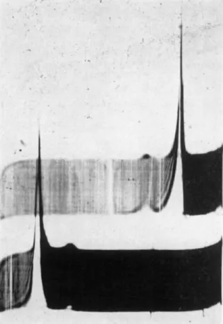
FIG. 1. Electrophoretic pattern of dissolved mitotic apparatus. Mitotic apparatus isolated by the alcohol-digitonin method were dissolved in 0.1 M salyrgan at pH 9.0. Run in phosphate buffer pH 7.5, ionic strength 0.2. Ascending boundary above.
Mobilities: major peak = 5.4 χ 10~Δ cm.2/volt sec; minor peak 10.7 X 10~4 cm.2/volt sec. Data of Zimmerman (a).
weight of 315,000 ( 5 ) and, according to amino acid analyses by Dr. J. D . Roslansky, has about two potential SH groups per weight unit of 20,000.
Thus, if 315,000 is the true weight of the "monomer" out of which the fibrous system is polymerized, each molecule has about 15 potential sites for forming an — S — S — bond to another of the same kind. There is a components, and we come out with a picture of surprising simplicity—a simplicity that may possibly be deceptive. It appears to be composed largely of one species of protein, judging by electrophoretic (Fig. 1) and ultracentrifugal characteristics (Fig. 2). This protein has a molecular
FIG. 2. Ultracentrifuge pattern of dissolved mitotic apparatus, prepared by alcohol- digitonin method and dissolved in 0.1 M salyrgan at pH 9.0. Concentration 4.2 mg./ml.; bar angle 50°. S20 for lighter major component = 3.67; for heavier com- ponent S20 = 8.56; Zimmerman (5).
second and smaller component, a ribonucleoprotein of higher charge and weight, that is of great interest in relation to the functions of the mitotic apparatus, and we may assume that other functionally significant species are present in still smaller quantities and that some may be lost in the course of the isolation.
2. T H E DIRECT ISOLATION OF THE MITOTIC APPARATUS FROM LIVING CELLS*
This problem of the direct isolation of the mitotic apparatus ( M A ) from living cells has been solved in recent months by the exploitation of the evidence that the coherence of the M A depends on — S — S — bonds.
*This method was developed in collaboration with Dr. J. M. Mitchison of the University of Edinburgh and Dr. Heitor Medina, Instituto de Biologia e Pesquisas Tecnologicas, Curitiba, Brazil. Dr. Mitchison's participation was aided by a travel grant from the Carnegie Fund and Dr. Medina worked as a fellow of the Rockefeller Foundation.
372 D A N I E L M A Z I A
But one must admit in advance that the chemical reasoning employed is as tenuous as the success of its application is real. Thinking that the structural stability of the mitotic apparatus depends on an oxidized state of the protein-S, and viewing the — S — S — (or other S-containing bonds) in a dynamic way, we consider the conditions that would favor the oxidized state. One would be the presence of an excess of some — S — S — compound that would "protect" the — S — S — of the mitotic apparatus against reduc- tion. Or we could imagine that such a compound was serving as a buffer against whatever unknown factor is tending to break the putative
— S — S — bonds in the MA. We need an — S — S — compound that will penetrate the cell readily, and will not be too toxic. There is a preliminary biological test of whether such a compound is effective. If it upholds the stability of the MA in vivo, it should tend to prevent the normal break- down of the MA at the end of division and should tend to "freeze" mitosis at telophase. For reasons that will become clearer, we struck upon dithiodi- glycol. We did indeed find that it tended to block the breakdown of the MA at telophase, and proceeded to test its usefulness for the direct isola- tion of the MA.
The method, briefly, is as follows. A suspension of sea urchin eggs is inseminated, and the fertilization membranes are removed by repeated passage of the just-fertilized eggs through fine bolting silk. The eggs are then washed repeatedly in Ca-free sea water to remove the "hyaline layer,"
and allowed to proceed to the time of division. When the population is at the desired stage, metaphase or anaphase, the eggs are transferred to the following medium: 1.0 M dextrose, 2 X 1 0 ~5M ethylene diamine tetra acetate ( E D T A ) , 0.15M dithiodiglycol ( D T D G ) . After allowing a few minutes for the D T D G to penetrate, the suspension is briskly shaken by hand. This is adequate to disperse the cytoplasm into a smooth particulate suspension, and we see that the mitotic apparatus has dropped out intact (Fig. 3A). The D T D G is absolutely essential. If it is omitted, we can ob- serve that the mitotic apparatus does come out as a unit, but it swells rapidly and vanishes from view. A nonelectrolyte medium is favorable for the easy dispersal of the cytoplasm without the aid of violent mechani- cal homogenizers. Dextrose has proved convenient, but we have worked with other media, especially dextrose-glycine mixtures. The E D T A com- pletes the removal of Ca, which is necessary to avoid aggregation of the particles.
FIG. 3 . Direct isolation of the mitotic apparatus from dividing sea urchin eggs (S. purpuratus), using dextrose-versene-dithiodiglycol medium at pH 6.2-6.3. A . High power view of final preparation; in dextrose-versene-dithiodiglycol. B. High power view of final preparation, after treatment with 2.5 X 10"* M CaCh, which sharpens the fibers and chromosomes.
374 DANIEL MAZIA
Once the MA have been set free, we concentrate them by centrifuga- tion. They are rather fragile, and we have encountered considerable diffi- culty in applying sufficiently elaborate centrifugal fractionation programs to obtain highly "pure" preparations of MA. The first question, of course, is whether we have isolated a truly "native" MA by this new procedure.
The ultimate test would be a functional one: the demonstration that the isolated mitotic apparatus will move the chromosomes, given the right conditions. So far, this test has failed, either because the apparatus has been altered in the course of isolation or because we have not yet found the right conditions. But we can at least show that the cells were alive at the instant when we broke them. If, instead of shaking the eggs in the isolation medium we return them to normal sea water, they are still ca- pable of completing division.
The stability and solubility properties of the MA isolated by the new method are different from those isolated after stabilization in cold alcohol.
The fibrous structure dissolves slowly in water and in dilute salt solutions, and very rapidly in 0.5 M or stronger salt solutions. The mitotic centers are not soluble under these conditions, and thus we are given a method for isolating these important structures. If we expose the preparation to C a+ + at concentrations as low as 2 X 1 0 ~4 M, they become extremely stable, a fact that we do not understand (Fig. 3 B ) .
The success of this procedure for isolating the MA directly from living, dividing cells, after several years of futile trials, rests entirely on the pre- diction—which can hardly be deemed more than a "hunch"—that the pres- ence of dithiodiglycol or something similar would "protect" the — S — S — bonding (or other bonding involving protein-S). Rather than emphasizing the strength of the individual — S — S — bond, it was imagined that the whole system was one in which such bonds were continually being opened and whose stability depended on the probability that a sufficient number would be closed at any given time. The stabilizing action of dithiodiglycol in the experimental situation, or of the unknown factors acting in the cell at the time of mitosis, was viewed as increasing this probability. All this is admittedly vague and its only merit is that it led to the desired result.
If this interpretation is untenable even in its vague form, we are required to account somehow for the stabilizing action of the dithiodiglycol, and this throws the problem, which could have rather deep biological implica- tions, into the ambit of this conference.
For the sake of the discussion, let me propose an alternative interpreta- tion of the stabilizing action of dithiodiglycol. This is merely that the dividing cell contains a proteolytic enzyme that is an SH enzyme, which is inactive in the presence of dithiodiglycol. If so, we might expect that once the mitotic apparatus was washed free from the rest of the cell
constituents, it would not be stable without benefit of D T D G . This is not the case. Even after purification in thç dextrose-DTDG medium, the MA begins to fall apart as soon as the D T D G is removed. If this hypothetical enzyme exists, it must be part of the structure of the MA and, moreover, would have the property of being reactivated merely upon removal of the D T D G and without the introduction of a reducing agent. But the alterna- tive hypothesis is directly testable by searching for proteolytic products after removal of the D T D G ; this has not yet been done.
In any event, the newer finding still implicates protein-S in the struc- ture of the mitotic apparatus. If the chemical interpretation is nebulous, the biological implications are clear. We shall have to ask how the living cell performs the task of stabilization and we shall see how we can manipu- late the mitotic process in vivo through the use of S agents.
III. The Fate of the "Glutathione Cycle"
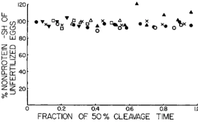
At the 1954 Symposium on Glutathione, a hypothesis to account for the formation of intermolècular — S — S — in the construction of the mi- totic apparatus was presented. This was based on the observation, dating back to the brilliant work of Rapkine (8) that the concentration of glu- tathione in the dividing cell tended to decline during the preparation for division and to rise during the growth of the mitotic apparatus. Accord- ing to the scheme then proposed, the oxidation of the glutathione (GSH) was related to the opening up of intramolecular — S — S — bonds in the protein units of which the fibers of the mitotic apparatus were to be made, and the reappearance of the GSH was related to the establishment of intermolècular —S—S—. In short, the cycle was one which converted intramolecular — S — S — to intermolècular —S—S—. At the time, we had confirmed Rapkine's data in a preliminary way. Recently, Dr. Eliza- beth F. Neufeld (9) has re-examined the glutathione of dividing cells (sea urchin eggs), taking greater precautions with the separation of protein, exposure of the material to oxidizing conditions, etc. In one group of ex- periments she used 2% trichloroacetic acid to separate the soluble SH, with determination of the SH by reduction of ferricyanide. She found that the GSH tended to disappear, presumably by oxidation, in TCA ex- tracts, but was much more stable when the TCA was neutralized to pH 6-7. In a second group of experiments, she used 3 % metaphosphoric acid to precipitate the proteins, and determined soluble SH by the nitroprusside method. If we examine the results (Fig. 4 ) , we see that there is no con- sistent fluctuation of soluble SH during the first division of the eggs of the sea urchin Strongylocentrotus purpuratus. What of Rapkine's cycle? The data given in Fig. 4 would seem to oppose the general applicability of
376 DANIEL MAZIA
Rapkine's findings about which there already has been some conflicting opinion, but the discrepancies may have been resolved by some recent work of Sakai and Dan (10). In a careful study, they have found that the fraction of the sea urchin egg that is extractable by TCA according to the method used by Rapkine does follow a time course through division
120 00"
X (3 CO Ο
1 LU 80 ί ^ 6 0
O =J
ο! [r 40
o u .
20
0.2 0.4 0.6 0.8
FRACTION OF 50% CLEAVAGE TIME
1.0
FIG. 4. Nonprotein SH during the first division of the eggs of the sea urchin Strongylocentrotus purpuratus. The symbols ψ , A, and Δ refer to experiments in which the eggs were extracted with trichloroacetic acid and analyzed by ferricyanide oxidation. The symbols # , Ο» and • refer to experiments in which the eggs were extracted with metaphosphoric acid and analyzed by the nitroprusside reaction.
rather like that attributed by him to glutathione. But the cycle is entirely attributable to a protein that is soluble in TCA. If they remove this pro- tein from their TCA extracts, the true nonprotein fraction remains un- changed through the course of division, just as Dr. Neufeld found.
Therefore, the Rapkine cycle may exist, but it is not referable to gluta- thione or other small SH compounds.
These results would permit us to retain the hypothesis that the as- sembly of the mitotic apparatus involves the transformation of intramo- lecular — S — S — to intermolecular —S—S—, the transformation being driven by a cycle of reduction and oxidation of another SH compound.
This would no longer be glutathione but the TCA-soluble protein. The hypothesis, however, remains just a hypothesis. We must recall, moreover, that even if the polymerization of protein molecules to form the mitotic spindle and asters does involve the transformation of intramolecular to intermolecular — S — S — bonds, such a mechanism does not necessarily demand a stoichiometrically equivalent change in other thiols of the cell.
As Huggins et al. (11) have shown, such a transformation can be driven catalytically by a small amount of —SH.
IV. — S H and the Functioning of the Mitotic Apparatus:
Blockage Experiments
The hypothesis that — S — S — bonds are involved in the structural assembly has shown its predictive power in leading us to a desirable isola- tion method, but we cannot say much about where these bonds fit into the picture. In earlier discussions, emphasis was placed on the possibility that the protein molecules were polymerized through — S — S — bonds. But we can imagine that the significance of the protein-S in the picture lies at the level of the individual molecule, somehow affecting its ability to interact with other molecules through bonds of another type. At the other extreme, we can imagine that the protein-S is involved at a higher level, in the arrangements that are responsible for microscopic fibrous order and the motor functions of the MA. For example, we could think that the macromolecules were polymerized by some other type of association, but were assembled in microscopic bundles by interactions through S. Some of the answers to these questions will come from further chemical studies, but we have first tried to ask them of the living cell, by purely biological experiments.
The experimental design is extremely simple. If the assembly of the MA in the cell involve — S — S — interactions, it should be possible to in- terfere with it by introducing an appropriate —SH compound. Such a compound could be viewed either as a competitor of protein SH or as a reducing agent for protein —S—S—. What is required is a substance that penetrates the cell readily and is not toxic at concentrations that would completely dominate the thiol picture in the living cell. For experiments on echinoderm eggs, these specifications are met perfectly by mercapto- ethanol. It's activity with respect to protein — S — S — has been well es- tablished by Olcott (12) and later workers. The two non-SH analogs, ethylene glycol and ethanol, are relatively harmless to cells at the con- centrations in which we are interested, even if the cheering powers of the latter are not evident in sea urchin eggs. From permeability data on the analogous substances it was predicted that mercaptoethanol would pene- trate the cells readily, and this proved to be so. It should be mentioned that considerations of permeability do restrict our choice of agents for these experiments, excluding those that are highly ionized at the pH of 7-8 at which we work.
The sea urchin eggs on which we have worked {Strongylocentrotus pur- puratus) take 90 minutes to go from fertilization through their first divi- sion at 18°. The crucial point is metaphase, which occurs at 75-80 minutes.
Before metaphase, the mitotic apparatus is being built up; at the end of
378 DANIEL MAZIA
metaphase it goes into action, separating the chromosomes toward the poles. When the eggs were exposed to mercaptoethanol at various times before division was expected, rather clear-cut results were obtained. Mer- captoethanol applied at any time before metaphase, and during metaphase, blocked their progress completely. The blockage was entirely reversible when the mercaptoethanol was removed. But, once the cells had reached a certain critical point, which we place at the end of metaphase or the very beginning of anaphase, the mercaptoethanol was completely ineffec- tive; it could no longer head off the division or even delay it. This is shown in Table I. At 75 minutes, some of the eggs had passed the critical
TABLE I
THE CRITICAL STAGE FOR BLOCKAGE OF DIVISION OF EGGS OF THE S E A URCHIN, Strongylocentrotus purpuratus, BY 0 . 0 7 5 M MERCAPTOETHANOL
Time after fertilization0 at which eggs were
transferred to 0 . 0 7 5 M mercaptoethanol Per cent divided at 8 5 minutes after fertilization
6 0 0
6 5 0
7 0 5
7 5 5 0
8 0 8 0
° Controls divide in 8 5 minutes at temperature of experiment (18.8°).
point while others had not reached it, and so we had split the population, whose synchrony is of course not perfect, into cells that were fully sensi- tive and those that were fully insensitive. It is the suddenness of the transition that is so striking, and perhaps it may be illustrated by com- parable data on another kind of echinoderm egg, that of the sand dollar Dendraster excentricus (13), in which division goes more rapidly and more synchronously (Table I I ) . The conclusion that we can draw is that mercaptoethanol is affecting some process involved in the build-up of the mitotic apparatus, but is inactive once the apparatus has entered its func- tional phase.
Before attempting any interpretation, let us pursue the character of the blocking action of mercaptoethanol in its biological aspect. Is the partially constructed MA broken down or merely arrested in its progress?
One answer can be obtained by taking advantage of the reversibility of the mercaptoethanol effect. The cells were blocked at a known time be- fore their expected division. When the mercaptoethanol is removed, how long does it take them to complete division? Table I I I gives the answer.
If cells are blocked at η minutes before division, they divide in η minutes
after return to normal conditions. They merely behave as though time had stopped for them while in the mercaptoethanol, even if blocked for an hour or more. It would appear that the mercaptoethanol did nothing but block the further build-up toward division.
But, if these superficial observations would lead us to believe that the mitotic apparatus was merely frozen in time, observations of its structure tell us quite another story. When we isolate the mitotic apparatus from blocked cells (14), we observe a most abnormal structure. Let us consider the case of cells blocked in early metaphase, a stage at which we have the best criteria of orderly structure (Fig. 5A). If we put these cells
T A B L E I I
THE CRITICAL STAGE FOR BLOCKAGE OF DIVISION OF EGGS OF THE SAND DOLLAR,
Dendraster excentricus, BY 0.1 M MERCAPTOETHANOL Time after fertilization0 at which eggs were
transferred to 0.1 M mercaptoethanol (minutes)
Per cent divided by 105 minutes after fertilization
19 0
24 0
28 0
32 0
36 18
40 80
44 95 +
48 95 +
α Normal time from fertilization to division = 50 minutes.
through our isolation procedure (in this case using the alcohol-digitonin method) we do recover the mitotic apparatus, but it has lost all of the exquisite organization that it possessed at the time the mercaptoethanol was applied (Fig. 5 B ) . The chromosomes are not well-aligned, and we do not see regularly aligned spindle fibers or astral rays. In fact, what we recover is a rather amorphous gel. Moreover, it takes only a few minutes of exposure to mercaptoethanol to bring about this condition of disarray.
How are we to reconcile these seemingly disastrous effects on the structure of the MA with the evidence of Table III, which tells us that the cell was merely arrested and not set back in its progress toward divi- sion. The answer is found by observing the recovery of the mitotic ap- paratus following the removal of the mercaptoethanol. We return the cell to sea water, wait a short while, 4-8 minutes, and then apply our isolation procedure. We find that the normal metaphase structure is completely re- stored in 4-8 minutes (Fig. 5C). The mitotic apparatus could not have been so drastically disorganized by the mercaptoethanol as appeared to
380 DANIEL MAZIA
T A B L E I I I
DIVISION TIMES OF EGGS REMOVED FROM 0 . 0 8 M MERCAPTOETHANOL AFTER VARIOUS PERIODS OF EXPOSURE (S. purpuratus)
Eggs placed in mercaptoethanol Total time from Column ( B ) 5 5 minutes after fertilization fertilization to minus
and exposed for": 5 0 % cleavage column ( A ) Excess delay
( A ) ( B )
0 (controls) 9 0 9 0
1 5 1 0 0 8 5 - 5
3 2 1 2 4 9 2 + 2
4 6 1 3 5 8 9 - 1
6 0 1 5 5 9 5 + 5
a Units are minutes, temperature 18.0.
the microscopist's eye. This eye is seeing only the highest level of organiza- tion, expressed as very regular arrays of fibers of microscopic dimensions.
A "loosening" or relaxation of this tight-appearing structure, a moderate decrease in the degree of orientation or condensation of submicroscopic fibrils, might, without much change in the more fundamental molecular arrangements, give the impression of disorganization that we do receive.
Obviously, the structure can readily tighten up again.
In a general way, then, the mercaptoethanol experiments do confirm the hypothesis that protein-S is implicated in the assembly of the mitotic apparatus. Under conditions of pH, etc., prevailing in the living cell, its action is to prevent the further buildup of the MA and to loosen but not to destroy the interactions that hold the mitotic apparatus together. The effects of mercaptoethanol on the structure of the mitotic apparatus sug- gest that the — S — S — bonds (or alternative bonds susceptible to SH) are involved in the higher level structural orientation of the M A (e.g., cross-linking of chains of molecules). They neither prove nor disprove the hypothesis that such bonds are also involved in the more primitive process of polymerizing the elementary molecules. The fact that the MA does not dissolve completely only confirms what we have suspected all along (3) : that other kinds of protein-to-protein interactions are important. This could have been deduced from the fact that the stability of the M A as isolated by our new method is also sensitive to ionic conditions (see p. 374).
FIG. 5. Effect of 0.075 M mercaptoethanol on sea urchin eggs, when applied at metaphase. In all cases the mitotic apparatus has been isolated by the alcohol-digi- tonin method for examination. A . Condition of mitotic apparatus just before mer- captoethanol was added. B . Mitotic apparatus isolated 10 minutes after exposure of eggs to mercaptoethanol. C. Recovery from effect of mercaptoethanol. Mitotic ap- paratus isolated 8 minutes after eggs were returned to sea water.
382 DANIEL MAZIA
Of utmost interest for the physiology of cell division is the fact that the MA becomes completely insensitive to the mercaptoethanol treatment just at the time when the chromosomes begin to move toward the poles. This could either reflect a change in the structure of the MA, rendering the
— S — S — groups inaccessible, or to a change in the intracellular environ- ment making the mercaptoethanol less active (e.g., by reducing the ratio S - / S H ) .
V. Protein-S in intercellular Adhesion: Experiments on Twinning When a cell has completed its division, the daughters may part com- pany and assume the destiny of independent individuals or they may re- main together as members of a multicellular community. Of course a multicellular organism involves a good deal more than the togetherness of the cells of which it is composed, but physical associations between the cells are predicates of its individuality. The most dramatic example of the relation between cell interaction and the individuation of the organism is seen in the divisions of the animal egg. When this goes normally, the daughters (blastomeres) remain together and form a single individual. If the daughters of the first division are separated, each can develop into a complete individual, and identical twins result. Reflecting on the thought that the number of souls produced by an act of procreation depends on how the daughter cells of an egg are stuck together, you may ask, justly, what does this have to do with proteins and sulfur?
That it has to do with proteins has been known for a long time. Em- bryologists have good reasons to think that in many cases the blastomeres are cemented together by what they call an extracellular cement, protein in nature. However, the view that they also communicate with each other through cytoplasmic bridges has not been rejected fully, and we are awaiting the verdict of the electron microscope to settle the question. Very little is known about the extracellular cements. They are so called because they may be removed without damage to the life of the individual cell.
Effective means of removing or at least weakening the extracellular ce- ments in invertebrate embryos are proteolytic enzymes and media that displace or chelate Ca+ + . The rigidity of the cements seems to depend on the presence of C a+ + , presumably combined with the protein.
In the studies of the action of mercaptoethanol on dividing eggs, we tested the full reversibility of its effects by following the eggs through development after they were returned to sea water. Unexpectedly it was discovered that those eggs which were exposed after they had passed metaphase, and divided while in the mercaptoethanol, gave rise to twin embryos! Naturally, the phenomenon was studied in some detail, and
it was found that only the eggs that were exposed during their actual cleavage produced twins. If the eggs were exposed too early, they were simply blocked, and when they divided upon return to sea water they produced single embryos. If they were exposed after they had completed division, it was too late for the mercaptoethanol to exert the twinning action. When the eggs were exposed during the first division and then again during the second, they produced quadruplets in fair yield! Table IV shows how the effect passes through a time of maximum efficacy, cor-
T A B L E I V
PRODUCTION OF TWIN EMBRYOS FROM Dendraster EGGS PLACED INTO 0.1 M MERCAPTOETHANOL AT VARIOUS TIMES AFTER FERTILIZATION
Time after fertilization when mercaptoethanol was introduced
(minutes)
Percent cleavage in mercaptoethanol
Per cent twin blastulae
35 50 50
38 90 85
41 95 + 90
44 95 + 45
47 95 + 30
50 95 + 8
53 95 + 8
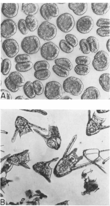
responding to the period when the cells are seen to be cleaving. Figure 6A shows the twin blastulae, still contained in their common membrane, and Fig. 6B shows the miniature later embryos (plutei) that develop from these blastulae. Perhaps it should be explained that these miniature em- bryos would be expected to develop into full-sized adults, once feeding and growth begin, although technical difficulties stand in the way of actually rearing them in the laboratory.
The "totipotence" of blastomeres of certain eggs is demonstrated classically by the fact that they produce complete individuals when sepa- rated physically. Conversely, the normal "regulation" to form a single individual is a consequence of the physical contacts or communications between blastomeres. With mercaptoethanol, we achieve by chemical means the same result as would be obtained by physical separation of the blastomeres at least in the case of the Dendraster egg. We are bound to look for evidence that we have in fact influenced the contacts or communi- cations between them. The effects of the mercaptoethanol on the physical adhesion between the sister cells are not obviously apparent in the normal case where the cells are restrained within a fertilization membrane, but if we remove the membrane we can show very clearly that we have influenced
384 DANIEL MAZIA
FIG. 6. A . Twin blastula produced by Dendraster eggs that have cleaved in 0.1 M mercaptoethanol. B. Miniature plutei resulting from twinning. The large plu- teus is derived from an egg that did not twin.
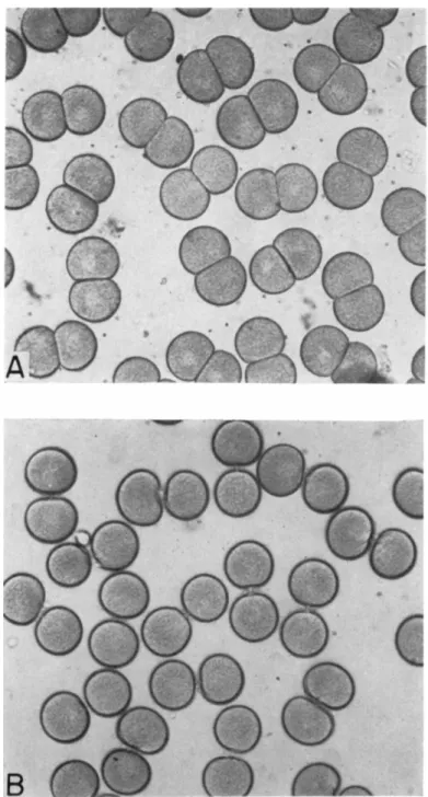
FIG. 7. The effect of mercaptoethanol on blastomere interaction in the cleavage of Dendraster egg. The fertilization membranes have been removed. A. Controls:
eggs have cleaved in sea water. B. Experimental: eggs were placed in 0.1 M mercapto- ethanol in sea water at 10 minutes before division and completed cleavage in this solution.
386 D A N I E L MAZIA
the surface contacts. Figure 7A shows how, in the control situations, the blastomere surfaces remain flatly apposed, even when the fertilization membrane has been removed. In Fig. 7B we see what happens when the eggs divide in mercaptoethanol. The surfaces do not adhere, and the blastomeres appear as spheres connected by a "stalk." Obviously we have interfered with some mechanism that normally keeps the new faces of the daughter cells in most intimate association as the furrow cuts the egg into two. This adhesion is generally attributed to a cementing matrix.
What remains to be seen is whether the surface apposition in itself is the sufficient condition of the regulation between the blastomeres or whether it in turn serves as a support for strands or bridges that serve in com- munication between them. Experiments such as these are hardly intended to solve the fundamental mysteries of totipotence and individuation, but only to reveal some of the crucial chemical conditions of the interactions.
The action of mercaptoethanol on the adhesion between the blasto- meres is superficially similar to what is observed when the cells are placed in a Ca-free medium, but the end effects are quite different. The effects of removal of Ca at the time of division are entirely reversible in the Dendraster egg, and twins are not produced. The reason for this is discussed in another paper (15).
AVe do see that we can interfere with the interaction by means of mer- captoethanol and can visualize the consequences of the interference. If the normal progress of cell division here includes the laying down of a rigid cement between the daughter cells, this act would seem to involve protein-to-protein interactions via the sulfur-containing groups. Again we encounter the fact that we may interfere with the process while it is taking place, but cannot reverse it once it has been completed (Table IV). The simplest explanation of this fact would invoke the "accessibility" of
—S—S:— groups to mercaptoethanol, but this might only be another way of saying that mercaptoethanol, under the conditions of the system, was more effective as a competitor of protein SH than as a reducing agent for
— S — S — bonds between proteins.
R E F E R E N C E S
1. D. Mazia, in "Glutathione" (S. Colowick, A. Lazarow, E. Racker, D. R. Schwarz, Ε. Stadtman, and H. Waelsch, eds.), p. 209. Academic Press, New York, 1954.
2. D. Mazia and K. Dan, Proc. Natl. Acad. Sei. U. S. 38, 826 (1952).
3. D. Mazia, Symposia Soc. Exptl. Biol. 9, 335 (1955).
4. D. Mazia, in "The Chemical Basis of Heredity" (W. McElroy and B. Glass, eds.), p. 169, Johns Hopkins Press, Baltimore, Maryland, 1957.
5. A. M. Zimmerman, Federation Proc. 17, 174 (1954).
6. M. Calvin, in "Glutathione" (S. Colowick, A. Lazarow, E. Racker, D. R. Schwarz, Ε. Stadtman, and H. Waelsch, eds.), p. 18. Academic Press, New York, 1954.
387 7. J. M. Swan, in this volume, see Section I. 1.
8. L . Rapkine, Ann. physiol. physicochim. biol. 7, 3 8 2 ( 1 9 3 1 ) . 9. E. Neufeld and D. Mazia, Exptl. Cell Research 13, 622 ( 1 9 5 7 ) . 10. H. Sakai and K. Dan, Exptl. Cell Research in press ( 1 9 5 9 ) . U. C. Huggins, D. F. Tapley, and Ε. V. Jensen, Nature 167, 5 9 2 ( 1 9 5 1 ) . 12. H. S. Olcott, Science 96, 5 4 ( 1 9 4 2 ) .
18. D. Mazia, Exptl. Cell Research 14, 4 8 6 ( 1 9 5 8 ) .
14. D. Mazia and A. M. Zimmerman, Exptl. Cell Research 15, 1 3 8 ( 1 9 5 8 ) . 15. D. Mazia, Biol. Bull. 114, 2 4 7 ( 1 9 5 8 ) .
Discussion
LINDLEY: I would like to ask if Dr. Mazia has any chemical studies on what hap- pens to SH groups in the normal process of division.
MAZIA: We do not have adequate chemical data on the protein SH of the mitotic apparatus during the division cycle as yet, but some very interesting information is being obtained by histochemical methods, involving specific staining reactions. I might cite a recent study by Kawamura and Dan (J. Biophys. Biochem. Cytol. in press) who observe, among other things, that the mitotic apparatus stains more in- tensely for protein SH than any other part of the dividing sea urchin egg. The inten- sity builds up as the mitotic apparatus is mobilized, and falls off in the later phases of the cycle. Using Bennet's method for staining protein SH, they obtain a striking pic- ture of the concentration of SH in the mitotic apparatus. The slide I have shown, a preparation made by Dr. John Roslansky employing the Barnett-Seligman procedure shows a similar concentration, but not so dramatically.
MADSEN : I was wondering what your evidence is for the fibers being held together by disulfide bonds because I was thinking that some of the things you did to dissolve them, such as PCMB and KCl might be more consistent with them being held to- gether by hydrogen-bonded sulfhydryl groups.
MAZIA : We have been aware of your interesting work on Phosphorylase and have cited it in a recent paper where we leave open the possibility that the protein-to-pro- tein links in the mitotic apparatus might involve bonds other than —S—S— bonds, even though the evidence for the participation of disulfide groups seems to be so strong at this time. Our difficulty in thinking about hydrogen-bonded SH groups has been, frankly, the fact that the chemists whom we have consulted have been so dis- couraging about the possibility. If you can assure us that such bonds are reasonable and can be strong enough to maintain a stable fibrous structure, we for our part must admit that we have no evidence that is inconsistent with your suggestion.
BENESCH : What is the situation with respect to the rise and fall of the so called
"soluble —SH" during mitosis?
MAZIA: Again that would be perfectly consistent with the disulfide bond formation but does not demand it.
BENESCH : Quite so, but it does not fit in with the concept that the fibers are held together purely by SH hydrogen bonds, because in that case why is there the rise and fall in soluble SH during the mitosis?
MAZIA : The correlation of the rise and fall of soluble SH with the formation of the mitotic apparatus does fit the concept of a disulfide-bonded system nicely, and it would be pleasing to preserve this concept. The point I have been making is that the present evidence for the participation of protein SH in some way seems to be com- pelling, but the evidence for —S—S— bonds specifically is less so. The latter is con-
388 DANIEL MAZIA
sistent with all we know about the solubility properties of the isolated mitotic ap- paratus and the stability of the apparatus in vivo, but does not exclude alternatives such as that suggested by Dr. Madsen. At one time, we thought that the solubilizing action of salyrgan and PCMB was a blow to the —S—S— picture, but now I take it that there is good evidence for heavy metal attack on —S—S—. That is correct, isn't it?
BENESCH: Yes.
MAZIA: Perhaps I should add that the organic mercurials solubilized the isolated mitotic apparatus in the range of pH 9 .
BENESCH: It is certainly likely that mercurials would catalyze the hydrolysis at this pH.
RIGGS: Have you any success in getting any of this material to form fibers or to form aggregates of any sort?
MAZIA: I can't say that Dr. Zimmerman, who did the studies with PCMB and salyrgan, observed decent fibers. But the material did aggregate as soon as the mer- curial compounds were dialyzed away. In fact, this was a major difficulty for him; he could not do ultraviolet spectrophotometry on these solutions because the solubilizing agents interfered.
FRAENKEL-CONRAT: In view of the present idea that disulfide and sulfhydryl are interchangeable—of a little bit of sulfhydryl going a long way and causing progressive changes—I wonder if we need a settlement of the argument? I wonder if one could not consider the presence in the system of both sulfhydryls and disulfides. I believe that most of the data would be explicable on this basis.
MAZIA : This is a point of view that we would like to develop a little more sharply in connection with the most recent work that I reported : the reversible stabilization of the native mitotic apparatus by dithiodiglycol. As you may recall, we can isolate it directly from the living cell in the presence of dithiodiglycol and it tends to fall apart as soon as we remove the latter. We have a kind of dynamic stability in the presence of an excess of the dithiodiglycol, as though it were holding the bonds of the mitotic apparatus in a more stable configuration. We think of it as increasing the probability that a pair of S atoms will be linked together as —S—S—. One isn't pleased with such a vague picture, but the more fluid conception of the relation be- tween SH and —S—S—, such as Dr. Fraenkel-Conrat has discussed, does help.
LORAND: I should like to ask Dr. Mazia, purely by analogy to clot formation in blood, if the mitotic apparatus prepared in the presence of calcium could be dispersed in urea?
MAZIA: The mitotic apparatus, isolated by the new method, becomes extraordi- narily insoluble after it has "seen" Ca ions in concentrations down to 2 X 10"* Μ, even though it is very unstable otherwise. After exposure to calcium, we have been able to dissolve the apparatus only in strong alkali.
LORAND: The analogy is quite striking. Next, I wonder how specific the effect of calcium is, for in the polymerization process leading to a blood clot, it cannot, for instance, be replaced by magnesium.
MAZIA: Magnesium is ineffective. That is, the mitotic apparatus is still reasonably soluble after exposure to Mg. To this extent, the effect of Ca is specific.
MAZUR: IS it possible to suggest that the explanation for the PCMB reaction is that you have sulfhydryls which are tied together, chelated via a metal, which is present. This metal can be replaced by the mercurial which has a very high affinity for the sulfhydryl. I am suggesting a metal such as iron. You do show the presence of sulfhydryl using staining compounds.
MAZIA: There is very good evidence of SH in the proteins of the mitotic appara- tus. A number of laboratories, including our own, have obtained unequivocal staining, using a variety of methods. In addition, we do have some analytical data on the iso- lated mitotic apparatus. Dr. Elizabeth Neufeld has done chemical estimations on the material isolated by the alcohol-digitonin method, and estimates that about two- thirds of the S is in the SH form and one-third in the —S—S— form. Drs. Zimmer- man and Neufeld have shown that the protein SH is readily oxidizable by gentle means, and that the mitotic apparatus becomes more difficult to dissolve as the oxi- dation proceeds.
MORALES: Will EDTA dissolve the mitotic apparatus?
MAZIA: We cannot dissolve the mitotic apparatus with EDTA after isolation by the alcohol-digitonin method, nor after isolation by the new method and exposure to low concentrations of Ca. The latter observation suggests that it binds Ca more strongly than does EDTA.