DEVELOPMENTAL BIOLOGY SUPPLEMENT 2 , 3 0 6 - 3 2 7 ( 1968 )
Plasticity of Neural Maturation
R. W. SPERRY
Division of Biology, California Institute of Technology, Pasadena, California 91109
INTRODUCTION
The development of the brain in many animals, and in man es
pecially, includes a long postnatal period during which the emergence of order in the still growing and differentiating cerebral nervous sys
tem depends not only on the forces of development, but also upon an interaction between developmental forces on the one hand and the effects of behavior and learning on the other. Brain development dur
ing this period—and this is a period that in man extends over 9 years as compared with the prenatal 9 months—exhibits a special kind of functional plasticity and a special potency for the long-term shaping of brain organization and behavior that is no longer apparent with the learning process alone after growth and maturation are finally completed.
While it is still growing, the brain seems to possess, for example, much greater facility in compensating for damaging injuries to the nervous system; it shows greater ability to correct certain dysfunctions like those associated with ocular squint; and special skills like language can be acquired much more easily during this period. Some of these organizational and reeducational accomplishments that are readily achieved by the still-developing brain can no longer be managed at all by the fully mature brain.
The mechanisms that underlie and are responsible for this extra functional plasticity of the still-developing brain remain entirely un
known. On the one hand, they would seem to relate to basic develop
mental events such as neuron differentiation, growth of neuron pro
cesses, selective membrane adhesion, protein synthesis, etc.; and on the other hand, they must relate also to the unknown chemical and cellu
lar mechanisms at the basis of learning, memory, imprinting, and the like.
306
These underlying mechanisms seem now rather remote and un
approachable. The problems involved are nevertheless of such supreme importance as to warrant our stretching a little to give them special attention. Unlike many aspects of brain development, the extra func
tional plasticity of postnatal growth is accessible to manipulation; it is a phase of brain development that one can do something about. For man especially the interaction between the forces of development and behavior during the prolonged period of infancy and childhood is critical for the shaping of the functional properties of the adult brain and hence of human nature generally. Answers to questions such as the following are at stake: To what extent can the preprogrammed processes of cerebral development be modified by function? Exactly how plastic is human nature? Can early function affect the structural patterning of the brain or just its physiology? Just what is it that a "head start" or early enrichment of experience can do for the cerebral cortex? Is the plasticity of learning and memory as seen in the adult a direct outgrowth or evolvement of the plasticity of neural develop
ment? In this connection, might not the developmental approach to the basis of learning and memory offer some special advantages and insight perhaps for solving the ancient problem of the memory trace?
The material that follows is pointed in the long run at these and many related questions. It starts much more specifically, however, with a review of some observations by Dr. Ronald Saul and myself (Saul and Sperry, 1968). These concern a patient who was first seen by Saul about one year ago at the Neuro-Medicine Service of the Los Angeles County General Hospital. The patient is a 20-year-old college girl in whom the entire corpus callosum was found to be missing as an anomaly of development. Absence of the callosum was discovered only incidentally in cranial X-rays taken when she became hospitalized following headaches and blackout for what was soon diagnosed to be acute hydrocephalus. With the installation of a Teflon tube ventriculo- atrial shunt and withdrawal of a progestational drug, she made a rapid recovery and returned to college where she is currently a sophomore in the Los Angeles City College system. She has been making an average scholastic record of B's and C's and during the past year has been working 20 hours a week as an office clerk at the college. Until the hydrocephalus developed and the absence of the corpus callosum was discovered, she had always been presumed by herself and by her family to be entirely normal. In an intelligence test (WAIS) adminis-
308 R. W . SPERRY
tered 2 months after surgery her verbal performance score was 96, and her verbal IQ, 111.
The missing structure in this patient's brain, the corpus callosum, constitutes of course the main channel for cross-communication be
tween the cerebral hemispheres. This great band of cross-connecting fibers is by far the largest single-fiber tract in the entire brain, and its 200 million or so fibers form a rich system of reciprocal right-left cross- connections for nearly all points in the neocortex of the two cerebral hemispheres. In the past 10 years or so we have learned a good deal about what happens when brains are deprived of this enormous system of fiber connections through surgical intervention—that is, after the system has once developed and has been in operation. The effects produced when the brain is deprived of this same system from the be
ginning through lack of development are quite another matter, and the results are much less clear. Accordingly, we were very much interested in the rare opportunity which this person afforded for making some direct, first-hand comparisons of the symptoms produced on the one hand by congenital absence and on the other by surgical section of this major brain structure. The degree to which functional deficits might be reduced in the congenital condition should give us a fairly direct measure of the functional plasticity of the developing brain. In the much-studied callosal system and its functions we might further
more have a model or handle for getting at some of the relatively in
accessible mechanisms that underlie the plasticity of neural maturation.
AGENESIS OF CORPUS CALLOSUM: ASYMPTOMATIC CASE Total absence of the corpus callosum in this person seems to be re
liably established in both the pneumoencephalographs and in the angiograms. Whether the much smaller anterior and hippocampal commissures are also missing cannot be said. Often this is the case, but it appears to depend on the timing of the developmental arrest
(Slager et al., 1957)—that is, whether it comes before or after the end of the fourth fetal month. In any case, most of the known symptoms with which we are acquainted from the surgical cases and which were used for comparison, appear to be attributable mainly to the huge corpus callosum itself.
The first evidence of the corpus callosum in the human fetus is seen in the third month as a thickening on the lamina terminalis just dorsal
to the anterior commissure (Hyndman and Penfield, 1937). The most anterior portion develops first, and its posterior enlargement is directly associated with the formation of the septum pellucidum and hippo- campal commissures. All three of these structures and the anterior commissure may be absent if the developmental error occurs before the beginning of the fourth month. Developmental arrest during the fourth month allows formation of the anterior commissure without the corpus callosum. If arrest occurs toward the end of the fourth month, the anterior commissure is apt to be present together with the anterior part of the corpus callosum and posterior extensions to a varying degree depending on the timing. All parts of the callosum and the associated interhemispheral commissures are laid down in outline form by about the start of the fifth month.
A record of average or slightly above average mental achievement with complete agenesis of the corpus callosum as seen in this patient is not common, but it is by no means unique. It is general textbook doctrine that total agenesis of the corpus callosum does not necessarily produce any disturbance of motility, coordination, general or special sensibility, reflexes, speech, or intelligence. In a detailed review of the literature ten years ago, Slager et al. (1957) found 16 such cases on record. All were adult patients with complete congenital absence of the callosum discovered at autopsy in whom no functional symptoms had been noticed. Cases with associated malformations of the brain and with varying degrees of mental retardation are, of course, seen in much larger numbers. Such cases with moderate to mild symptoms have been easier to obtain and observe; most of the truly asymptomatic cases, however, have not been discovered until autopsy. It is logical to assume, as Slager points out, that only a small fraction of the truly asymptomatic cases ever come to light. Accordingly, the discovery of the present patient in her sophomore year in college with no known symptoms and with average grades, and her availability for behavioral testing represented a most exceptional opportunity.
In connection with the possible absence of symptoms in this con
dition, it must be remembered that it was the prevailing doctrine in neurology for many years, until the mid-1950's, that no important be
havioral deficits are found in man correlated with either congenital absence or with complete surgical section of the corpus callosum. This was aptly summarized in McCulloch's classic statement cited by
310 K. W . SPERR Y
Bremer et al. (1956) that the only demonstrated function for the corpus callosum seems to be the transmission of epileptic seizures from one to the other hemisphere.
This no longer applies today, of course, since we have been able in the past 12 years or so with specialized tests to demonstrate a large number of distinct functional roles for the great cerebral commissure (Ettlinger, 1965; Sperry, 1958, 1961, 1968a). These have been inferred entirely, however, from the effects of surgical transection, not agenesis, first in cats and monkeys and, more recently, in a series of human pa
tients (Gazzaniga et al, 1967; Sperry, 1966, 1968b; Sperry et al, 1968).
These latter are patients of Philip Vogel and Joseph Bogen of Los Angeles, patients in whom the forebrain commissures were surgically sectioned to control severe intractable epileptic seizures (Bogen et al., 1965).
HEMISPHERIC DECONNECTION SYNDROME
The nature of the deconnection symptoms as seen in the surgical patients can be summarized quickly as follows : In general the two sep
arated hemispheres continue to function at a high level, but each op
erates in isolation with respect to most of the so-called gnostic or higher functions of the brain. In other words, the commissurotomized person or animal behaves as if each of the two separated hemispheres has an independent mind of its own with no conscious connection be
tween the two. Each hemisphere seems to have its own private sen
sations, perceptions, mental images and ideas, its own volitional and learning experiences, and each has its own separate chain of memories.
All of these are cut off from the corresponding mental activities in the opposite hemisphere.
This bisection of the normally unified stream of conscious awareness into two largely independent mental systems remains remarkably in
conspicuous, however, so far as ordinary daily behavior goes. It also goes undetected in most of the standard neurological and psychological tests, which presumably is why the symptoms of the corpus callosum remained undefined for so long. For example, the human patients with surgical section of all the forebrain commissures, continue to read the newspaper and books and to watch television, etc., with no particular complaints about peculiarities in their visual perception. Yet when we test them more carefully under conditions in which each hemisphere gets restricted information not available to the other hemisphere, it
then becomes evident that these people have, in effect, not one inner visual world any longer, but actually two separate and independent inner visual worlds—one serving the right half-field of vision and the other the left half-field. Each of the disconnected hemispheres is quite capable on its own of good visual perception, learning, and memory, intermodal and other mental associations, etc. Because speech and writing are lateralized to one hemisphere, these people are able to talk to us only about the inner visual world for the right half-field of vision.
The visual experiences of the other hemisphere have to be expressed nonverbally as by manual pointing to words, pictures, matching ob
jects, etc.
Under the conditions of ordinary behavior these people readily com
pensated for this condition by rapid scanning movements of the eyes.
It is similarly characteristic of most of the observed symptoms of hem
isphere deconnection that the symptoms tend not to show up under ordinary conditions, requiring for their demonstration some restricted lateralization of sensory input and/or other measures to obtain separate and asymmetric cerebral activity, along with methods for the separate readout from the left and right hemispheres.
In view of all the foregoing it thus seemed highly possible that cross- integrational deficits might really be present in this and the other asymptomatic patients but had simply gone undetected. As with the surgical cases, perhaps we needed only to apply the appropriate tests and the symptoms of hemisphere deconnection would become appar
ent. Accordingly, we brought the patient into the laboratory and put her through one after another of our tests for hemisphere deconnection.
These are test tasks that normal human subjects perform without diffi
culty, but on which the patients with surgical section of the callosum have been found either to fail completely or show severe impairment (Sperry, 1968a). The findings in the agenesis patient were consistent throughout and might as well be forecast at this point: In marked contrast to the surgical patients, the agenesis subject passed every one of these tests with flying colors showing not one of the numerous symp
toms of cerebral disconnection exhibited by the surgical cases under exactly the same testing conditions.
SURGICAL-CONGENITAL DIFFERENCES
Let us look now at some samples of the kinds of interhemispheric functions that were involved in these tests, in other words, at the kinds
312 R. W . SPERRY
of interhemispheral functional deficits for which a developing brain seems able to compensate, but not a brain in which development is completed. Most of the functions in question can be described for con
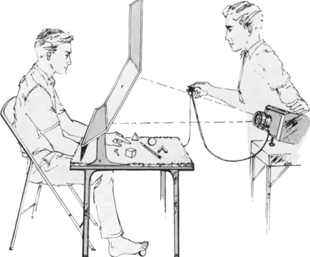
venience with reference to the simple general testing unit illustrated in Fig. 1. The slanting shield on the table and the table's surface serve to screen out vision in tests involving functions of the hands and feet.
In tests involving the lateralization of vision, the subject centers his
FIG. 1. Apparatus for testing the functions of the corpus callosum.
gaze with one eye closed, on a designated fixation point on the trans
lucent screen. The visual stimuli on a 2 X 2 slide projector are back- projected at 1/10 second or less, too fast for eye movements to get the stimuli into the wrong half-field of vision.
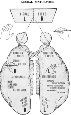
In Fig. 2, we have a schematic summary of the results as seen in the surgical patients. This will serve also as a general reminder that the neuroanatomical pathways of the brain are such that the image of things viewed through the left half-field of either eye is projected across into the right hemisphere and vice versa. The same applies for
\ VISUAL I HELD ■":■'/
V ί Α » /
FIG. 2. Schematic outline of functional representation in the cerebral cortex as seen in patients with surgical section of the forebrain commissures.
things perceived by touch or by stereognosis, with the left and right hands or with the feet. Note further that speech, writing, and calcula
tion along with the main language centers as shown, are all repre
sented along with the right hand and the right half visual field in the same, the left, hemisphere. This left hemisphere is the dominant hemisphere for right handers. All the surgical patients that we have studied have been predominantly right-handed. The agenesis patient, on the other hand, is left-handed and somewhat ambidextrous. This is a finding that is common among patients with agenesis, and it has been suggested that the presence of the corpus callosum may be im
portant for the development of strong cerebral dominance (Smith, 1966). In applying our tests to the agenesis patient we accordingly
314 R. W. SPERRY
simply reversed the tests from left to right. As will be seen, however, it made little difference to this patient on which side the stimuli were applied.
When the split brain surgical patients are confronted with visual stimuli consisting of letters, numbers, words, colors, geometric figures, irregular shapes, or pictures, flashed to the left and right half visual fields, the surgical subjects report with no special difficulty whatever stimuli appear on the right side. At the same time they consistently deny having seen any of the left field stimuli excepting possibly just a flash of light. With further analysis it becomes clear that the subjects are telling us only a half truth with respect to the left-field stimuli in the sense that the hemisphere that is talking to us speaks for itself alone when it says that it did not see anything on the left side. That the stimuli in the left half field are in fact seen very well by the other, the nontalking hemisphere, is shown by the fact that when the sub
ject is encouraged to use his left hand to point to matching pictures or to find by touch matching objects among a collection of items screened from view, we find that the left hand can consistently select the correct item that corresponds to the left field stimulus. After the subject has correctly retrieved by blind touch a left-field stimulus using the left hand, and is then asked what it is that he has selected and is still holding, he seems to have no idea of what the item is and can only make wild guesses. It is quite evident from the verbal comments throughout tests of this kind that the left hemisphere does not know what has been going on in the right hemisphere, and as we see later in other tests, it is also true the other way around. Nothing of the sort occurred when the same kinds of tests were administered to the pa
tient with agenesis of the callosum. She had no difficulty in describing verbally with normal facility anything that was presented on either side of the visual field.
If we flash words to the surgical patients so that they extend across the vertical midline, the commissurotomy patients are able to read off only the part of the word that falls in the right half field of vision. The agenesis patient in the same situation reads off the whole word or the double word without hesitation about as rapidly as a normal person, and can also write out the full word with either hand. Series of words were used with many syllables in common like al-ign, re-ign, al-ly, re-ly, etc., such that one half of the word could not be used to cue off the other half, and even the pronunciation of one half of the word
might depend on the other half. When such words were presented in
dividually at 1/50 second, half of the word in the left field of vision and half in the right, the agenesis patient was able to read aloud the whole word promptly and surely.
Similar results obtained for stimulation of left and right hands: Ob
jects placed in the right hand are identified by touch in the normal manner by the surgical subjects, but the same items presented to the left hand cannot be named or described in speech or writing. These same items, however, if removed from the left hand, can be selectively retrieved by touch from among a large collection of other objects. Such retrievals have to be performed by the same hand with which the test object was initially identified. Cross-retrieval with the opposite hand consistently fails in the surgical patients. When the subject tries to use the wrong hand in such tests, each object may be correctly identified by the exploring hand, but the hemisphere that is recognizing and processing this information does not know what it is looking for and the informed hemisphere that knows what it is looking for gets no feedback from the exploring hand, so the two processes never get to
gether and the performance fails. The agenesis patient, on the other hand, readily named items placed in either hand and had no difficulty retrieving with the right hand objects identified by the left hand or left visual field and vice versa.
If we use a stylus to stimulate a point on a particular joint of a par
ticular finger of one hand that is held palm up and is screened from sight, the surgical patient can find the stimulated point on the same hand, but unlike a normal subject he is quite unable to find a corre
sponding mirror point on the opposite hand and is also unable to localize verbally the point of stimulation if it is on the subordinate hand. The agenesis patient had no trouble performing this task in either direction and, like the normal subject, was able to report ver
bally the correct locus of the stimulus on either hand. Also if a par
ticular hand posture like those shown in the chart on the right of Fig.
3 is flashed to the left or the right visual half field, the surgical subject can copy the sample posture with the hand on the same side but fails when he tries to use the hand on the opposite side. Similarly, if the sample finger posture is forcibly impressed by the examiner on one hand of the subject, the surgical patient is not able as a rule to mimic the same posture with the opposite hand, which you recall is repre
sented in the other hemisphere. These and similar test responses give
316 R. W . SPERRY
FIG. 3. Procedures for testing cross integration that involves hand postures (see text).
further evidence that the two disconnected hemispheres in the surgical patients are not communicating. By contrast the agenesis patient has no special trouble with any of these tasks applied in any combination.
In double retrieval tests two objects are placed simultaneously, one in each hand, or two objects are pictured one in each half visual field, and then the same two test items are scrambled for blind retrieval by touch among a collection of other objects, all of which are screened from sight. The two hands of the surgical patient will then work in sequence or in parallel to explore through the pile of items and usu
ally each hand will come up successfully with that particular object that was initially perceived by the same hand or by the corresponding half visual field. In this double retrieval process, either hand may commonly pick up, identify and reject the very same item for which the patient is simultaneously searching with his opposite hand. This double retrieval performance in the surgical subject is thus like two separate people searching through the collection of objects with no cooperation or communication between them.
In trying to interpret the performance of the surgical patient in this and in similar test responses, we find it to be generally less confusing if we do not try to think of the responses of these people as being per
formed by a single individual any longer, but try instead to think always in terms of two independent mental systems proceeding in right
and left hemispheres separately. There is nothing to suggest that any similar interpretation would apply to the congenital patient. In this last test, for example, involving double right and left retrieval, the agenesis subject immediately picked up with either hand whichever of the two test items was first encountered—that is, the two hands were used interchangeably.
If pairs of numbers are presented visually, one to the left and one to the right half-field, or if cut-out plastic numerals are placed in the right and left hands ior identification by touch, the congenital patient can integrate the right and left input so as to read off the sum or the product just as rapidly as an average normal control subject. Somehow the numeric, like the verbal information, when projected into the separated hemispheres, is rapidly cross-integrated much as in the normal brain. Similarly if one to four fingers are tapped in both of the subject's outstretched hands screened from sight, the sum or product can be reported with average speed by the congential patient, but not at all by the surgical patients.
DEVELOPMENTAL COMPENSATION: GENERALIZED PHENOMENON The foregoing is sufficient to illustrate the nature of the inter- hemispheric functions involved and the kind of behavioral compen
sations that the developing brain is somehow able to achieve under these conditions. Other patients with callosal agenesis for whom be
havioral studies have been reported, although not subjected to quite the same kinds of tests and having mild to moderate complications, would appear also to have achieved significant compensation over and above the condition seen after surgical section (Jeeves, 1965; Russell and Reitan, 1955; Solursh et al., 1965). It would not be surprising that with further, more refined tests or tests of different design we may eventually succeed in finding in this patient consistent, perhaps more subtle, commissural deficits.
The marked difference between the functional effects of early con
genital and late surgical elimination from the brain of the same system of fiber connections is of greater interest and significance, however, demonstrating as it does the kind of functional compensation that it is possible to achieve in the still-developing but not in the fully devel
oped brain. A comparable difference between congenital absence and adult removal is reported to apply to the cerebellum. The entire right or left half of the cerebellum may fail to develop without producing
318 R. W. SPERRY
marked functional symptoms (Dow and Moruzzi, 1958). There is the oft-cited case of the brain that showed up at autopsy in the Harvard Pathology Laboratory lacking all but a minute remnant of the cere
bellum ( Sidman, 1968 ). The cerebellum, of course, is the master organ for balance, steadiness, equilibrium, and precise motor control of vol
untary movement. When the history of this individual was investigated it was found that he had been making his living as a window washer in skyscrapers. Corresponding removal of the same cerebellar tissue from an adult would be radically disabling.
Much the same can be said for hemispherectomy. Surgical removal in the adult of one whole hemisphere, as for a glioma, produces ordi
narily a severe and lasting hemiplegia with loss of both sensory and motor functions in the affected limbs. The outcome is much less in
capacitating if the hemisphere that is removed has had long-standing damage, atrophy, or dysfunction extending back into infancy or early childhood—in other words, if there has been a long period during de
velopment when the healthy hemisphere has had to compensate for the missing functions of the damaged hemisphere (Ueki, 1966). The kind of developmental compensation that we see in callosal agenesis would thus appear to be representative of a widespread and general
ized property of the developing brain. "As the twig is bent, so grows the tree," would seem to have smoewhat more than just "hearsay" ap
plication for the developmental patterning of behavior and the func
tional shaping of brain properties.
The nature of the underlying mechanisms that are responsible and their limitations and potentialities for the determination of adult ca
pacity in the human brain, have yet to be determined. The develop
mental properties in question are of wide and profound concern for problems in many disciplines ranging from social science and psychia
try to the physiology of learning, neurogenesis, and molecular biology.
With this in mind we might now look further at the agenesis of the corpus callosum, asking what kind of underlying developmental changes could account for the ability of a brain, totally lacking its callosal cross-connections, to perceive two different syllables flashed separately into right and left visual half-fields, put them together mentally and to read them off as a single word with normal speed.
This feat is easily accomplished with the callosum intact, but it has been totally impossible where the corpus callosum has been sectioned surgically.
POSSIBLE EXPLANATORY FACTORS
Judging from anatomical studies of the brains of other asymptomatic cases with agenesis of the corpus callosum, it would seem that we can rule out as an explanation the deflected or aberrant growth of the callosal fibers through roundabout routes to their former or to other crossed destinations. We can also rule out as an explanation, the for
mation of new systems of commissural cross-connections at other lo
cations. Microscopic examination of brains with total agenesis of the corpus callosum by Slager et al., (1957), suggests that in uncom
plicated cases the two hemispheres may be essentially normal and ex
hibit a typical cytoarchitecture except for the missing commissures.
Other evidence from studies of nerve growth and regeneration (Sperry, 1965) indicates that, in general, the fiber patterns and con
nections in the brain seem to be preprogrammed genetically within rather narrow specifications. Growing axon fibers of different brain cell types cannot be expected to take off across country through the central nervous system into strange channels and into strange synaptic zones. The growth properties that determine the course and connec
tions of the developing brain fibers seem to be rather strictly controlled by refined cytochemical specificities that in turn are predetermined genetically.
Compensatory enlargement of other commissures can also be ruled out from anatomical studies with the exception of the anterior com
missure. The anterior commissure is formed in close association with the corpus callosum and though often absent in agenesis of the callo
sum, it may also be somewhat enlarged ( Reeves and Courville, 1938 ).
Its presence and possible hypertrophy in the present patient up to IK times normal size cannot be excluded. The extra fibers, however, would be at most 1-2% of the missing callosal system. Although anterior com
missure hypertrophy might be a contributing factor in this patient, we must look much further for a full explanation.
The possibility of localized compensatory changes in the axonal or dendritic connections within the fine structure of the cortex has not been excluded. However, it also remains possible that there may be no critical network changes involved at all; the underlying changes could all be physiological and chemical, within the normally existing morphological networks developmentally patterned under genetic control. This also applies to normal learning and memory in the adult.
320 R. W . SPEKRY
Pure learning in the absence of something additional correlated with growth and maturation is ruled out by the evidence, otherwise re
education after surgical removal should soon eliminate the symptoms and make the functional picture similar to that with congenital ab
sence. The evidence continues to point rather to some unknown result
ant of function working in combination with development, something that we might label for convenience "the plasticity of neural matura
tion, nature unknown." Whether this is best interpreted as an influence of function Upon the growth process, or as an influence of the develop
mental state that allows the mechanisms of learning to proceed more effectively and more extensively, remains to be determined.
Turning now to other approaches, we can see one explanatory pos
sibility that would go a long way toward explaining the lack of many of the symptoms in the congenital condition, namely, a bilateral de
velopment of language including speech in both hemispheres. The degree to which language is lateralized in different people is subject to considerable variation in the normal population ( Millikan and Dar- ley, 1967 ). In a small fraction of people speech seems to be represented bilaterally. Seven out of 44 left-handed epileptic patients, who took the Waada test involving intracarotid injection of sodium amytal into one hemisphere, were found to have speech as well as language com
prehension represented in both hemispheres (Milner et al., 1964). This figure is probably high for the normal population, but if speech can be managed bilaterally in these persons with the callosum intact, it is reasonable to infer that it is more likely to be present bilaterally under the more demanding conditions of agenesis of the corpus callosum.
Cerebral organization of language is apparently bilateral in most normal individuals in early childhood. If the language centers in the major hemisphere are destroyed, or even if the whole hemisphere is removed during early childhood, the language functions can be taken over by the opposite hemisphere ( Obrador, 1964 ). This is not so, how
ever, after about age 10 or 11. After this cut-off point unilateral brain damage may cause severe language problems. Cortical organization of speech is itself a good model for the analysis of plasticity in neural maturation except that the circuitry is more vague and less approach
able in certain respects than that mediating callosal functions.
Approximately the same age appears to be critical also for the cor
rection of amblyopia caused by ocular squint. If not treated by about age 9 the visual problems can no longer be satisfactorily corrected. The
subject tends to lose vision in the suppressed eye and to depend en
tirely on the leading eye (Phillip, 1965). Studies of experimentally produced squint (Hubel and Wiesel, 1965) indicate that changes oc
cur in the effectiveness with which one or the other or both eyes fire individual cortical neurons in the visual area. It is important to note that both eyes and optic nerves function in squint, but the way in which the optic input is attended to centrally determines the functional effectiveness with which the ingrowing optic radiation fibers affect or engage the cortical· neurons. It is reasonable to assume that the same kind of extra central attention to a variety of fiber systems that help compensate for the missing callosum may similarly enhance the func
tional potency of these compensatory systems.
Another likely factor that would help to account for the observed functional compensation in the agenesis patient is an increased bi- lateralization at the cortical level of the functional representations of the hands, feet, and body generally. This would involve simply an en
hancement of function in the ipsilateral somatic systems of the brain on both the sensory and motor sides. Just such an enhancement in the ipsilateral functions is indeed evident in cases of hemispherectomy, when one compares the results obtained with and without long
standing damage going back to birth or early childhood. It is clear that the commissurotomy patients in various testing situations attempt to use these residual ipsilateral functions for all they are worth, and there is no reason to assume that the same would not be the case from the beginning with congenital absence of the callosum.
In the visual system as well, some of the simpler, more crude mid- brain features of visual perception appear to project ipsilaterally in the surgical patients, crossing presumably at the midbrain collicular level. Under favorable testing conditions the surgical subjects are sometimes able to report crude features of visual stimuli in the left half-field like simple movement, position, direction, gross size differ
ences (Trevarthen, 1968). We would expect utilization of these nor
mally weak ipsilateral components of vision as in the case of somesthesis, to be enhanced in various ways, for example, in strength
ened associations with the language functions on both sides.
Another form of compensation is suggested by the gradual improve
ments in test performances occurring with time and practice in the surgical patients, particularly in the younger ones (Sperry, 1968b).
These appear to involve increased sophistication in the use of ipsi-
322 R. W. S PERRY
lateral information but also self-education in the use of various strategies and cross-cuing devices, for example, the use of subvocal speech, conjugate eye movements, and implicit acceptance-rejection responses for getting information from one to the other side. There re
mains a further possibility that supernormal development of the ipsi- lateral and other systems might be aided further by denervation effects.
The degree of innervation of a terminal tissue in which a growing fiber system makes synapses can have a definite influence in stimulating or inhibiting further growth of invading fibers (Weiss, 1960). The ab
sence of the vast system of callosal fibers normally innervating the cortex could upset the balance of controls to favor a richer innervation of the affected neurons by related fiber systems that share these corti
cal areas. It is to be expected that the partially denervated cortical neurons on which the missing callosal fibers normally would have ter
minated should acquire greater-than-normal responsiveness to their remaining neural associations. The ipsilateral and related systems con
cerned with cross integration might be selectively favored over other systems in this respect, not only from the standpoint of functional re
inforcement, but because of the pattern of flow in the callosal cross- connections (Sperry, 1962).
In these several kinds of factors which may reinforce one another in combination, one can conceive in outline form an explanation for the kind of functional compensation achieved with congential absence of the callosum. It is important to note that none of the explanatory possibilities that have been mentioned call for the establishment of new fiber or cell systems which are not already provided for in the mechanisms of normal growth. So far as it can be seen at present, the explanation would seem to rely more on the absence of disuse during development than on the stimulated growth and addition of novel brain elements. This fits the old and widely supported observation that brains tend to remain remarkably immutable in their size and struc
ture in the face of tremendous variations in the uses to which they are put in the lifetimes of different individuals. According to available evidence on neural maturation and on adult learning the functional changes in brain capacity effected through learning and memory seem to be achieved without gross structural additions. They would seem to be better conceived as occurring within the limitations of the genetically determined morphology rather than as renovations of, or additions thereto.
The general conclusion to which we are led here may seem to be directly contradicted in what are undoubtedly the most extensive and detailed series of experiments on this problem. These are the well- known studies of Rosenzweig (1966) and associates at Berkeley in which it would appear that raising animals, rats and mice specifically, in "enriched" environments causes a significant thickening of the cerebral cortex up to as much as 6%. The issue is critical here and calls for some comment. One should remember that the term "enriched" is only relative; in this case it is by comparison to a standard cage ex
istence in the laboratory. If one were a rat or a mouse he might justly feel that the so-called enriched condition consisting of large social cages equipped with novelty items is actually somewhat on the im
poverished side compared to the normal free-swinging rodent life in the wild in which normal development of the rodent brain evolved and on at least a semblance of which, it may still be somewhat de
pendent for full expression of the cortical growth potential of the species. To a rat or a mouse it might appear that the above experi
ments have dealt not with "enrichment" so much as with degrees and kinds of functional deprivation, and even behavioral stress. The three rearing conditions that were compared, were (a) growing up in solitary confinement in small cages with solid walls, (b) the inter
mediate standard laboratory cage existence, and (c) the more nearly normal "enriched" condition. Measurements of the "enriched" cortex have not been shown to exceed those of the normal "wild" brain.
Actually the cerebral cortex of the severely impoverished Berkeley rat that has spent his entire life since weaning in solitary confinement, measures up remarkably well on the whole to that of his enriched brothers. It is mainly in the occipital or visual area that the major differences are found, and even this selectively sensitive, late-develop
ing occipital region tends gradually to catch up in its growth to the
"enriched" dimensions, provided the impoverished animals are allowed to live into late middle age. The effects of the different rearing con
ditions are most marked in younger stages during the period when cortical growth is still rapid. Thereafter the retarded cortex tends gradually to make up the difference.
The same studies have shown that if the monotony ( and the stress? ) of the impoverished solitary cell existence is relieved for only 2 hours per day the adverse effects on cortical growth are cancelled. There are indications in the data that if the daily monotony of the impoverished
324 R. W. SPERRY
existence is relieved for only 15 minutes of handling during transfer to and from cages, this in itself is sufficient to cancel the growth dif
ferences. It is thus not inconceivable that the observed effects on cortical growth in these studies are mediated, not by nerve excitation per se as long assumed, but rather through secondary, hormonal, metabolic or other changes in blood chemistry. Such a possibility is further suggested in that the differential effects in the visual area of the cortex are observed even among rats raised in the dark or blinded by enucleation of the eyes ( Krech et al., 1963 ). In summary it remains possible that these important studies deal with growth effects pri
marily rather than with learning and memory storage in the cortex as long supposed. If so, the findings would properly belong not so much within the province of psychology where they have been mainly communicated in the past, as within the scope of interest and expertise of this society.
As for any attempt to relate these findings in rodents to primates, especially to man, one can only say that such incomplete evidence as is available has failed, as yet, to show that enrichment of experience in infancy and early childhood beyond average care and maintenance will increase cortical dimensions or even intellectual potential (see Skeels, 1966). Adverse extremes in early experience, on the other hand, seem to have demonstrable and even striking effects, at least on social behavior and personality. More subtle intellectual effects may be found in time; but in any case, more evidence along all these lines is much needed.
To return to the more specific questions at hand concerning con
genital absence of the corpus callosum, it should be mentioned before closing that we have emphasized here only the more positive side of the picture—that is, the remarkable extent to which compensation for cross-integration was achieved by this patient in specific test per
formances on which the surgical patients break down. On the other side, it should be pointed out that the records on such cases indicate in general that people with congenital absence of the corpus callosum do not show more than mediocre intellectual capacity. Our present patient, making an average grade record as a college sophomore, would seem to be one of the more mentally advanced cases on record.
She is enrolled, however, in the two-year City Junior College system, not in the State College or University system. Further, she has majored in something like home economics that seems not to demand more
than average mental ability. Having failed to demonstrate specific cross-integrational deficits, we have turned now to more generalized tests of perceptuomotor abilities following Jeeves' (1965) report that his callosal agenesis patients are subnormal on such tasks. In tests like stringing beads or putting pegs into holes or matching jigsaw pieces using left and right hands together, our patient has scored distinctly below normal controls in the few measurements made to date. Whereas the compensatory properties of the developing brain are seen to be remarkable in many respects, one should not be left with the impression that they do not have their inherent limits.
Most of the compensatory changes suggested above to account for the unimpaired test performances in spite of agenesis of the corpus callosum, would serve not so much to reinstate cross-communication between the congenitally separated hemispheres as to make each hemisphere more self-sufficient. The changes would tend mainly to put the total sensory information and the central processing into one or both hemispheres, thereby obviating the need for interaction be
tween the two. Whereas each hemisphere by itself might be more fully capable than if separated by surgery, the combined action of the two under these conditions would be little greater than either one alone and hence inferior for complex tasks to the integrated action of the two hemispheres normally interconnected through the corpus callosum. It should be obvious, however, that all this is still quite speculative and that we have not succeeded in arriving at any definite answers. In all these problems we are still little beyond the introduc
tory stage of trying to recognize and express testable questions and to formulate and refine the issues.
ACKNOWLEDGMENTS
The work of the author and his associates has been supported by a grant from the National Institute of Mental Health ( N o . MH-3372) of the U. S. Public Health Service and the F . P. Hixon Fund of the California Institute of Technology.
REFERENCES
BOGEN, J. E., FISHER, E. D., and VOGEL, P. J. ( 1 9 6 5 ) . Cerebral commissurotomy:
A second case report. J. Am. Med. Assoc. 194, 1328-1329.
BREMER, F., BRIHAYE, J., and ANDRE-BALISAUX, G. ( 1 9 5 6 ) . Physiologie et
pathologie du corps calleux. Arch. Suisses Neurol. Neurochir. Psychiat. 78, 3 1 - 87.
Dow, R. S., and MORUZZI, G. (1958). "The Physiology and Pathology of the Cerebellum." Univ. of Minnesota Press, Minneapolis, Minnesota.
326 R. W . SPERRY
ETTLINGER, G., éd. ( 1965 ). "Functions of the Corpus Callosum," Ciba study group No. 20. Churchill, London.
GAZZANIGA, M. S., BOGEN, J. E., and SPERRY, R. W. (1967). Dyspraxia following division of the cerebral commissures. Arch. Nenrol. 16, 606-612.
HUBEL, D. H., and WIESEL, T. N. (1965). Binocular interaction in striate cortex of kittens reared with artificial squint. /. Neurophysiol. 28, 1041-1059.
HYNDMAN, O. R., and PENFIELD, W. (1937). Agenesis of the corpus callosum.
Arch. Nenrol Exptl. Psychiat. 37, 1251.
JEEVES, M. A. (1965). Agenesis of corpus callosum—physiopathological and clini
cal aspects. Troc. Australian Assoc. Neurol. 3, 41-48.
KRECH, D., ROSENZWEIG, M. R., and BENNETT, E. C. (1963). Effects of complex environment and blindness on rat brain. Arch. Neurol. 8, 403-412.
MILLIKAN, C. H., and DARLEY, F . L., eds. (1967). "Brain Mechanisms Under
lying Speech and Language." Grune & Stratton, New York.
MILNER, B., BRANCH, C , and RASMUSSEN, T. ( 1 9 6 4 ) . Observations on cerebral dominance. Ciba Found. Symp. Disorders Language pp. 200-214. Little, Brown, Boston, Massachusetts.
OBRADOR, S. (1964). Nervous coordination after hemispherectomy in man. In
"Cerebral Localization and Organization" (G. Schaltenbrand and C. H. Wool- sey, eds.), pp. 133-155. Univ. Wisconsin Press, Madison, Wisconsin.
PHILIPP, J. (1965). Recent thoughts on strabismic amblyopia. Surgery 20, 3 1 6 - 320.
REEVES, D. L., and COURVILLE, C. B. (1938). Complete agenesis of corpus cal
losum: report of four cases. Bull. Los Angeles Neurol. Soc. 3, 169-181.
ROSENZWEIG, M. R. (1966). Environmental complexity, cerebral change, and be
havior. Am. Psychol. 2 1 , 321-332.
RUSSELL, J. R., and REITAN, R. M. (1955). Psychological abnormalities in agenesis of corpus callosum. J. Nervous Mental Disease 121, 205-214.
SAUL, R., and SPERRY, R. W. (1968). Absence of commissurotomy symptoms with agenesis of the corpus callosum. Neurology 18, 307.
SIDMAN, R. ( 1968 ). Personal Communication.
SKEELS, H. M. (1966). Adult status of children with contrasting early life exper
iences. Monograph Soc. Res. Child Develop. 31, 1-65.
SLAGER, V. T., KELLY, A. B., and WAGNER, J. A. ( 1 9 5 7 ) . Congenital absence of the corpus callosum. New Engl. J. Med. 256, 1171-1176.
SMITH, A. (1966). Speech and other functions after left (dominant) hemispherec
tomy. J. Neurol. Neurosurg. Psychiat. 29, 467^171.
SOLURSH, L. P., MARGULIES, A. I., ASHEM, B., and STASIAK, E. A. ( 1 9 6 5 ) . The
relationship of agenesis of the corpus callosum to perception and learning. J.
Nervous Mental Disease 141, 180-189.
SPERRY, R. W. (1958). Physiological plasticity and brain circuit theory. In "Bio
logical and Biochemical Bases of Behavior" ( H. F. Harlow and C. W. Woolsey, eds.), pp. 401-421. Univ. Wisconsin Press, Madison, Wisconsin.
SPERRY, R. W. (1961). Cerebral organization and behavior. Science 133, 1749- 1757.
SPERRY, R. W. (1962). Orderly function with disorderly structure. In "Principles
of Self-organization', ( H . Von Foerster and G. W. Zopf, eds.), p p . 279-289.
Pergamon, London.
SPERRY, R. W . (1965). Embryogenesis of behavioral nerve nets. In
"Organogenesis" (R. L. DeHaan and H. Ursprung, eds.), p p . 161-186. Holt, New York.
SPERRY, R. W . (1966). Brain bisection and mechanisms of consciousness.
In "Brain and Conscious Experience" (J. C. Eccles, e d . ) , pp. 298-313. Springer, New York.
SPERRY, R. W . (1967). Split-brain approach to learning problems. In "The Neurosciences: A Study Program" (G. C. Quarton, T. Melnechuk, and F . O.
Schmitt, eds.), pp. 714-722. Rockefeller Univ. Press, New York.
SPERRY, R. W. (1968a). Mental unity following surgical disconnection of the cerebral hemispheres. Harvey Lectures Ser. 62, 293-323.
SPERRY, R. W . ( 1968b ). Hemisphere deconnection and unity in conscious aware
ness. Am. Psychol. in press.
SPERRY, R. W., GAZZANIGA, M. S., and BOGEN, J. E. ( 1 9 6 8 ) . Function of neo-
cortical commissures: Syndrome of hemisphere deconnection. In "Handbook of Clinical Neurology" ( P . J. Vinken and G. W . Bruyn, eds.). North Holland, Amsterdam ( in press ).
TREVARTHEN, C. (1968). Personal communication.
UEKI, K. ( 1966 ). Hemispherectomy in the human with special reference to the preservation of function. In "Progress in Brain Research" (J. P. Schade and T.
Tokizane, eds.) Vol. 21B, pp. 285-338. Elsevier, New York.
WEISS, P. (1960). Nervous system: Neurogenesis. In "Analysis of Development"
(B. H. Willier, P. A. Weiss, and V. Hamburger, eds.), pp. 346-401. Saunders, Philadelphia, Pennsylvania.