Clinics in Surgery
Minimal Invasive Transnasal Endoscopic Removal of Intracranial Foreign Body after Airbag Deployment
OPEN ACCESS
*Correspondence:
Nimrod Kovacs, Department of Neurosurgery, University of Szeged, Hungary, E-mail: kovacs_nimrod@yahoo.com Received Date: 08 Feb 2021 Accepted Date: 08 Mar 2021 Published Date: 26 Mar 2021 Citation:
Kovacs N, Zsolt V, Toth-Molnar E, Foldi J, Gyura E, Bella Z, et al. Minimal Invasive Transnasal Endoscopic Removal of Intracranial Foreign Body after Airbag Deployment. Clin Surg.
2021; 6: 3111
Copyright © 2021 Kovacs N. This is an open access article distributed under the Creative Commons Attribution License, which permits unrestricted use, distribution, and reproduction in any medium, provided the original work is properly cited.
Case Report
Published: 26 Mar, 2021
Abstract
Introduction: Airbag induced injuries such as skull and cervical spine fractures, epidural and subdural hematomas, atlanto-occipital dislocations or brainstem lacerations are already documented in published literature, however, no previous case have been published about a penetrating foreign body (a coin) of the skull base following airbag deployment. On the other hand, removal of an intracranial foreign body is very dangerous and difficult, or even if it possible and necessary requires open surgery in most of the cases. In this article we present the minimal invasive, Transnasal removal of a coin from the intracranial, frontonasal region using high-resolution endoscopy combined with image-guided navigation.
Case Report: A 59-year-old male was brought to the emergency department by ambulance after being involved in a car accident. He suffered a penetrating injury by a coin that was placed on the car’s airbag at the moment of the accident. Upon the airbag being deployed the foreign body entered the skin through the right lower eyelid, crossing the orbital cavity, ethmoid cells, sphenoid sinus and the anterior part of the planum sphenoidale (roof of the sphenoid sinus) at an equal distance of 2 mm from the two internal carotid arteries, extending into the intracranial space, without injuring the pituitary stalk and the chiasm. We proceeded to remove the coin endoscopically using a Transnasal transseptal transsphenoidal approach under general anesthesia. The dura was closed with a multilayer skull base reconstruction technique using two layers of abdominal free fat and nasal septal mucoperiosteal flap. A lumbar drain was inserted for 9 days to divert the flow of the CSF, and to decrease the pressure at the leak site and to allow it to heal. There were no postoperative complications, nor CSF rhinorrhea. The patient was discharged 10 days after the operation.
Conclusion: To our knowledge, this is the first case in published literature of a penetrating foreign body of the skull base, extending into the intracranial cavity following airbag deployment. Secondly, in some dedicated cases, a minimal invasive endoscopic approach should be considered as an alternative to anterior craniotomy if access is possible when foreign bodies from the skull base area need to be removed. This procedure is efficient, safe and minimally invasive.
Keywords: Intracranial foreign body; Coin; Airbag induced injury; Endoscopic approach; Skull base reconstruction
Kovacs N1*, Zsolt V2, Toth-Molnar E3, Foldi J4, Gyura E4, Bella Z5 and Barzo P1
1Department of Neurosurgery, University of Szeged, Hungary
2Department of Traumatology, University of Szeged, Hungary
3Department of Ophthalmology, University of Szeged, Hungary
4Department of Anesthesiology and Intensive Therapy, University of Szeged, Hungary
5Department of Otorhinolaryngology, University of Szeged, Hungary
Introduction
Airbags act as an energy-absorbing medium between a vehicle’s occupants and steering wheel, headliner and windshield. They became widespread in the 1990s and ever since then they are an essential part of an automobile. Modern vehicles may feature up to 12 airbag modules in various locations. Although these security measures decrease the severity of injuries related to motor vehicle accidents, published literature demonstrates that they can lead to serious eye and head injuries [1,2].
Penetrating foreign bodies of the skull base and paranasal sinuses, especially of the ethmoidal and sphenoidal sinuses, are very rare [3-5] and may pose complications due to the potential major vessel and nerve injury, intracranial hemorrhage, cerebral contusion, consequential edema, focal neurological deficits, infections and hydrocephalus [6]. Surgical removal is associated with an increased risk due to the surrounding anatomical structures such as the internal carotid artery,
which makes sphenoidal sinus injures a potentially life-threatening occurrence [7,8]. Because of this most of the foreign bodies penetrating the anterior fossa and skull base are removed via anterior craniotomy, which carries a significantly higher perioperative risk. Endoscopic removal of foreign bodies from the nose and paranasal sinuses has been previously performed, however, the endoscopic approach has rarely been used in case of intracranial foreign bodies [19,10].
In our institute, we have been using a neuroendoscope procedure for around thirty years. At the beginning only III ventricle fenestration and tumor biopsy were performed, but as our experience has grown, we have supplemented our repertoire with endonasal transsphenoidal pituitary surgery, and finally we have transplanted the endoscopic approach for the management of the larger and more complex tumors invading the Sinonasal area and the skull base during the last 10 years [11-13]. With the aim of minimizing the invasivity, the potential complications and the unsatisfactory cosmetic outcomes, we took the courage to remove the intracranial foreign body endoscopically.
Based on this, the authors hereby present the successful, Transnasal endoscopic removal of a foreign body which was impacted in the skull base, “decapitating” the sphenoid sinus following airbag deployment during a car accident. The multidisciplinary surgical management was carried out using high-resolution endoscopy and image-guided navigation by a team consisting of neurosurgeons and otorhinolaryngologists.
Case Presentation
Patient history
A 59-year-old male was brought to the emergency department by ambulance after being involved in a car accident. Respecting the polytrauma protocol, the patient immediately underwent a whole- body CT scan, which revealed a circular shaped foreign body (28 mm
× 30 mm × 2 mm) impacted in the medial, frontonasal area above the planum sphenoidale, extending into the intracranial space. The scan also revealed right periorbital swelling, free air and blood in the right orbit, fracture of the medial and inferior wall of the right orbit and ethmoidal labyrinth. Intracranial free air was observed frontally on the right side (Figures 1-3).
Based on the reconstruction given by the CT images, a metal foreign body might have entered through the skin via the lower eyelid
(marked with arrow), crossing the eyeball, orbit, the sphenoid sinus and the planum sphenoidale. There was no sign of vascular, brain and meningeal injury.
The patient was under the influence of alcohol, with a Glasgow Coma Scale of 14/15.
Figure 4 during the physical examination, right periorbital
Figure 1: 3D reconstruction of the axial CT scan, showing a coin-shaped foreign body in sphenoidal sinus extended into intracranial space.
Figure 2: 3D reconstruction of the axial CT scan, showing a coin-shaped foreign body in sphenoidal sinus extended into intracranial space.
Figure 3: Three plane reconstruction (axial, coronal and sagittal) showing a probably metallic foreign body.
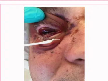
Figure 4: Preoperative photograph of the patient demonstrating right periorbital ecchymosis, conjunctival hemorrhage, suffusion and a 2 cm laceration of the lower eyelid.
suffusion, 2-cm laceration on the lower eyelid, massive subconjunctival hemorrhage and severe perforating ocular injury could be detected.
On slit lamp examination, structures of the anterior segment couldn’t be recognized due to the severe, destructing perforating trauma.
Hemophtalmus could also be diagnosed with the partial collapse of the globe. The visual acuity was decreased to no light perception and the eye movements were limited to supraduction and adduction.
There was no impairment of visual acuity of the left eye, direct pupillary light reflex was normal and eye movements were without limitations on that side. The rest of the cranial nerve functions were intact. No external bleeding or liquor leakage was observed during
the evaluation of the patient.
After excluding all other possible traumatic injuries, the patient was admitted to the neurosurgery department for further treatment.
Prophylactic antibiotic therapy was started with ceftriaxone.
Considering the entry wound in the right infraorbital region and the close contact with the internal carotid arteries, optic nerves and sphenoid sinus, a multidisciplinary team meeting, composed of neurosurgery specialists, otorhinolaryngologists, ophthalmologists, and the radiologist was essential to provide with the best possible surgical treatment.
A CT scan was performed using an ultrafast 256-slice Philips Brilliance iCT, the slice thickness was 1 mm with a total of 220 slices. Images were exported as DICOM files to a Medtronic Stealth Station S8 navigation system. Access to the target was visualized in multiplanar image reconstruction (Figure 5).
Endoscopic procedure
The sphenoid sinus exploration was performed via a Transnasal, transseptal endoscopic (30 degrees, 4 mm, 170 mm, Storz) approach.
Following Xylometazoline surface anemization (topical instillation), the posterior third of the septum was infiltrated with 2% Lidocaine + adrenaline, and the mucoperiosteum was raised. On the right side, a vertical incision was made at the height of the anterior pole of the middle turbinate, the mucoperiosteum was elevated from the vomer below, and the lamina perpendicular above (Figure 6). The bony septum was pierced and both sphenoidal ostia were identified.
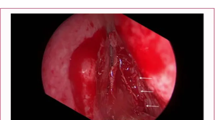
The sphenoidal rostrum was removed to give a broad view of the sphenoidal plain (Figure 7).
With suction and with a tough respiratorium, keeping the mucoperiosteum in sight, the horizontal (coin) foreign body was exposed (Figure 8,9). Exploration was made slowly and cautiously not
Figure 5: High-resolution endoscope (STORZ), Medtronic StealthStation S8 Navigation connected with endoscope and Zeiss Kinevo neurosurgical microscope. An intraoperative photograph shows the endonasal transsphenoidal endoscopy-assisted procedure. Fortunately, the use of microscope was not necessary.
Figure 6: Vertical incision on the septum, creating a muco-periosteal flap on the right and the left side.
Figure 7: View of rostrum sphenoidale with the right and left ostiums after removing the vomer and perpendicular plate of ethmoid bone.
Figure 8: Intraoperative endoscope image of the foreign body (Hungarian 200-forint coin).
Figure 9: The foreign body (coin).
to injure the frontonasal anatomical structures especially the internal carotid artery, the circle of Willis and the optic nerves or the optic chiasm. To do so, we avoided fast movements in cranial and posterior directions. With Rosen's curette, the front edge of the coin was tilted and removed with a Weil handle.
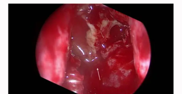
Although the planum sphenoidale and the dura were extensively destroyed, and even the damaged frontobasal brain surface was visible (Figure 10) no cerebrospinal liquor (CSF) leakage was detected. The suspected CSF leak pathway was closed by local edema.
Laceration, i.e., the absence of free and removable wound edges made the reconstruction of the skull base difficult. Therefore, the defect was closed with just abdominal fat tissue in two layers (intracranial giant- bath plug and overlay extracranial intrasphenoidal support) (Figure 11) and overlaid with the nasal mucoperiosteum in septal line as a third layer. No fibrin glue was used. Moistened, gel-coated, balloon inflated nasal tampons were introduced in both nostrils to compress the mucosal flap against the defect, for 7 days. However, this last step isn’t necessary for small defects. Due to overlay closure, ceftriaxone, vancomycin and a spinal drain was applied for 7 days to prevent nasal liquor rhea and consequential meningitis. After removal of the tampons the endoscopic control examination revealed good wound healing and no SCF leakage. The patient was discharged 10 days after the operation without any postoperative complication.
Discussion
Intracranial metallic foreign bodies occur as a result of accidents, self-harm or iatrogenic incidents and may have significant potential morbidity and mortality [8,14]. Removal of the foreign body is indicated to prevent further complications, such as central nervous system infection, CSF leakage, injuries to the nerves and vessels [8,9]. Traditionally these foreign bodies were removed
Figure 10: View of the planum sphenoidale and skull base after the foreign body removal.
Figure 11: "Giant" bath-plug technique using free abdominal fat.
via craniotomy; however, this approach increases the risk for perioperative complications. Foreign bodies close to the base of the skull can be considered for endoscopic removal, depending on the experience of the surgeon and most importantly on the availability of a collaborative multidisciplinary team [9]. Given that our institute had these endowments, in contrast with the traditional approach, we opted for an endoscopic procedure and successfully removed the coin using the Transnasal transseptal transsphenoidal endoscopic technique. As a result of the minimally invasive intervention, which was relatively short and uncomplicated, the patient was able to leave the hospital quickly.
Endoscopic surgery has the benefits of decreased morbidity, decreased length of hospitalization, less blood loss, no remaining visible scars and has been used to treat lesions of the anterior skull base [4].
After the foreign body is removed, the bony defect needs to be closed endoscopically to prevent meningitis and CSF leakage. In the case of multilayer dura closure when intracranial intradural and extradural layers are used, it is not recommended to reduce the liquor pressure because it leads to slippage of the layers. In the case of inlay or overlay, however, it is the increased liquor pressure that can push the graft away from the CSF leaks. Nota Bene reducing liquor pressure increases the risk of ascending infection.
However, due to the indication of unusual endoscopic surgery and the significantly damaged and presumably infected wound, we faced significant difficulty with the closure of the frontobasal defect.
Traditionally the skull base is reconstructed with a multilayer technique using fascia lata and abdominal free fat; however in our case due to the mechanism of the injury which resulted in brain contusion, the lack of intact dura and bone edges made the closing more difficult [15].
Normally, abdominal free fat is another feasible option for grafting material during endoscopic skull base reconstruction [16]. Because the widely accepted multilayer technique requires larger access in order to be spread out over the intracranial surface and we did not want to cause more damage and complications we decided to use only bath-plug technique supplemented with nasal mucoperiosteal flap [17]. With the help of the two-layer abdominal fat tissue (intracranial giant bath plug and overlay extracranial intrasphenoidal support) and the mobilized nasal mucoperiosteum from caudally we finally achieved our goal of the multiple layer wound closure. This latter was also supported with gel-coated, balloon inflated tampons intranasally, but this is usually not necessary. To promote wound cleansing and to avoid the formation of local abscess no fibrin glue was used. After the patient was healed, with no complication, we believe that closure of the anterior skull base using “Giant” bath-plug together with nasal mucoperiosteal flap is an easy-to-use and also reliable skull base reconstruction technique especially in contaminated, traumatic skull base injuries.
Periorbital trauma may result in orbital hemorrhage with the risk of developing the sight threatening orbital compartment syndrome. The increased orbital pressure can damage the optic nerve by direct compression, or by causing ocular ischemia via decreased perfusion from compromised vascular flow. Immediate lowering of the intraorbital pressure can be reached by acute canthotomy and cantholysis. It should be noted; however, that the procedure is contraindicated in case of open globe injury since it can aggravate the damage of the eye by enhancing the prolapse of the intraocular content. Taken together, although without decompression,
irreversible vision loss may occur due to the increased orbital pressure and early recognition and prompt treatment is essential to prevent vision loss, careful ophthalmological examination is indispensable in order to exclude open globe injury.
In our case, unfortunately, the destruction of the eye was so extensive that it made it impossible to attempt the restoration of the anatomical integrity of the eyeball [18-20].
Conclusion
In this paper we presented a successful endoscopic Transnasal removal of a coin pierced into the sphenoidal sinus that extended into the intracranial space. Due to the potential life-threatening complications, foreign bodies in the sphenoid sinus and skull base should be treated. Recently, with the rising popularity of endoscope, alternative approaches to craniotomy should be considered if access is possible, as it is an efficient, safe and minimally invasive procedure.
However, closure of the anterior skull base in these cases might be difficult; using “Giant” bath-plug together with nasal mucoperiosteal flap is an easy to use and also reliable skull base reconstruction technique especially in contaminated, traumatic skull base injuries.
References
1. Lee WB, O’Halloran HS, Pearson PA, Sen HA, Reddy SH. Airbags and bilateral eye injury: Five case reports and a review of the literature.
2001;20(2):129-34.
2. Wallis LA, Greaves I. Injuries associated with airbag deployment. Emerg Med J.2002;19(6):490-3.
3. Krause H-R, Rustemeyer J, Grunert R. Fremdkörper in den Nasennebenhöhlen. Mund Kiefer Gesichtschir. 2002;6(1):40-4.
4. Casler JD, Doolittle AM, Mair EA. Endoscopic surgery of the anterior skull base. Laryngoscope. 2005;115(1):16-24.
5. Mladina R, Hat J, Klapan I, Heinzel B. An endoscopic approach to metallic foreign bodies of the nose and paranasal sinuses. Am. J. Otolaryngol.
1995;16(4):276-9.
6. Verret DJ, Defatta R, Ducic Y. Transorbital penetration of the skull base with an occult foreign body. Am J Emerg Med. 2005;23(7):901-2.
7. Feichtinger M, Zemann W, Kärcher H. Removal of a pellet from the left orbital cavity by image-guided endoscopic navigation. Int J Oral Maxillofac Surg. 2007;36(4):358-61.
8. Kitajiri S, Tabuchi K, Hiraumi H. Transnasal bamboo foreign body lodged in the sphenoid sinus. Auris Nasus Larynx. 2001;28(4):365-7.
9. Thomas S, Daudia A, Jones NS. Endoscopic removal of foreign body from the anterior cranial fossa. J Laryngol Otol. 2007;121(8):794-5.
10. Bhattacharyya N, Wenokur RK. Endoscopic management of a chronic ethmoid and sphenoid sinus foreign body. Otolaryngol Head Neck Surg.
1998;118(5):687-90.
11. Barzo P, Zador Z, Bodosi M, Bella Z, Jambor D, Fulop B, et al. Combined minimally invasive supraciliary and transfacial approach for large tumors with skull base and sinonasal involvement. World Neurosurg. 2018;109:1- 9.
12. Bella Z. Endoscopic posterior transseptal pituitary surgery - Advances in surgical technique to complete endoscopic tumor removal based on analysis of 61 cases..
13. Fülöp B, Bella Z, Palágyi P, Barzó P. Endoscopic removal of tuberculum sellae meningeoma through endonasal transsphenoidal approach.
Ideggyogy Sz. 2016;69(3–4):133-8.
14. Yildirim AE, Divanlioglu D, Cetinalp NE, Ekici I, Dalgic A, Belen AD.
Endoscopic endonasal removal of a sphenoidal sinus foreign body extending into intracranial space. Ulus Travma Acil Cerrahi Derg.
2014;20(2):139-42.
15. Daele JJM, Goffart Y, Machiels S. Traumatic, iatrogenic and spontaneous Cerebrospinal Fluid (CSF) leak: Endoscopic repair. B-ENT. 2011;7 Suppl 17:47-60.
16. Lam K, Luong AU, Yao WC, Citardi MJ. Use of autologous fat grafts for the endoscopic reconstruction of skull base defects: Indications, outcomes, and complications. Am J Rhinol Allergy. 2018;32(4):310-7.
17. Wormald PJ, McDonogh M. The bath-plug closure of anterior skull base cerebrospinal fluid leaks. Am. J. Rhinol. 2003;17(5):299-305.
18. Smock WS. Road traffic accidents, airbag-related injuries and deaths. In:
Payne-James J, editor. Encycl. Forensic Leg. Med. [Internet]. Oxford:
Elsevier; 2005 [cited 2020 Mar 19]. p. 1–11.
19. Effectiveness of Air Bags. NEJM. 1992.
20. Munich SA, Fenstermaker RA, Fabiano AJ, Rigual NR. Cranial base repair with combined vascularized nasal septal flap and autologous tissue graft following expanded endonasal endoscopic neurosurgery. J Neurol Surg A Cent Eur Neurosurg. 2013;74(2):101-8.