rapid communications
Li fi leucel, a Tumor-In fi ltrating Lymphocyte Therapy, in Metastatic Melanoma
Amod A. Sarnaik, MD1; Omid Hamid, MD2; Nikhil I. Khushalani, MD1; Karl D. Lewis, MD3; Theresa Medina, MD3; Harriet M. Kluger, MD4; Sajeve S. Thomas, MD5; Evidio Domingo-Musibay, MD6; Anna C. Pavlick, DO, MBA7; Eric D. Whitman, MD8;
Salvador Martin-Algarra, MD, PhD9; Pippa Corrie, PhD, FRCP10; Brendan D. Curti, MD11; Judit Ol ´ah, MD, DSc12; Jose Lutzky, MD13; Jeffrey S. Weber, MD, PhD7; James M. G. Larkin, MD, PhD14; Wen Shi, MD, PhD15; Toshimi Takamura, BA, BS15; Madan Jagasia, MD15; Harry Qin, PhD15; Xiao Wu, PhD15; Cecile Chartier, PhD15; Friedrich Graf Finckenstein, MD15; Maria Fardis, PhD, MBA15;
John M. Kirkwood, MD16; and Jason A. Chesney, MD, PhD17
abstract
PURPOSE Effective treatment options are limited for patients with advanced (metastatic or unresectable)melanoma who progress after immune checkpoint inhibitors and targeted therapies. Adoptive cell therapy using tumor-infiltrating lymphocytes has demonstrated efficacy in advanced melanoma. Lifileucel is an autologous, centrally manufactured tumor-infiltrating lymphocyte product.
METHODS We conducted a phase II open-label, single-arm, multicenter study in patients with advanced melanoma who had been previously treated with checkpoint inhibitor(s) and BRAF6MEK targeted agents.
Lifileucel was produced from harvested tumor specimens in central Good Manufacturing Practice facilities using a streamlined 22-day process. Patients received a nonmyeloablative lymphodepletion regimen, a single infusion of lifileucel, and up to six doses of high-dose interleukin-2. The primary end point was investigator-assessed objective response rate (ORR) per RECIST, version 1.1.
RESULTS Sixty-six patients received a mean of 3.3 prior therapies (anti–programmed death 1 [PD-1] or programmed death ligand 1 [PD-L1]: 100%; anticytotoxic T-lymphocyte-associated protein-4: 80%;
BRAF6MEK inhibitor: 23%). The ORR was 36% (95% CI, 25 to 49), with two complete responses and 22 partial responses. Disease control rate was 80% (95% CI, 69 to 89). Median duration of response was not reached after 18.7-month median study follow-up (range, 0.2-34.1 months). In the primary refractory to anti– PD-1 or PD-L1 therapy subset, the ORR and disease control rate were 41% (95% CI, 26 to 57) and 81% (95%
CI, 66 to 91), respectively. Safety profile was consistent with known adverse events associated with non- myeloablative lymphodepletion and interleukin-2.
CONCLUSIONLifileucel demonstrated durable responses and addresses a major unmet need in patients with metastatic melanoma with limited treatment options after approved therapy, including the primary refractory to anti–PD-1 or PD-L1 therapy subset.
J Clin Oncol 00. © 2021 by American Society of Clinical Oncology Creative Commons Attribution Non-Commercial No Derivatives 4.0 License
INTRODUCTION
The treatment of advanced (unresectable or meta- static) melanoma with immune checkpoint inhibitors (ICI) and targeted oncogenic pathway inhibition with BRAF and MEK inhibitors has improved patient outcomes.1-7 Forty percent to 65% of patients with advanced melanoma have primary resistance to ICI.8-11Of those with initial disease control, 30%-40%
develop acquired resistance.8,12Approximately 15% to 20% ofBRAF V600 mutation-positive patients fail to respond to targeted therapy initially,13and only 22%
remain progression-free at 3 years.14Although primary resistance is lower in patients treated with programmed
death 1 (PD-1) blocking antibody plus anticytotoxic T-lymphocyte–associated protein 4 (CTLA-4) therapy, 36% of patients discontinue therapy because of treatment-emergent adverse events (TEAEs), with 88%
developing immune-related adverse events (irAEs), many of these being persistent.10Patients progressing after anti–PD-1 therapy, anti–PD-1 plus anti–CTLA-4 therapy, and targeted agents have limited options.15-17 Only 4%-10% of these patients have objective re- sponses to chemotherapy, with a limited median overall survival (OS) of 7 months.15,16,18,19There are no treat- ment options with approval based on data from patients with advanced melanoma who have progressed after
ASSOCIATED CONTENT Data Supplement Protocol Author affiliations and support information (if applicable) appear at the end of this article.
Accepted on March 31, 2021 and published at ascopubs.org/journal/
jcoon May 12, 2021:
DOIhttps://doi.org/10.
1200/JCO.21.00612
one line of ICI therapy (forBRAFwild-type tumors), or two lines of therapy (forBRAFV600 mutation-positive tumors).
In addition, patients recurring with advanced melanoma after adjuvant anti–PD-1 therapy for high-risk disease rep- resent an emerging unmet need.20-22
Adoptive cell therapy with tumor-infiltrating lymphocytes (TIL) offers a potential therapeutic option for metastatic melanoma, although it has not been studied extensively in the ICI era.23-25TIL are enriched with polyclonal T cells with diverse antigen specificity.26 Extraction of a fragment of tumor followed by ex vivo expansion removes TIL from the hostile tumor microenvironment and reduces the immu- nosuppressive effects of intratumoral regulatory T cells.
Expansion of TIL ex vivo rejuvenates the cells, yielding billions of such cells to be infused back into the patient.
Melanoma is characterized by a high mutational burden27 and highly individualized neoantigens.28A cellular therapy product that can address the broad nature of neoantigens and the unique array from each patient would lead to the possibility of a tailored response. Lifileucel (LN-144) is an autologous TIL therapy that uses tumor-tissue T cells ca- pable of recognizing tumor antigens and being expanded ex vivo while maintaining the heterogeneous repertoire of T cells, using a centralized manufacturing process. We report the safety and efficacy of lifileucel, a one-time cel- lular therapy, in patients with advanced melanoma who have progressed on ICI and BRAF inhibitors (ifBRAFV600 mutation-positive).
METHODS Trial Conduct
The study was approved by the institutional review board at each site and was conducted in accordance with the Declaration of Helsinki and Good Clinical Practice guide- lines of the International Conference on Harmonization. All patients provided written informed consent. The study was
designed and sponsored by Iovance Biotherapeutics, Inc.
All authors discussed, analyzed, and interpreted the re- sults, and vouch for the accuracy and completeness of the data analyses and adherence to the Protocol (online only).
All authors contributed to this study and the writing of the manuscript. Professional medical writing or editorial as- sistance was paid for by the sponsor.
Patients and Study Design
The parent study (ClinicalTrials.gov identifier:NCT02360579) consisted of multiple cohorts (Data Supplement, online only). Cohort 2 data are reported here. Patients were enrolled from April 2017 to January 2019 at 26 sites (see the Data Supplement for investigator list).
Patients had unresectable or metastatic melanoma (stage IIIC or IV) with confirmed radiologic progression. Patients must have progressed following one or more prior systemic therapies including a PD-1–blocking antibody and ifBRAF V600 mutation-positive, a BRAF 6 MEK inhibitor. Key eligibility criteria are detailed in the Data Supplement.
Patients with a history of irAEs were eligible, as outlined in the Data Supplement.
At least one resectable lesion (or aggregate of lesions) measuring a minimum of 1.5 cm in diameter postresection was required. Resected tumor was processed in a protocol- specified manner and shipped to a Good Manufacturing Practice facility in the provided tumor procurement kit. The optimized manufacturing conditions involved a centralized 22-day process, resulting in a cryopreserved product (Data Supplement). Lifileucel (LN-144) was shipped to the clinical sites after meeting prespecified release criteria.
Patients received a nonmyeloablative lymphodepleting (NMA-LD) regimen with cyclophosphamide (60 mg/kg) once daily for 2 days followed byfludarabine (25 mg/m2) once daily for 5 days. A single infusion of lifileucel (13109 – 150 3 109 cells) was thawed and administered after approximately 24 hours from the last dose offludarabine. A CONTEXT
Key Objective
This study evaluated the efficacy and safety of lifileucel, a one-time, autologous tumor-infiltrating lymphocyte (TIL) product, in patients with metastatic melanoma who had progressed on standard immune checkpoint inhibitors (ICI) and targeted therapies (if applicable), who otherwise have limited treatment options. Notably, chemotherapy post-ICI shows poor response rates (4%-10%).
Knowledge Generated
Sixty-six patients received lifileucel infusion with.13109TIL cells. Lifileucel was efficacious with an objective response rate of 36%, and a median duration of response that is not reached at 18.7-month median study follow-up.
Relevance
Lifileucel represents a significant improvement in the treatment of advanced melanoma, particularly in the post-ICI patient population, which is an expanding population. The study contributes to the advancement in TIL therapy through a centrally standardized manufacturing approach for autologous TIL, allowing broader patient access.
short course of bolus interleukin (IL)-2 (600,000 IU/kg) was infused every 8-12 hours for up to six doses, starting within 3-24 hours of completing lifileucel infusion.
End Points and Assessments
The primary objective was to evaluate the efficacy of a single infusion of lifileucel in patients with unresectable or metastatic melanoma using investigator-assessed objective response rate (ORR) by RECIST v1.1.29 Secondary end points included duration of response (DOR), disease control rate (DCR), OS, and safety. Efficacy assessments started at week 6. Subsequent efficacy, adverse event (AE), and serious AE (SAE) assessment schedules are outlined in the Data Supplement.
Statistical Analysis
The analyses for efficacy and safety were conducted on the full analysis set (FAS), defined as patients from cohort 2 who received lifileucel that met manufacturer’s specifica- tions, including a cell dose 1 3109– 1503109. The planned sample size was 60 based on estimation of ORR using the maximum half-width of the two-sided 95% CI of,13.2% when ORR is expected to be 20%-50%. This was considered meaningful, assuming that the historical response rate of similar patients after chemotherapy is 10%.15,30 The FAS consisted of 66 patients because of rapid enrollment.
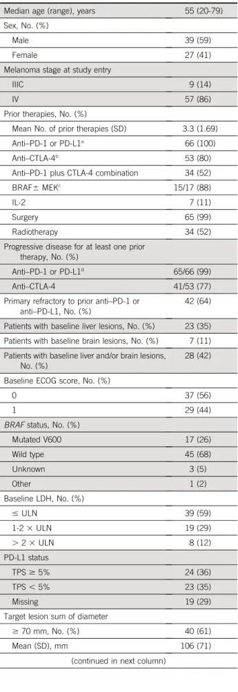
The ORR was analyzed as a binomial proportion with two- sided 95% CI estimated based on the Clopper-Pearson TABLE 1. Patient Demographics and Baseline Characteristics
Characteristic Cohort 2 (N566)
Median age (range), years 55 (20-79)
Sex, No. (%)
Male 39 (59)
Female 27 (41)
Melanoma stage at study entry
IIIC 9 (14)
IV 57 (86)
Prior therapies, No. (%)
Mean No. of prior therapies (SD) 3.3 (1.69)
Anti–PD-1 or PD-L1a 66 (100)
Anti–CTLA-4b 53 (80)
Anti–PD-1 plus CTLA-4 combination 34 (52)
BRAF6MEKc 15/17 (88)
IL-2 7 (11)
Surgery 65 (99)
Radiotherapy 34 (52)
Progressive disease for at least one prior therapy, No. (%)
Anti–PD-1 or PD-L1d 65/66 (99)
Anti–CTLA-4 41/53 (77)
Primary refractory to prior anti–PD-1 or anti–PD-L1, No. (%)
42 (64)
Patients with baseline liver lesions, No. (%) 23 (35) Patients with baseline brain lesions, No. (%) 7 (11) Patients with baseline liver and/or brain lesions,
No. (%)
28 (42)
Baseline ECOG score, No. (%)
0 37 (56)
1 29 (44)
BRAFstatus, No. (%)
Mutated V600 17 (26)
Wild type 45 (68)
Unknown 3 (5)
Other 1 (2)
Baseline LDH, No. (%)
#ULN 39 (59)
1-23ULN 19 (29)
.23ULN 8 (12)
PD-L1 status
TPS$5% 24 (36)
TPS,5% 23 (35)
Missing 19 (29)
Target lesion sum of diameter
$70 mm, No. (%) 40 (61)
Mean (SD), mm 106 (71)
(continued in next column)
TABLE 1. Patient Demographics and Baseline Characteristics (continued)
Characteristic Cohort 2 (N566)
No. of target and nontarget lesions (at baseline)
.3, No. (%) 51 (77)
Mean (SD) 6 (2.7)
Median (range) 5 (2-14)
Median time from stop of anti–PD-1 or PD-L1 to TIL infusion (range), months
4.8 (1.6-56.5)
Abbreviations: CTLA-4, cytotoxic T-lymphocyte–associated protein 4; ECOG, Eastern Cooperative Oncology Group; IL, interleukin; LDH, lactate dehydrogenase; PD-1, programmed cell death protein 1; PD- L1, programmed death-ligand 1; SD, standard deviation; TIL, tumor- infiltrating lymphocytes; TPS, tumor proportion score; ULN, upper limit of normal.
aIncludes pembrolizumab, nivolumab, durvalumab, and atezolizumab.
bIncludes ipilimumab and tremelimumab.
cOne patient received only BRAF inhibitor. Two patients were enrolled under an earlier protocol version that did not requireBRAF V600 mutation-positive patients to receive BRAF6MEK inhibitors.
Percentage is calculated based on number of patients who wereBRAF V600E- or V600K-mutated and received a BRAF inhibitor (dabrafenib or vemurafenib)6a MEK inhibitor (trametinib or cobimetinib).
dOne patient discontinued anti–PD-1 therapy because of toxicity and then progressed on interval therapy before enrollment.
exact method. Time-to-event efficacy end points were estimated using the Kaplan-Meier product limit method, and two-sided corresponding 95% CIs were based on log- log transformation. Safety data were reported descriptively.
All statistical analyses were conducted using the Statistical Analysis System (SAS) version 9.4.
RESULTS
Patients and Treatment
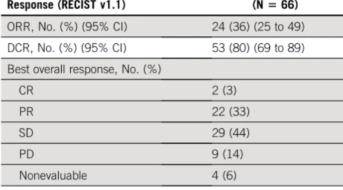
Seventy-eight patients underwent tumor resection. Sixty-six patients received lifileucel (LN-144) infusion with.13109 but,1503109TIL cells and comprised the FAS. Three patients either did not receive TIL or received,13109TIL cells, whereas nine patients could not be treated because of other causes (Data Supplement).Table 1 details the de- mographics and baseline characteristics. Patients had received a mean of 3.3 lines of prior therapies (range, 1-9 lines). All patients had received prior anti–PD-1 or anti– programmed death ligand 1 (PD-L1) therapy, and 53 (80%) had received prior anti–CTLA-4 therapy. Fifty-two percent of the patients had received concurrent CTLA-4 plus PD-1 blockade. Notably, 99% had progressed on prior anti–PD-1 or PD-L1 therapy, and 77% had progressed on prior anti–CTLA-4 therapy. Overall, 42 patients (64%) had a best response of progressive disease to initial anti–PD-1 or PD-L1 therapy (primary refractory subset). Of the 17 pa- tients who were BRAF V600 mutation-positive, 88% had received BRAF6MEK inhibitors. Forty patients (61%) had a baseline target lesion sum of diameters (SOD)$70 mm, 51 (77%) patients had more than three target and nontarget lesions at baseline, and 27 (41%) had baseline lactate dehydrogenase levels higher than institutional upper limit of normal. Overall, patients had a high tumor burden at TABLE 2. Efficacy Outcomes by Investigator Assessment
Response (RECIST v1.1)
Cohort 2 (N566) ORR, No. (%) (95% CI) 24 (36) (25 to 49) DCR, No. (%) (95% CI) 53 (80) (69 to 89) Best overall response, No. (%)
CR 2 (3)
PR 22 (33)
SD 29 (44)
PD 9 (14)
Nonevaluable 4 (6)
Median DOR, months (range) Not reached (2.2-26.91) NOTE.1, censored.
Abbreviations: CR, complete response; DCR, disease control rate;
DOR, duration of response; ORR, objective response rate; PD, progressive disease; PR, partial response; SD, stable disease.
CR PR SD PD
1 2 3 4 5 7 8 44 9 10 11 45 13 14 16 46 12 47 15 49 50 17 18 51 48 19 6 20 21 53 54 23 55 52 63 28 27 26 31 32 25 57 33 36 38 59 40 60 61 35 37 41 24 58 29 34 62 39 42 22 30 43
Patient
% Change From Baseline
80
60
40
Best Overall Response
20
0
–20 –30 –40
–60
–80
–100
A
FIG 1. Change in tumor burden of target lesions, response by subgroup, and response assessment in individual patients. (A) Waterfall plot depicting BOR as assessed by investigator and the best change from baseline in the SOD of the target lesions (per RECIST v1.1 criteria) in the FAS. A change of2100% from baseline is presented for CR assessment that includes lymph node lesions that resolved to,10 mm. The horizontal dashed line indicates a 30% reduction in the tumor burden in the target lesions. Twelve patients had an increase in the SOD of the target lesions, whereas 50 patients had a decrease in the SOD of the target lesions. Thirty patients (two CR, 22 PR, and six SD) had.30% reduction in the SOD of the target lesions. Three patients had no post-TIL assessments because of early death. One patient had no post-TIL assessment because of start of new anticancer therapy before day 42. (continued on next page)
baseline (mean SOD for the target lesions: 106 mm); 28 patients (42%) had liver and/or brain lesions at baseline.
The harvested tumor was collected from a variety of sites, such as skin, lymph nodes, liver, lung, peritoneum, mus- culoskeletal sites, breast, and other organs. The median
number of cyclophosphamide andfludarabine doses were 2 (range, 1-2) and 5 (range, 2-5), respectively. The mean number of TIL cells infused was 27.3 3 109 (range, 1.23 109 to 99.5 3109). The median number of IL-2 doses administered was 5.5 (range, 1-6).
Subgroup
Overall Age group, years
< 65 t65
Prior anti–CTLA-4 use Yes
No
BRAF mutation status V600- or V600K-mutated Nonmutated
PD-L1 status (TPS t1% v < 1%)
t1%
< 1%
PD-L1 status (TPS t5% v < 5%)
t5%
< 5%
Baseline ECOG 0
t1
Baseline target lesion sum of diameters, mm
< 70 t70
Patients with baseline brain and/or liver lesion Time from stop of anti-PD-1 or PD-L1 to TIL infusion
d median (4.8 months)
> median (4.8 months) Patients with baseline liver lesion
B
Baseline lactate dehydrogenase
n/N ORR 95% Cl 24.9 to 49.1 36.4
24/66
23.6 to 51.0 12.8 to 64.9 36.5
35.7 19/52
5/14
23.1 to 50.2 13.9 to 68.4 35.8
38.5 19/53
5/13
18.4 to 67.1 21.7 to 49.6 41.2
34.7 7/17 17/49
20.8 to 53.8 10.9 to 69.2 36.1
36.4 13/36
4/11
18.8 to 59.4 16.4 to 57.3 37.5
34.8 9/24 8/23
27.1 to 60.5 12.7 to 47.2 43.2
27.6 16/37
8/29
33.4 to 73.4 12.7 to 41.2 53.8
25.0 14/26 10/40
15.9 to 52.4 32.1
9/28
20.4 to 54.9 20.4 to 54.9 36.4
36.4 12/33 12/33
0 20 40 60 80 100
ORR (95% Cl)
16.4 to 57.3 34.8
8/23
23.4 to 55.4 20.3 to 66.5 0.3 to 52.7 38.5
42.1 12.5 15/39
8/19 1/8 d ULN
1-2 × ULN
> 2 × ULN
FIG 1. (Continued). (B) Forest plot for ORR (FAS) by subgroup per investigator assessment using the RECIST v1.1 criteria. 95% CI is calculated using the Clopper-Pearson Exact test. (continued on next page).
Efficacy
Sixty-six patients received a lifileucel infusion of$13109 TIL cells. At the data cutoff of April 23, 2020 (median follow- up of 18.7 months [range, 0.2-34.1 months]), the in- vestigator-assessed ORR was 36% (95% CI, 25 to 49) and the DCR was 80% (95% CI, 69 to 89) (Table 2), with 2 (3%) complete responses (CRs), 22 (33%) partial re- sponses (PRs), and 29 (44%) patients showing stable disease (SD). Sixty-two patients (94%) had a baseline and at least one postbaseline radiologic assessment. Of the four patients in the FAS who did not undergo postbaseline assessment, three had died of disease, and one received an additional line of systemic therapy; all were considered as not evaluable for best overall response. Of the evaluable patients, 50 (81%) had a reduction in tumor burden (Fig 1A). Data Supplement details the percentage change in target SOD from baseline over time in patients who achieved a confirmed response. Response to lifileucel was observed regardless of age, prior anti–CTLA-4 use,BRAF mutation status, PD-L1 status as measured by tumor proportion score, baseline Eastern Cooperative Oncology Group performance status, tumor burden (assessed by lactate dehydrogenase elevation above upper limit of normal and target lesion SOD at baseline), presence of liver and/or brain lesions at baseline, and timing of prior PD-1 therapy (Fig 1B).
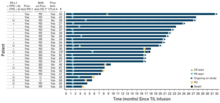
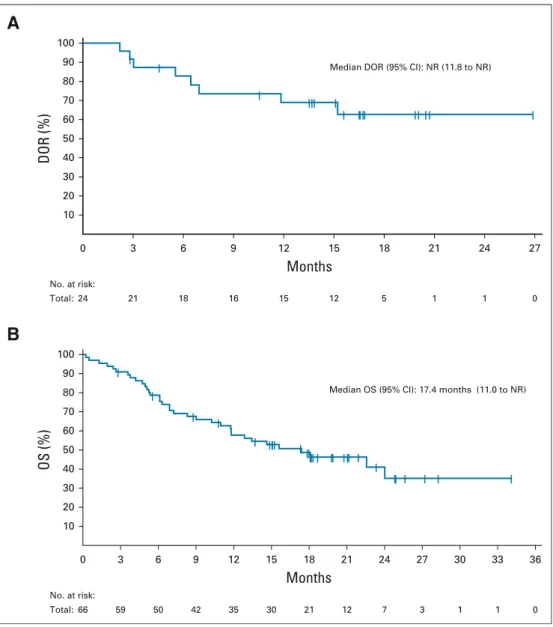
Median time from lifileucel infusion to best response was 1.4 months (range, 1.3-8.7 months). Time to response for individual patients is illustrated in Figure 1C; 19 of 24 patients achieved response by the time of first planned assessment (6 weeks after lifileucel infusion). Only 25% of patients had progressed after achieving a response. The median DOR has not been reached (95% CI, 11.8 months to not reached) (Fig 2A) with a 1-year DOR of 69% (95% CI, 46 to 84). The median OS was 17.4 months (95% CI, 11.0 to not reached;Fig 2B). Of the patients who had SD and PR or CR, 38% and 92% patients, respectively, had an OS$1 year. Progression-free survival for the FAS is shown in the Data Supplement, and duration of SD in individual patients is outlined in the Data Supplement.
An efficacy analysis was performed for the primary- refractory subset (42 patients primary refractory to anti– PD-1 or PD-L1 therapy). The ORR was 41% (95% CI, 26 to 57), with 2 CRs (5%) and 15 PRs (36%), and the DCR was 81% (95% CI, 66 to 91). Seventeen (41%) of these patients had SD, and five (12%) had progressive disease; three patients were nonevaluable. Median DOR was not reached for this subpopulation.
Thirty-four (52%) patients received anti–PD-1 plus anti– CTLA-4 combination therapy, either as frontline (n515, 23%), or after failing frontline therapy (n519, 29%). The
0 1 2 3 4 5 6 7 8 9 10 11 12 13 14 15 16 17 18 19 202122 23 24 25 26 27 28 29 30 31 32 43
42 29 27 25 37 39 26 24 34 30 35 22b 62 58 57 40 61 36 41 32 59 55 28 Yes Yes No Yes Yes Yes Yes No No Yes Yes No Yes Yes Yes Yes No Yes Yes Yes Yes Yes Yes Yes Yes
Yes Yes Yes Yes Yes Yes Yes Yes Yes Yes Yes Yes Yes Yes Yes Yes Yes Yes Yes Yes Yes Yes Yes
PD PD PD PD PD PD PD PD PR PD PD PD SD SD PD PD SD PD PD PD U U PD PR
−
− U +
− +
− +
− + U + U
− +
− + U
− + U U U +
# Prior Anti- CTLA-4 Prior
Anti-PD-1 BOR on Prior Anti-PD-1a PD-L1
+ (TPS t5)/
− (TPS < 5)
Time (months) Since TIL Infusion
Patient
CR start PR start Ongoing on study PD
Death
C
FIG 1. (Continued). (C) Swimmer’s plot showing time tofirst response, duration of response, and time on efficacy assessment in confirmed responders by investigator per RECIST v1.1 criteria. Among 24 responders, 12 (50%) showed ongoing response to lifileucel, six (25%) had progressed, two (8%) had died, three (13%) started a new anticancer therapy, and one patient discontinued assessment because of relocation.aBOR is best overall response on prior anti-PD-1 immunotherapy.bFor patient 22, a CR was not confirmed; therefore, the BOR with lifileucel for this patient was PR. Causes of death: patient 22: possible pulmonary embolism; patient 41: failure to thrive. BOR, best overall response; CR, complete response; CTLA-4, cytotoxic T-lymphocyte–associated protein 4; ECOG, Eastern Cooperative Oncology Group; FAS, full analysis set; ORR, objective response rate; PD, progressive disease; PD-1, programmed death 1; PD-L1, programmed death-ligand 1; PR, partial response; SD, stable disease; SOD, sum of diameters; TIL, tumor-infiltrating lymphocytes; TPS, tumor proportion score; U, unknown; ULN, upper limit of normal.
ORRs for lifileucel in these two subsets were 33% (5/15) and 32% (6/19), respectively. The ORRs for lifileucel in patients with primary resistance (n517) or acquired re- sistance (n511) to anti–PD-1 plus anti–CTLA-4 combi- nation therapy were 35% (6/17) and 27% (3/11), respectively. Details of these patients who responded to lifileucel are outlined in the Data Supplement.
Exploratory analyses of product-specific characteristics, including levels of phenotypic markers of T-cell lineage, memory subset, youth, activation or exhaustion, or trafficking (Data Supplement), did not demonstrate
association with response. Tumor burden reductions were seen across the continuum of cell doses (Data Supplement).
Safety
All patients experienced at least one TEAE, with the most common ($30%) grade 3 or 4 TEAEs being thrombocyto- penia (82%), anemia (56%), febrile neutropenia (55%), neutropenia (39%), hypophosphatemia (35%), leukopenia (35%), and lymphopenia (32%) (Table 3), consistent with the toxicity profile of NMA-LD and IL-2. Fatal TEAEs occurred in two patients—1 death was because of intra-abdominal tumor
Median DOR (95% CI): NR (11.8 to NR)
No. at risk:
Total:
0 3 6 9 12 15 18 21 24 27
24 21 18 16 15 12 5 1 1 0
Months
10 20 30 40 50 60 70 80 90 100
DOR (%)
A
Median OS (95% CI): 17.4 months (11.0 to NR)
No. at risk:
Total:
0 3 6 9 12 15 18 21 24 27 30 33 36
66 59 50 42 35 30 21 12 7 3 1 1 0
Months
10 20 30 40 50 60 70 80 90 100
OS (%)
B
FIG 2. (A) The Kaplan-Meier curve for DOR in confirmed responders who achieved a PR or better. The DOR is measured from the time point at which the initial measurement criteria are met for a PR or CR, whichever occurred first, until thefirst date that PD or death occurred. (B) The Kaplan-Meier curve for OS in the full analysis set. OS was defined as the time (in months) from the start date of lifileucel infusion to death because of any cause. Patients who were alive at the time of data cutoff had their event times censored on the last date of their known survival status.
The median OS was 17.4 months (95% CI, 11.0 to NR), with 1-year OS of 58% (95% CI, 45 to 69). CR, complete response; DOR, duration of response; NR, not reached; OS, overall survival; PD, progressive disease; PR, partial response.
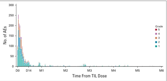
hemorrhage reported as possibly related to TIL, and one was because of acute respiratory failure assessed as not related to TIL by the investigator. The incidence of TEAEs, including grade 3 or 4 TEAEs, decreased rapidly over time (Fig 3) with no lifileucel-related SAEs reported after 6 months, and no recurrence of irAEs related to prior ICI. Tumor harvest AEs related to surgery are outlined in the Data Supplement.
DISCUSSION
Treatment options for patients with advanced melanoma who progress after treatment with ICI and BRAF6MEK inhibitors are limited. Cytotoxic chemotherapy has shown poor response rates,15,16,18,19 with a limited median OS of 7 months.15 Many of the patients in our study had exhausted all approved therapy (mean lines of prior ther- apy, 3.3). The encouraging antitumor activity of lifileucel observed in our study addresses a major unmet need in patients with advanced melanoma after progression on ICI, and targeted agents if indicated.
Lifileucel, a one-time cellular therapy, represents a significant improvement in the treatment of advanced melanoma, par- ticularly the current post-ICI patient population. First, lifileucel demonstrated an ORR of 36%, meeting the study primary end point in a patient population that had failed frontline anti–PD-1 therapy, the current standard of care. This is noteworthy because prior TIL therapy studies were conducted in the pre- ICI era, or enrolled a very small population of patients who had received prior anti–PD-1 therapy.23-25,31A previous cohort 2 analysis has demonstrated a high concordance rate of 89.4%
between the Independent Review Committee–assessed and investigator-assessed ORR.32 Second, at a median 18.7- month follow-up, the median DOR has not been reached, emphasizing the durability of lifileucel responses in a heavily pretreated post-ICI patient population with a high baseline tumor burden. Third, the efficacy of lifileucel was equivalent agnostic of PD-L1 status, BRAF mutation status, or prior anti–CTLA-4 therapy. Lifileucel was effi- cacious in the subset of patients who were primarily re- fractory to anti–PD-1/PD-L1 therapy, demonstrating an ORR of 41% and a DCR of 81% in this subgroup. Fur- thermore, lifileucel demonstrated similar ORR in patients who received anti–PD-1 plus anti–CTLA-4 combination as a frontline therapy (33%) or after failing frontline therapy (32%).
TIL recognize multiple tumor-specific neoantigens,33which may be required for response in solid tumors with high mutational burden. Removal from the hostile microenvi- ronment and ex vivo expansion enable TIL to evade a broad array of immunosuppressive mechanisms. Indeed, both downregulation of PD-1 expression and restored func- tionality were reported for ex vivo expanded TIL.34,35 By TABLE 3. TEAEs Occurring in$20% of Patients
Preferred Term, No. (%)
Cohort 2 (N566)
Any Grade Grade 3 or 4 Grade 5 No. of patients reporting at least one TEAE 66 (100) 64 (97) 2 (3)a
Thrombocytopenia 59 (89) 54 (82) 0
Chills 53 (80) 4 (6) 0
Anemia 45 (68) 37 (56) 0
Pyrexia 39 (59) 11 (17) 0
Neutropeniab 37 (56) 26 (39) 0
Febrile neutropenia 36 (55) 36 (55) 0
Hypophosphatemia 30 (46) 23 (35) 0
Leukopeniab 28 (42) 23 (35) 0
Fatigue 26 (39) 1 (2) 0
Hypotension 24 (36) 7 (11) 0
Lymphopeniab 23 (35) 21 (32) 0
Tachycardia 23 (35) 1 (2) 0
Alopecia 19 (29) 0 0
Increased AST 19 (29) 0 0
Decreased appetite 19 (29) 1 (2) 0
Diarrhea 19 (29) 1 (2) 0
Hypokalemia 17 (26) 2 (3) 0
Hypoxia 17 (26) 10 (15) 0
Peripheral edema 17 (26) 1 (2) 0
Rash 17 (26) 3 (5) 0
Hypocalcemia 16 (24) 3 (5) 0
Hypomagnesemia 16 (24) 0 0
Increased weight 16 (24) 1 (2) 0
Increased ALT 15 (23) 2 (3) 0
Nausea 15 (23) 0 0
Increased blood alkaline phosphatase 14 (21) 2 (3) 0
Dyspnea 14 (21) 3 (5) 0
Hypoalbuminemia 14 (21) 3 (5) 0
Maculopapular rash 14 (21) 6 (9) 0
Vomiting 14 (21) 0 0
Constipation 13 (20) 0 0
Pruritus 13 (20) 0 0
Abbreviations: AE, adverse event; TEAE, treatment-emergent adverse event; TIL, tumor-infiltrating lymphocytes.
aOne death was because of intra-abdominal hemorrhage reported as possibly related to TIL, and one was because of acute respiratory failure assessed as not related to TIL by the investigator.
bAll patients had grade 4 laboratory abnormality per the Common Terminology Criteria for Adverse Events v4.03 for leukopenia, neutropenia, and lymphopenia during the treatment-emergent period. Only clinically significant laboratory abnormalities per investigators were reported as AEs.
contrast, ICI target only a limited number of pathways in situ. Additionally, in vitro culture results in large-scale expansion of TIL, potentially increasing the number of tumor-specific T cells available for tumor targeting after adoptive transfer. The T cells comprising the TIL product are recovered directly from the tumor tissue, a site enriched for T-cell clones that are able to recognize patient-specific tumor antigens.36,37 As a result, a polyclonal product is obtained that has the potential to target multiple relevant antigens, addressing (1) the ability to identify the unique spectrum of patient-specific tumor antigens38; (2) the heterogeneous nature of solid tumors39; and (3) immune escape through antigen loss.40Finally, substantial fractions of TIL-derived T cells were shown to persist for at least 6 weeks,41consistent with the memory phenotype of the majority of the T cells comprising the product.35 These varied mechanisms and TIL properties likely contribute to the antitumor efficacy of lifileucel.
The tumors were harvested with minimal surgical morbidity, although 58% were extranodal or nonskin/subcutaneous lesions. A small subset of enrolled patients (12%) could not be treated because of progression, death, or other causes.
TEAEs occurred during or immediately after NMA-LD or IL-2 administration and were generally transient, with no new lifileucel-associated SAEs reported after 6 months.
Although patients were hospitalized for NMA-LD and IL-2 administration, lifileucel is a one-time cellular therapy with durable responses, as demonstrated by an ongoing re- sponse in 50% of responders at a median follow-up of 18.7 months. In addition, the safety profile indicates that
lifileucel is a viable option for patients who are not eligible for ICI because of prior significant irAEs, as it is not as- sociated with recrudescence of irAEs.
Single-center studies conducted at NCI23,31 have been important in laying the groundwork for TIL therapy in pa- tients with advanced melanoma but were limited to a few centers with dedicated on-site cell therapy facilities. Al- though lifileucel centralized manufacturing required ship- ping of the tumor samples, TIL could be manufactured in 96% of patients. The present multicenter study constitutes a significant advance by successfully demonstrating the feasibility of a centrally standardized manufacturing ap- proach for TIL therapy, which allows for broadened patient access, whereas cryopreservation of lifileucel provides flexibility in treatment scheduling in the real-world clinical setting.
In summary, lifileucel, a first-in-class centrally manufac- tured autologous TIL cell therapy, was efficacious and demonstrated durable responses in heavily pretreated patients and represents a potential new standard of care for patients with advanced melanoma following failure of ICI and targeted therapy. Patients with advanced melanoma who have failed anti–PD-1 therapy (and BRAF 6 MEK inhibitors ifBRAFV600 mutation-positive), irrespective of baseline tumor characteristics, should be considered for the one-time lifileucel therapy as second-line therapy (third-line if BRAF V600 mutation positive) if they have performance status and organ function adequate for ad- ministration of lymphodepleting chemotherapy and a shortened course of IL-2. The US Food and Drug
Grade 5 4 3 2 1 300
250
200
150
100
50
0
D0 D14 M1 M2 M3 M4 M5 M6
Time From TIL Dose
No. of AEs
FIG 3. AEs over time. The distribution of onset of AEs starting from lifileucel infusion until 6 months postinfusion is shown. A TEAE was defined as any AE with onset after start of lifileucel through day 30 postinfusion. All occurrences of AEs were counted if a patient experienced a new onset of the same AE at different timepoints. If multiple records were reported on the electronic case report form because of toxicity grade decrease of the same AE that had not resolved, then the event was counted once with the highest grade reported. Overall, 24 AEs were reported post month 6 until data cutoff date, which are not shown in the histogram. No SAEs related to lifileucel were reported post month 6. AE, adverse event; D, day; M, month; SAE, serious adverse event; TEAE, treatment-emergent adverse event; TIL, tumor- infiltrating lymphocytes.
Administration has granted lifileucel a Regenerative Med- icine Advanced Therapy designation, Orphan Drug des- ignation, and a Fast Track designation for advanced melanoma. Based on these encouraging results, an ad- ditional cohort has been fully enrolled, using Independent
Review Committee–assessed ORR for registration pur- poses. Given the favorable risk-benefit profile of lifileucel, its role earlier in the disease course and in combination with ICI is being investigated in melanoma, as well as additional studies in other metastatic solid malignancies.
AFFILIATIONS
1H. Lee Moffitt Cancer Center, Tampa, FL
2The Angeles Clinic and Research Institute, A Cedars Sinai Affiliate, Los Angeles, CA
3University of Colorado Cancer Center—Anschutz Medical Campus, Aurora, CO
4Yale University School of Medicine, Smilow Cancer Center, New Haven Hospital, New Haven, CT
5University of Florida Health Cancer Center at Orlando Health, Orlando, FL
6Division of Hematology, Oncology and Transplantation, University of Minnesota, Minneapolis, MN
7Laura and Isaac Perlmutter Cancer Center, NYU Langone Medical Center, New York, NY
8Atlantic Health System Cancer Care, Morristown, NJ
9Cl´ınica Universidad de Navarra, Pamplona, Spain
10Cambridge University Hospitals NHS Foundation Trust— Addenbrooke’s Hospital, Cambridge, United Kingdom
11Earle A. Chiles Research Institute at Robert W. Franz Cancer Center, Providence Cancer Institute, Portland, OR
12University of Szeged—Albert Szent-Gy ¨orgyi Health Center, Szeged, Hungary
13Mount Sinai Comprehensive Cancer Center, Miami, FL
14Royal Marsden NHS Foundation Trust, London, United Kingdom
15Iovance Biotherapeutics Inc, San Carlos, CA
16Hillman Cancer Center, University of Pittsburgh Medical Center, Pittsburgh, PA
17James Graham Brown Cancer Center, University of Louisville, Louisville, KY
CORRESPONDING AUTHOR
Amod A. Sarnaik, MD, Department of Cutaneous Oncology, Moffitt Cancer Center, 10920 N. McKinley Dr, 4th Floor, Tampa, FL 33612;
e-mail: Amod.Sarnaik@moffitt.org.
PRIOR PRESENTATION
Presented in part at the annual meeting of the Society for Immunotherapy of Cancer (SITC), November 6-10, 2019, National Harbor, MD; annual meeting of the Society of Melanoma Research (SMR), November 20-23, 2019, Salt Lake City, UT; and the ASCO 2020 Virtual Annual Meeting, May 29-June 1, 2020.
SUPPORT
Supported by Iovance Biotherapeutics Inc. NIH grant that was funded during the clinical trial 5K23CA178083-03, A.A.S.
Iovance Biotherapeutics sponsored the C-144-01 trial, provided the trial drugs, and collaborated with the authors on the trial design and on the collection, analysis, and interpretation of the data. Medical writing support, funded by Iovance Biotherapeutics (with specific direction from authors), was provided by Swati Ghatpande, PhD, of Second City Science (Vaniam Group LLC).
CLINICAL TRIAL INFORMATION
NCT02360579
AUTHORS’DISCLOSURES OF POTENTIAL CONFLICTS OF INTEREST
Disclosures provided by the authors are available with this article at DOI https://doi.org/10.1200/JCO.21.00612.
AUTHOR CONTRIBUTIONS
Conception and design:Amod A. Sarnaik, Harriet M. Kluger, Eric D.
Whitman, Wen Shi, Xiao Wu, Friedrich Graf Finckenstein, Maria Fardis, Jason A. Chesney
Provision of study materials or patients:Nikhil I. Khushalani, Karl D. Lewis, Theresa Medina, Evidio Domingo-Musibay, Anna C. Pavlick, Salvador Martin-Algarra, Pippa Corrie, Brendan D. Curti, Jose Lutzky, Jeffrey S.
Weber, James M. G. Larkin, Cecile Chartier
Collection and assembly of data:Amod A. Sarnaik, Omid Hamid, Nikhil I.
Khushalani, Karl D. Lewis, Theresa Medina, Harriet M. Kluger, Sajeve S.
Thomas, Evidio Domingo-Musibay, Anna C. Pavlick, Eric D. Whitman, Salvador Martin-Algarra, Pippa Corrie, Brendan D. Curti, Judit Ol ´ah, Jose Lutzky, Jeffrey S. Weber, Wen Shi, Toshimi Takamura, Xiao Wu, Cecile Chartier, John M. Kirkwood, Jason A. Chesney
Data analysis and interpretation:Amod A. Sarnaik, Omid Hamid, Nikhil I.
Khushalani, Harriet M. Kluger, Sajeve S. Thomas, Evidio Domingo- Musibay, Eric D. Whitman, Salvador Martin-Algarra, Pippa Corrie, Brendan D. Curti, James M. G. Larkin, Wen Shi, Madan Jagasia, Harry Qin, Xiao Wu, Cecile Chartier, Friedrich Graf Finckenstein, Maria Fardis, John M. Kirkwood, Jason A. Chesney
Manuscript writing:All authors Final approval of manuscript:All authors
Accountable for all aspects of the work:All authors
REFERENCES
1. Hodi FS, O’Day SJ, McDermott DF, et al: Improved survival with ipilimumab in patients with metastatic melanoma. N Engl J Med 363:711-723, 2010 2. Robert C, Ribas A, Schachter J, et al: Pembrolizumab versus ipilimumab in advanced melanoma (KEYNOTE-006): Post-hoc 5-year results from an open-label,
multicentre, randomised, controlled, phase 3 study. Lancet Oncol 20:1239-1251, 2019
3. Larkin J, Chiarion-Sileni V, Gonzalez R, et al: Five-year survival with combined nivolumab and ipilimumab in advanced melanoma. N Engl J Med 381:
1535-1546, 2019
4. Chapman PB, Hauschild A, Robert C, et al: Improved survival with vemurafenib in melanoma with BRAF V600E mutation. N Engl J Med 364:2507-2516, 2011 5. Robert C, Grob JJ, Stroyakovskiy D, et al: Five-year outcomes with dabrafenib plus trametinib in metastatic melanoma. N Engl J Med 381:626-636, 2019 6. Ascierto PA, Dummer R, Gogas HJ, et al: Update on tolerability and overall survival in COLUMBUS: Landmark analysis of a randomised phase 3 trial of
encorafenib plus binimetinib vs vemurafenib or encorafenib in patients with BRAF V600-mutant melanoma. Eur J Cancer 126:33-44, 2020
7. McArthur GA, Johnson DB, Larkin J, et al: 5-year survival update of cobimetinib plus vemurafenib BRAF V600 mutation-positive advanced melanoma: Final analysis of the coBRIM study. Presented at the 16th International Congress of the Society for Melanoma Research, Salt Lake City, UT, November 20-23, 2019
8. Mooradian MJ, Sullivan RJ: What to do when anti-PD-1 therapy fails in patients with melanoma. Oncology (Williston Park) 33:141-148, 2019
9. Gide TN, Wilmott JS, Scolyer RA, et al: Primary and acquired resistance to immune checkpoint inhibitors in metastatic melanoma. Clin Cancer Res 24:
1260-1270, 2018
10. Larkin J, Chiarion-Sileni V, Gonzalez R, et al: Combined nivolumab and ipilimumab or monotherapy in untreated melanoma. N Engl J Med 373:23-34, 2015 11. Wolchok JD, Chiarion-Sileni V, Gonzalez R, et al: Overall survival with combined nivolumab and ipilimumab in advanced melanoma. N Engl J Med 377:
1345-1356, 2017
12. Hamid O, Robert C, Daud A, et al: Five-year survival outcomes for patients with advanced melanoma treated with pembrolizumab in KEYNOTE-001. Ann Oncol 30:582-588, 2019
13. Czarnecka AM, Bartnik E, Fiedorowicz M, et al: Targeted therapy in melanoma and mechanisms of resistance. Int J Mol Sci 21:4576, 2020
14. Long GV, Flaherty KT, Stroyakovskiy D, et al: Dabrafenib plus trametinib versus dabrafenib monotherapy in patients with metastatic BRAF V600E/K-mutant melanoma: Long-term survival and safety analysis of a phase 3 study. Ann Oncol 28:1631-1639, 2017
15. Goldinger SM, Lo S, Hassel JC, et al: The utility of chemotherapy after immunotherapy failure in metastatic melanoma: A multicenter case series. J Clin Oncol 36, 2018 (suppl; abstr e21588)
16. Weichenthal M, Ugurel S, Leiter UM, et al: Salvage therapy after failure from anti-PD-1 single agent treatment: A study by the German ADOReg melanoma registry. J Clin Oncol 37, 2018 (suppl; abstr 9505)
17. Buchbinder EI, Dutcher JP, Daniels GA, et al: Therapy with high-dose Interleukin-2 (HD IL-2) in metastatic melanoma and renal cell carcinoma following PD1 or PDL1 inhibition. J Immunother Cancer 7:49, 2019
18. Larkin J, Minor D, D’Angelo S, et al: Overall survival in patients with advanced melanoma who received nivolumab versus investigator’s choice chemotherapy in CheckMate 037: A randomized, controlled, open-label phase III trial. J Clin Oncol 36:383-390, 2018
19. Ribas A, Puzanov I, Dummer R, et al: Pembrolizumab versus investigator-choice chemotherapy for ipilimumab-refractory melanoma (KEYNOTE-002): A randomised, controlled, phase 2 trial. Lancet Oncol 16:908-918, 2015
20. Eggermont AMM, Blank CU, Mandala M, et al: Adjuvant pembrolizumab versus placebo in resected stage III melanoma. N Engl J Med 378:1789-1801, 2018 21. Weber J, Mandala M, Del Vecchio M, et al: Adjuvant nivolumab versus ipilimumab in resected stage III or IV melanoma. N Engl J Med 377:1824-1835, 2017 22. Weber JS, Del Vecchio M, Mandala M, et al: Adjuvant nivolumab (NIVO) versus ipilimumab (IPI) in resected stage III/IV melanoma: 3-year efficacy and
biomarker results from the phase 3 CheckMate 238 trial. Ann Oncol 30:v533-v563, 2019
23. Rosenberg SA, Yang JC, Sherry RM, et al: Durable complete responses in heavily pretreated patients with metastatic melanoma using T-cell transfer im- munotherapy. Clin Cancer Res 17:4550-4557, 2011
24. Radvanyi LG, Bernatchez C, Zhang M, et al: Specific lymphocyte subsets predict response to adoptive cell therapy using expanded autologous tumor- infiltrating lymphocytes in metastatic melanoma patients. Clin Cancer Res 18:6758-6770, 2012
25. Ellebaek E, Iversen TZ, Junker N, et al: Adoptive cell therapy with autologous tumor infiltrating lymphocytes and low-dose Interleukin-2 in metastatic melanoma patients. J Transl Med 10:169, 2012
26. Maeurer MJ, Gollin SM, Martin D, et al: Tumor escape from immune recognition: Lethal recurrent melanoma in a patient associated with downregulation of the peptide transporter protein TAP-1 and loss of expression of the immunodominant MART-1/Melan-A antigen. J Clin Invest 98:1633-1641, 1996 27. Alexandrov LB, Nik-Zainal S, Wedge DC, et al: Signatures of mutational processes in human cancer. Nature 500:415-421, 2013
28. Schumacher TN, Schreiber RD: Neoantigens in cancer immunotherapy. Science 348:69-74, 2015
29. Eisenhauer EA, Therasse P, Bogaerts J, et al: New response evaluation criteria in solid tumours: Revised RECIST guideline (version 1.1). Eur J Cancer 45:
228-247, 2009
30. Weber JS, D’Angelo SP, Minor D, et al: Nivolumab versus chemotherapy in patients with advanced melanoma who progressed after anti-CTLA-4 treatment (CheckMate 037): A randomised, controlled, open-label, phase 3 trial. Lancet Oncol 16:375-384, 2015
31. Goff SL, Dudley ME, Citrin DE, et al: Randomized, prospective evaluation comparing intensity of lymphodepletion before adoptive transfer of tumor- infiltrating lymphocytes for patients with metastatic melanoma. J Clin Oncol 34:2389-2397, 2016
32. Sarnaik A, Khushalani N, Chesney J, et al: Safety and efficacy of lifileucel (LN-144) tumor infiltrating lymphocyte therapy in metastatic melanoma patients after progression on multiple therapies—Independent review committee data update. J Immunother Cancer 8, 2020 (abstr P865)
33. Lu YC, Yao X, Crystal JS, et al: Efficient identification of mutated cancer antigens recognized by T cells associated with durable tumor regressions. Clin Cancer Res 20:3401-3410, 2014
34. Thommen DS, Koelzer VH, Herzig P, et al: A transcriptionally and functionally distinct PD-11CD81T cell pool with predictive potential in non-small-cell lung cancer treated with PD-1 blockade. Nat Med 24:994-1004, 2018
35. Simpson-Abelson MR, Hilton F, Fardis M, et al: Iovance generation-2 tumour-infiltrating lymphocyte (TIL) product is reinvigorated during the manufacturing process. Ann Oncol 31:S645-S671, 2020 (suppl 4)
36. Rosenberg SA, Packard BS, Aebersold PM, et al: Use of tumor-infiltrating lymphocytes and interleukin-2 in the immunotherapy of patients with metastatic melanoma. A preliminary report. N Engl J Med 319:1676-1680, 1988
37. Cohen CJ, Gartner JJ, Horovitz-Fried M, et al: Isolation of neoantigen-specific T cells from tumor and peripheral lymphocytes. J Clin Invest 125:3981-3991, 2015
38. Schumacher TN, Scheper W, Kvistborg P: Cancer neoantigens. Annu Rev Immunol 37:173-200, 2019
39. Wolf Y, Bartok O, Patkar S, et al: UVB-induced tumor heterogeneity diminishes immune response in melanoma. Cell 179:219-235.e21, 2019
40. Orlando EJ, Han X, Tribouley C, et al: Genetic mechanisms of target antigen loss in CAR19 therapy of acute lymphoblastic leukemia. Nat Med 24:1504-1506, 2018
41. Gontcharova V, Suzuki S, Simpson-Abelson MR, et al: Persistence of cryopreserved tumor-infiltrating lymphocyte product lifileucel (LN-144) in C-144-01 study of advanced metastatic melanoma. Cancer Res 79, 2019 (abstr LB-069)
n n n